Allocation and Fixation Procedures - Vanderbilt University Medical
advertisement
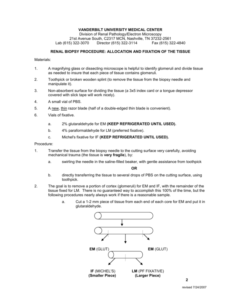
VANDERBILT UNIVERSITY MEDICAL CENTER Division of Renal Pathology/Electron Microscopy 21st Avenue South, C2317 MCN, Nashville, TN 37232-2561 Lab (615) 322-3070 Director (615) 322-3114 Fax (615) 322-4840 RENAL BIOPSY PROCEDURE: ALLOCATION AND FIXATION OF THE TISSUE Materials: 1. A magnifying glass or dissecting microscope is helpful to identify glomeruli and divide tissue as needed to insure that each piece of tissue contains glomeruli. 2. Toothpick or broken wooden splint (to remove the tissue from the biopsy needle and manipulate it). 3. Non-absorbent surface for dividing the tissue (a 3x5 index card or a tongue depressor covered with slick tape will work nicely). 4. A small vial of PBS. 5. A new, thin razor blade (half of a double-edged thin blade is convenient). 6. Vials of fixative. a. 2% glutaraldehyde for EM (KEEP REFRIGERATED UNTIL USED). b. 4% paraformaldehyde for LM (preferred fixative). c. Michel's fixative for IF (KEEP REFRIGERATED UNTIL USED). Procedure: 1. Transfer the tissue from the biopsy needle to the cutting surface very carefully, avoiding mechanical trauma (the tissue is very fragile), by: a. swirling the needle in the saline-filled beaker, with gentle assistance from toothpick OR b. 2. directly transferring the tissue to several drops of PBS on the cutting surface, using toothpick. The goal is to remove a portion of cortex (glomeruli) for EM and IF, with the remainder of the tissue fixed for LM. There is no guaranteed way to accomplish this 100% of the time, but the following procedures nearly always work if there is a reasonable sample. a. Cut a 1-2 mm piece of tissue from each end of each core for EM and put it in glutaraldehyde. EM (GLUT) IF (MICHEL'S) (Smaller Piece) EM (GLUT) LM (PF FIXATIVE) (Larger Piece) 2 revised 7/24/2007 If more than one core is obtained, handle each according to this protocol. If a very small amount of tissue is obtained, allocate in regard to the clinical setting. Ordinarily, it is preferable to allocate the tissue for EM and IF, since some LM information can also be obtained from the tissue studied in EM. IF is most important in the diagnosis of anti-GBM disease and IgA nephropathies. EM of a single glomerulus will usually permit a diagnosis in patients with immune complex disease. EM is necessary for diagnosis of Alport's/thin basement membranes. When you have the available tissue, proceed to divide the tissue for processing. The optimal biopsy will have at least 20 glomeruli. Take a piece off each end containing at least 1 glomerulus and put in 2% glutaraldehyde for a total of 4 glomeruli, (if the biopsy has at least 2 good cores), for EM. Next cut a piece for IF that has at least 4 to 6 glomeruli from one of the cores and put in Michel's. That should leave you at least 10 glomeruli for Light Microscopy to be placed in 4% paraformaldehyde. 3. 4. Put the allocated tissue into appropriate fixative: (guidelines for optimal division of tissue on page 1). a. 2% glutaraldehyde for EM. b. 4% paraformaldehyde (PF) for LM. c. Michel's solution for IF. Label vials. Tighten securely using appropriately color coded specimen tops. Pack to avoid breakage. Enclose data and clinical summary. revised 7/24/2007
