Oral Pathology - Jordan University of Science and Technology
advertisement
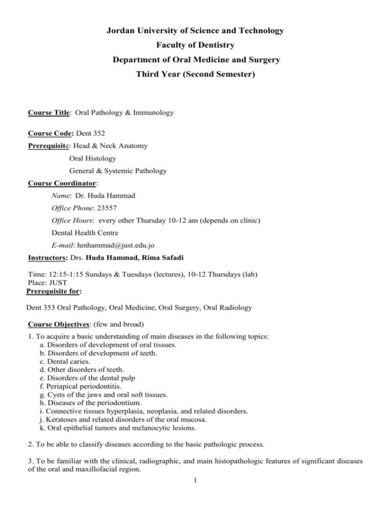
Jordan University of Science and Technology Faculty of Dentistry Department of Oral Medicine and Surgery Third Year (Second Semester) Course Title: Oral Pathology & Immunology Course Code: Dent 352 Prerequisite: Head & Neck Anatomy Oral Histology General & Systemic Pathology Course Coordinator: Name: Dr. Huda Hammad Office Phone: 23557 Office Hours: every other Thursday 10-12 am (depends on clinic) Dental Health Centre E-mail: hmhammad@just.edu.jo Instructors: Drs. Huda Hammad, Rima Safadi Time: 12:15-1:15 Sundays & Tuesdays (lectures), 10-12 Thursdays (lab) Place: JUST Prerequisite for: Dent 353 Oral Pathology, Oral Medicine, Oral Surgery, Oral Radiology Course Objectives: (few and broad) 1. To acquire a basic understanding of main diseases in the following topics: a. Disorders of development of oral tissues. b. Disorders of development of teeth. c. Dental caries. d. Other disorders of teeth. e. Disorders of the dental pulp f. Periapical periodontitis. g. Cysts of the jaws and oral soft tissues. h. Diseases of the periodontium. i. Connective tissues hyperplasia, neoplasia, and related disorders. j. Keratoses and related disorders of the oral mucosa. k. Oral epithelial tumors and melanocytic lesions. 2. To be able to classify diseases according to the basic pathologic process. 3. To be familiar with the clinical, radiographic, and main histopathologic features of significant diseases of the oral and maxillofacial region. 1 4. To be able to make a differential diagnosis of oral lesions based on the knowledge received in the above-mentioned topics. 5. To be aware of the relative significance and basic management of each disease process. 6. To acquire a basic understanding of immunology. Learning Outcomes: (many and specific) Successful completion of this theoretical course should lead to the following learning outcomes: Knowledge and Understanding (student should) - Be able to classify diseases according to the basic pathologic process. - Be familiar with the clinical, radiographic, and main histopathologic features of significant diseases of the oral and maxillofacial region. - Be able to make a differential diagnosis of oral lesions based on the knowledge received in the abovementioned topics. - Be aware of the relative significance and basic management of each disease process. - Have a basic understanding of immunology and the range of immunological reaction which could affect dental patients. Skills (intellectual, and manual) Successful completion of this clinical or lab course, the student should gain the following skills. 1. Be able to classify diseases according to the basic pathologic process. 2. Be able to make a differential diagnosis of oral lesions based on the knowledge of clinical and radiographic features of significant diseases of the oral and maxillofacial region in the above-mentioned topics. 3. Be aware of the relative significance and basic management of each disease process. 4. Have a basic understanding of immunology and the range of immunological reaction which could affect dental patients. Teaching methods: Duration: 14 weeks, (39 contact hours in total, including lab) Lectures: 29 hours, 2 hour per week ( including 1-hour midterm exam) clinical : Laboratory: 5 lab sessions, 2 hours each, given after lecture blocks according to subject. Modes of assessment: The examination will comprise two formal written examinations in the form of: 1. A midterm examination of multiple choice questions. The mark will be 40%, inclding 5 marks for contiuous assessment based on participation and attendance. 2 2. Final exam of MCQs including lab material . The mark will be 60%. Attendance policy: Students are expected to attend more than 90% of lectures or labs. Course Content & Weight: Theory No. of Lecture title Material covered lectures 2 Disorders of development of oral soft Cleft lip and cleft palate tissues and maxillofacial bones Commissural & paramedian lip pits Double lip and Ascher syndrome Fordyce granules Leukoedema Miroglossia Maroglossia Ankyloglossia Lingual thyroid nodule Fissured tongue Hairy tongue Varicosities Caliber-persistent artery Lateral soft palate fistulas Coronoid hyperplasia Condylar hyperplasia Codylar hypoplasia Bifid condyle Exostosis Torus palatinus Torus mandibularis Eagle syndrome Stafne defect Palatal cysts of the newborn Nasolabial cyst Epidermoid cyst Dermoid cyst Thyroglossal duct cyst Branchial cleft cyst Oral lymphoepithelial cyst Hemihyperplasia Progressive hemifacial atrophy Segmental odontomaxillary dysplasia Crouzon syndrome Apert syndrome Mandibulofacial dysostosis Disorders of development of teeth 2 Disturbances in number of teeth: hypodontia, anodontia, & related syndromes hyperdontia Disturbances in size of teeth: 3 macrodonia and microdontia. Disturbances in form of teeth: Germination Fusion ontism concrescence accessory cusps dens invaginatus ectopic enamel taurodontism hypercementosis accessory roots dilaceration Disturbances in structure of teeth: -disturbances in structure of enamel: localized causes (local infection or trauma, enamel opacities) generalized causes (chronological hypolasias, congenital syphilis, fluorosis, amelogenesis imperfecta) - disturbances in structure of dentin: dentinogenesis imperfecta dentinal dysplasia metabolic disturbances affecting dentinogenesis regional odontodysplasia Non-carious disorders of teeth Disorders of eruption and shedding Premature eruotion, natal, and neonatal teeth Retarded eruption Premature loss Persistence of deciduous teeth Impaction of teeth Reimpaction of teeth Non-bacterial loss of tooth substance Attrition Abrasion Erosion Abfraction Resorption Discoloration of teeth: Extrinsic staining Changes in structure or thickness of dental hard tissues Diffusion of pigments after formation of hard tissues Diffusion of pigments during formation of hard tissues Transplantation and reimplantation of teeth Root fracture Age changes 1 4 Disorders of the dental pulp 1 Pulpitis: Clinical features Etiology Histopathology Pulp polyp Effects of cavity preparation and restorative materials Healing of pulp Pulp calcification Pulp necrosis Age changes in the pulp Periapical periodontitis Etiology Acute periapical periodontitis Chronic periapical periodontitis (periapical granuloma) sequelae Acute periapical abscess and spread of inflammation Etiology and microbiology Routes of spread Cellulitis Ludwig’s angina 1 Cysts of the jaws and oral soft tissues Classification and incidence of cysts of the jaws Odontogenic cysts:definition and origins Radicualr cysts Dentigerous and eruption cysts Odontogenic keratocysts Gingival cyst Lateral periodontal cyst\paradental cyst Glandular odontogenic cyst Non-odontogenic cysts Nasopalatine duct cyst Nasolabial cyst Median cyst of the palate Non-epithelialized primary bone cysts Solitary bone cyst Aneurysmal bone cyst Stafne idiopathic bone cavity Cysts of the soft tissues Salivary mucoceles Dermoid and epidermoid cysts Lymphoepithelial cyst Thyroglossal cyst 3 3 Connective tissues hyperplasia, neoplasia, and related disorders Connective tissue hyperplasia Epulides Pyogenic granuloma Fibroepithelial polyp Denture irritation hyperplasia 5 Papillary hyperplasia of the palate Connective tissue neoplasms and allied conditions Tumors of fibrous tissue Tumors of adipose tissue Tumors of vascular tissue Tumors of peripheral nerves Tumors of muscle The granular cell tumor Lymphomas Classification Definition of histopathologic terms Hereditary conditions White sponge nevus Leukoedema Traumatic keratoses Mechanical Chemical Thermal Leukoplakia Incidence Clinical features etiological factors Pathology, epithelial dysplasia Prognosis Dermatological causes of white patches Lichen planus Lupus erythematosus Keratoses and related disorders of the oral mucosa 3 Oral epithelial tumors and melanocytic lesions Squamous cell papilloma and other benign lesions associated with HPV Squamous cell papilloma Verruca vulgaris Condyloma acuminatum Squamous cell carcinoma Epidemiology Etiological factors Oncogenes and tumor suppressor genes Clinical presentation Pathology Prognosis Verrucous carcinoma Carcinoma in situ Premalignant lesions and conditions Exfoliative cytology Basal cell carcinoma Melanocytic nevi and melanoma 3 Miscellaneous Disorders of the Oral Mucosa 1 Fordyce’s Granules Sublingual Varices Lingual Tonsil, Foliate Papillitis Geographic Tongue (Migratory Glossitis) Orofacial Granulomatosis Crohn’s Disease 6 Immunology Sarcoidosis Pyostomatitis Vegetans Progressive Systemic Sclerosis (Scleroderma) Verruciform Xanthoma Oral Submucous Fibrosis Amyloidosis Oral Pigmentation Superficial staining of the mucosa Black hairy tongue Foreign bodies Amalgam tattoo Melanin pigmentation-developmental causes Heavy metal salts Melanin pigmentation-acquired causes (Addison’s disease, pulmonary disease, HIV infection, chronic inflammation & lichen planus, drug-induced, idiopathic oral melanotic macule, lentigo simplex) sialadenitis Neoplastic causes (melanoma in situ and melanoma) Other endogenous pigments Age Changes in the Oral Mucos The immune system The innate immune system Complement Cells of the immune system The immune response Antibodies Cytokines Antigen processing and presentation T-helper subsets Activation of macrophages B-cell activation Target cell killing Regulation of the immune response Immunological memory Immunologic diseases: Classification of immunologic diseases Immune deficiency diseases Allergy (hypersensitivity) Autoimmunity Allergic versus nonallergic reactions Most frequent and serious problems Atopic allergies: hay fever & asthma Anaphylactic reactions, urticaria Cytotoxic reactions: transfusion reactions, erythroblastosis fetalis Delayed hypersensitivity reaction: contact dermatitis Immune deficiencies Symptoms, signs, and tests Electrophoresis Immunodiffusion Skin tests Coomb’s test 7 Tissue immunofluorescence Antinuclear antibody test Specific diseases Immune deficiency diseases:AIDS Anaphylactic-atopic allergies (type I): allergic rhinitis & asthma, urticaria, and angioedema, systemic anaphylaxis, gastrointestinal food allergies Cytotoxic hypersensitivities (type II) Erythroblastosis fetalis Blood transfusion reactions Autoimmune hemolytic anemia & thrombocytopenia Immune complex, or arthus type, hypersensitivities (type III) Arthus reaction Serum sickness Glomerulonephritis Polyarteritis nodosa Delayed, or cell-mediated, hypersensitivities (type IV) Contact dermatitis Infections manifest primarily as delayed hypersensitivity reactions Graft rejection Autoimmune diseases Systemic lupus erythematosus Laboratory: No. of Practical requirement Skills gained labs Developmental disorders - To be familiar with the clinical, radiographic, and main histopathologic features of some developmental disorders of the oral and maxillofacial region and teeth. - To be able to make a differential diagnosis of developmental oral lesions. - To be aware of the relative significance and basic management of each developmental disease process. Non-carious disorders of teeth, disorders of dental pulp, periapical periodontitis - To be familiar with the clinical, radiographic, and main histopathologic features of non-carious disorders of teeth, disorders of the dental pulp, and periapical periodontitis - To be able to make a differential diagnosis of these lesions. - To be aware of the relative significance and basic management of each disease process. Cysts of the jaws and oral soft tissues - To be familiar with the clinical, radiographic, and main histopathologic features of some cysts of the jaws and oral soft tissues - To be able to make a differential diagnosis of cysts of 1 1 1 8 the jaws and oral soft tissues - To be aware of the relative significance and basic management of each cyst. Connective tissue hyperplasia, neoplasia, and related disorders - To be familiar with the clinical and main histopathologic features of oral connective tissue hyperplasia, neoplasia, and related disorders - To be able to make a differential diagnosis of these lesions. - To be aware of the relative significance and basic management of each disease process. Epithelial pathology - To be familiar with the clinical, and main histopathologic features of some epithelial disorders of the oral and maxillofacial region. - To be able to make a differential diagnosis of epithelial oral lesions. - To be aware of the relative significance and basic management of each epithelial disease process. Miscellaneous disorders - To be familiar with the clinical, radiographic, and main histopathologic features of some miscellaneous disorders of the oral and maxillofacial region covered in the lecture. - To be able to make a differential diagnosis of thesedisorders. - To be aware of the relative significance and basic management of each disorder. 1 1 1 Feedback: Concerns or complaints should be expressed in the first instance to the course instructor. If no resolution is forthcoming then the issue should be brought to the attention of the Department Chair and if still unresolved to the Dean. Questions about the material covered in the lecture, notes on the content of the course, its teaching and assessment methods can be also sent by e-mail. References and Supporting Material: Textbook: 1. Oral Pathology by Soames & Southam, Oxford Uiversity Press, 3rd edition 1998, reprints 1999, 2001. 2. Oral Pathology by Soames & Southam, Oxford University Press, 4th edition 2005. Since this edition was released after commencement of the course, students are given the option of using either edition depending on availability in bookstores. 3. Oral and maxillofacial Pathology by Neville et al, Saunders, 2nd edition 2002, (Developmental Disorders) Useful websites: http://www.dental.mu.edu/oralpath/opgloss.html http://www.usc.edu/hsc/dental/opath/ http://www.forsyth.org/oralpathology/ http://www.library.vcu.edu/tml/oralpathology/ 9 http://www.mednet.gr/pim/oralpath.htm http://www.uiowa.edu/~oprm/AtlasWIN/AtlasFrame.html http://aaomp.org/ http://abomp.org/links.htm http://www.lib.uiowa.edu/hardin/md/dent.html http://www.usc.edu/hsc/dental/opfs/index.html http://www.mic.ki.se/MEDIMAGES.html#DentistryandOralHealth 10

