Supplementary Materials Methods: Cell culture and cell proliferation
advertisement
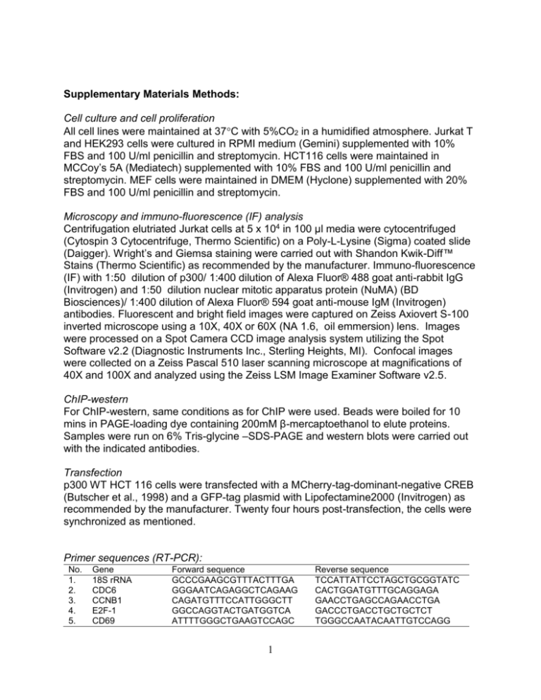
Supplementary Materials Methods: Cell culture and cell proliferation All cell lines were maintained at 37C with 5%CO2 in a humidified atmosphere. Jurkat T and HEK293 cells were cultured in RPMI medium (Gemini) supplemented with 10% FBS and 100 U/ml penicillin and streptomycin. HCT116 cells were maintained in MCCoy’s 5A (Mediatech) supplemented with 10% FBS and 100 U/ml penicillin and streptomycin. MEF cells were maintained in DMEM (Hyclone) supplemented with 20% FBS and 100 U/ml penicillin and streptomycin. Microscopy and immuno-fluorescence (IF) analysis Centrifugation elutriated Jurkat cells at 5 x 10⁴ in 100 µl media were cytocentrifuged (Cytospin 3 Cytocentrifuge, Thermo Scientific) on a Poly-L-Lysine (Sigma) coated slide (Daigger). Wright’s and Giemsa staining were carried out with Shandon Kwik-Diff™ Stains (Thermo Scientific) as recommended by the manufacturer. Immuno-fluorescence (IF) with 1:50 dilution of p300/ 1:400 dilution of Alexa Fluor® 488 goat anti-rabbit IgG (Invitrogen) and 1:50 dilution nuclear mitotic apparatus protein (NuMA) (BD Biosciences)/ 1:400 dilution of Alexa Fluor® 594 goat anti-mouse IgM (Invitrogen) antibodies. Fluorescent and bright field images were captured on Zeiss Axiovert S-100 inverted microscope using a 10X, 40X or 60X (NA 1.6, oil emmersion) lens. Images were processed on a Spot Camera CCD image analysis system utilizing the Spot Software v2.2 (Diagnostic Instruments Inc., Sterling Heights, MI). Confocal images were collected on a Zeiss Pascal 510 laser scanning microscope at magnifications of 40X and 100X and analyzed using the Zeiss LSM Image Examiner Software v2.5. ChIP-western For ChIP-western, same conditions as for ChIP were used. Beads were boiled for 10 mins in PAGE-loading dye containing 200mM β-mercaptoethanol to elute proteins. Samples were run on 6% Tris-glycine –SDS-PAGE and western blots were carried out with the indicated antibodies. Transfection p300 WT HCT 116 cells were transfected with a MCherry-tag-dominant-negative CREB (Butscher et al., 1998) and a GFP-tag plasmid with Lipofectamine2000 (Invitrogen) as recommended by the manufacturer. Twenty four hours post-transfection, the cells were synchronized as mentioned. Primer sequences (RT-PCR): No. 1. 2. 3. 4. 5. Gene 18S rRNA CDC6 CCNB1 E2F-1 CD69 Forward sequence GCCCGAAGCGTTTACTTTGA GGGAATCAGAGGCTCAGAAG CAGATGTTTCCATTGGGCTT GGCCAGGTACTGATGGTCA ATTTTGGGCTGAAGTCCAGC 1 Reverse sequence TCCATTATTCCTAGCTGCGGTATC CACTGGATGTTTGCAGGAGA GAACCTGAGCCAGAACCTGA GACCCTGACCTGCTGCTCT TGGGCCAATACAATTGTCCAGG 6. 7. 8. 9. 10. 11. 12. FOS (-28.9 kb) FOS (-19.3 kb) FOS (-81.9 kb) FOS (+84.6 kb) *FOS (-6.3 kb) * FOS (-0.22 kb) FOS eRNA GCCCATATCATCAGCCACTT TCCTACCGATTACAGCCCAG CACTTCCTCTTTCCCCAGTG CCTCCAGTGGTTTCATCACC TCAATGCTTTGAAGCACGTC CCCCCTAAGATCCCAAATGT CCCTGACAATTATGGCCAAC CCACCCCACTTGGTTGTATC ATATGGAAAGGCCTCCGAGT TGCTCCAAGGTAAGACAGGG GTGGGGACTCAATAAGGGGT CTTGTGTGGCTCTCTGGTCA GTCGCGGTTGGAGTAGTAGG CAGGTCCTGAGCCAACATCT Additional primer sequences are as reported in Byun et al., 2009. * Mouse locus Primer sequences (3C): No. 1. 2. 3. 4. 5. 6. 7. 8. Locus FOS (-26.6kb) FOS (-23.8 kb) FOS (-10.1 kb) FOS (-6.21 kb) FOS (-3.59 kb) FOS (+6.0 kb) HBB (-4.18 kb) HBB (+5.07 kb) Sequence ACCCGGATGAGATAATGTGC AAATGCCCTTCCCACCTACT TTTGCAGCGTCCATACAAAG CCCGAGGTGGGAGAGTAGAC CCACAGGGAGAGTGCAAAGT TCCTCTCCTGGTCCCTGTTA ATGTCCCATCCAGGTGATGT GTGGGTGCAGGACAGTAGGT Antibodies: Protein pol II & p300 Brd4 Brg1 Cohesin (SMC1) p-Cohesin (p-SMC1,S966) CREB p-CREB (Ser133) ELK-1 p- ELK-1 (Ser383) p-ERK (phospho-p44/42 MAPK (Erk1/2) (Thr202/Tyr204) c-FOS H3 Lys 9,14 acetylation (acetylated histone H3) H3 Lys 18 acetylation (H3K18ac) H3 Lys 27 acetylation (H3K27ac)) H4 Lys 5, 8, 12, 16 acetylation (acetylated histone H4) Histone H2A.Z Histone H3 2 Source Byun et al., 2009 Jang et al., 2005 & Bethyl Lab. Wurster & Pazin, 2008 Bethyl Lab. Bethyl Lab. Santa Cruz Cell Signaling Millipore Cell Signaling Santa Cruz Santa Cruz Cell Signaling Catalog no. Santa Cruz Millipore sc-52 06-599 Abcam Abcam Millipore ab1191 ab4729 06-598 Abcam Millipore ab4174 06-755 A301-985A A300-055A A300-050A sc-186 9197 06-519 9191 sc-355 sc-8406 4370 H3 Lys 4 mono-methylation (H3K4Me1) H3 Lys 4 tri-methylation (H3K4Me3) H3 Lys 27 tri-methylation (H3K27Me3) LaminB MED1 (TRAP220) MED17 NuMA TBP (TFIID) TORC2 Abcam Millipore Millipore Santa Cruz Santa Cruz Santa Cruz BD Biosciences Santa Cruz Calbiochem 3 ab 8895 07-473 07-449 sc-6217 sc-8998 sc-12453 610561 sc-273 ST1099 References: Byun,J.S., Wong,M.M., Cui,W., Idelman,G., Li,Q., De,S.A., Bilke,S., Haggerty,C.M., Player,A., Wang,Y.H., Thirman,M.J., Kaberlein,J.J., Petrovas,C., Koup,R.A., Longo,D., Ozato,K., and Gardner,K. (2009). Dynamic bookmarking of primary response genes by p300 and RNA polymerase II complexes. Proc. Natl. Acad. Sci. U. S. A. 106, 19286-19291. Jang,M.K., Mochizuki,K., Zhou,M., Jeong,H.S., Brady,J.N., and Ozato,K. (2005) The bromodomain protein Brd4 is a positive regulatory component of P-TEFb and stimulates RNA polymerase II-dependent transcription. Mol Cell 19, 523–534. Wurster,A.L. and Pazin,M.J. (2008). BRG1-mediated chromatin remodeling regulates differentiation and gene expression of T helper cells. Mol. Cell Biol. 28, 7274-7285. 4

