Kidney, 2003-2004 - The Pathology Guy
advertisement

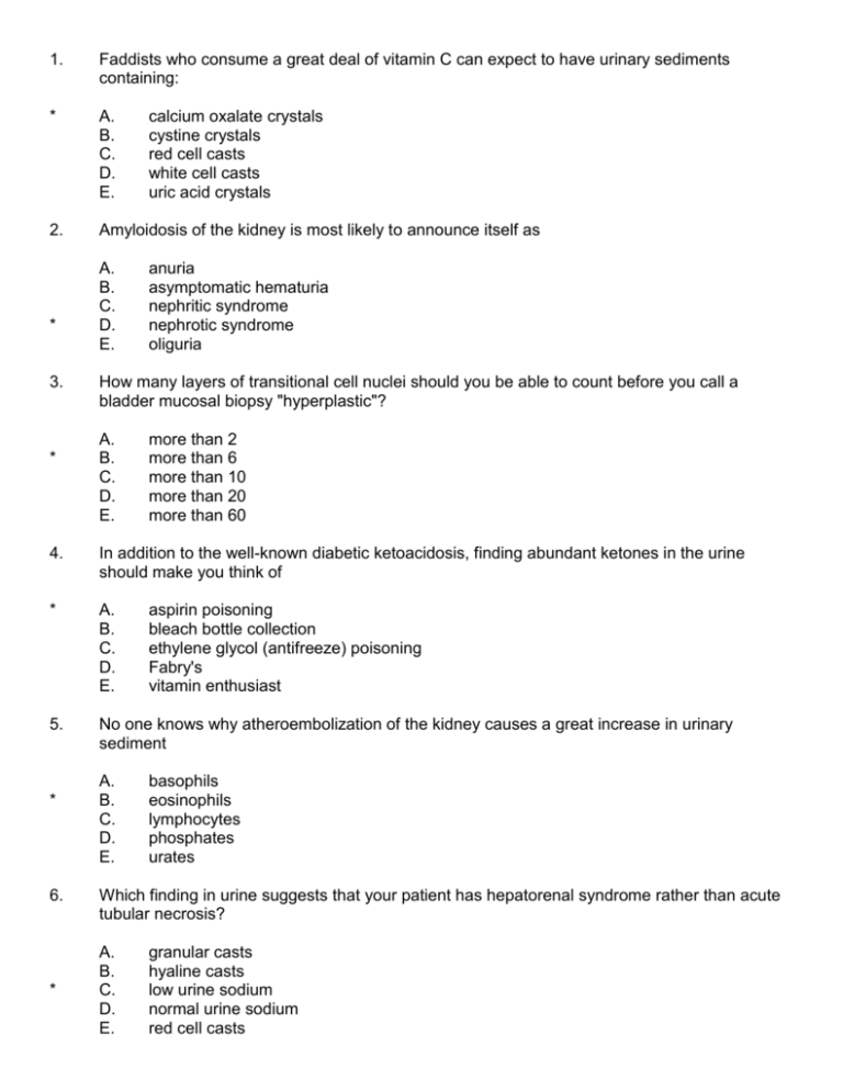
1. Faddists who consume a great deal of vitamin C can expect to have urinary sediments containing: * A. B. C. D. E. 2. Amyloidosis of the kidney is most likely to announce itself as * 3. * A. B. C. D. E. calcium oxalate crystals cystine crystals red cell casts white cell casts uric acid crystals anuria asymptomatic hematuria nephritic syndrome nephrotic syndrome oliguria How many layers of transitional cell nuclei should you be able to count before you call a bladder mucosal biopsy "hyperplastic"? A. B. C. D. E. more than 2 more than 6 more than 10 more than 20 more than 60 4. In addition to the well-known diabetic ketoacidosis, finding abundant ketones in the urine should make you think of * A. B. C. D. E. 5. No one knows why atheroembolization of the kidney causes a great increase in urinary sediment * 6. * A. B. C. D. E. aspirin poisoning bleach bottle collection ethylene glycol (antifreeze) poisoning Fabry's vitamin enthusiast basophils eosinophils lymphocytes phosphates urates Which finding in urine suggests that your patient has hepatorenal syndrome rather than acute tubular necrosis? A. B. C. D. E. granular casts hyaline casts low urine sodium normal urine sodium red cell casts 7. Common to all the hemolytic-uremic syndromes: * A. B. C. D. E. 8. A patient with anti-tubular basement membrane antibodies demonstrated on fluorescence very likely has these as a result of damage to endothelium pigment casts causing renal failure recessive inheritance resorption droplets in proximal tubules red cell membrane lesions * A. B. C. D. E. 9. Acute tubular necrosis plus fusion of foot processes suggests renal disease caused by * A. B. C. D. E. 10. cryoglobulinemia Godpasture's lupus old bacterial infection Sjogren's chronic mild ischemia cyclophosphamide hantavirus hepatorenal syndrome non-steroidal anti-inflammatory agents It is almost impossible to get necrosis of the entire cortical regions of both kidneys in the absence of * A. B. C. D. E. 11. Hyalinization of both the afferent and efferent arteries of the glomeruli is most suggestive of * 12. * A. B. C. D. E. atheroembolization bacterial infection disseminated intravascular coagulation other evidence of viral infection type II or III immune injury common hypertension diabetes hemolytic-uremic syndrome malignant hypertension vasculitis Rapidly-progressive glomerulonephritis without significant antibody demonstrable by immunofluorescence in the glomeruli is most suggestive of A. B. C. D. E. anti-GBM disease bacterial endocarditis BK polyomavirus Puumalavirus epidemic nephropathy vasculitis 13. * Patients with thin GBM disease usually have A. B. C. D. E. both nephritic and nephrotic syndromes lifelong hematuria without other problems nephritic syndrome only nephrotic syndrome only renal cysts with normal urine 14. Which is LEAST LIKELY to give striking onionskin intimal hyperplasia of the small renal arteries / arterioles? * A. B. C. D. E. 15. Mercury nephropathy features striking coagulation necrosis of the * A. B. C. D. E. diabetes hemolytic-uremic syndrome malignant hypertension radiation damage scleroderma distal tubules mesangial cells ("mesangiolysis") podocytes proximal tubules urothelium 16. Medullary sponge kidney is most noteworthy as a cause of * A. B. C. D. E. 17. Which is NOT a probable risk factor of renal cell carcinoma? * A. B. C. D. E. kidney stones nephritic syndrome nocturia unexplained hematuria unexplained proteinuria end-stage kidney disease exposure to hair dyes tobacco smoking von Hippel-Lindau disease none of these are risk factors 18. The "natural weight loss" remedy "Aristolochia" causes * A. B. C. D. E. bladder cancer calcium stones renal cell carcinoma uric acid stones Wilms' tumor 19. TWO PHOTOS. Patient found to have red cell casts in the urine. Which is the most likely cause? * A. B. C. D. E. 20. TWO PHOTOS. Autopsy kidney. What caused these changes? * A. B. C. D. E. acute post-streptococcal glomerulonephritis bacterial endocarditis IgA nephropathy membranoproliferative glomerulonephritis type I Wegener's granulomatosis atheroembolization chronic pyelonephritis common high blood pressure hemolytic-uremic syndrome malignant hypertension 21. ONE PHOTO. Urinary sediment showing * A. B. C. D. E. 22. ONE PHOTO. Two kidneys from the same patient, with a normal kidney in-between (from a different person of similar age, of course). What's the diagnosis? * A. B. C. D. E. calcium oxalate crystals candida red cell casts squamous epithelial cells white cell casts acquired dialysis cystic disease autosomal dominant polycystic kidney disease autosomal recessive polycystic kidney disease metastatic carcinoma to the kidney tuberous sclerosis with angiomyolipomas 23. TWO PHOTOS. Glomerulus. What is the diagnosis? * A. B. C. D. E. 24. ONE PHOTO. Kidney from a young child. What is the diagnosis? * A. B. C. D. E. diffuse proliferative glomerulonephritis membranous glomerulonephritis minimal change disease thin GBM disease no pathology acquired dialysis cystic disease autosomal dominant polycystic kidney disease autosomal recessive polycystic kidney disease cystic dysplasia medullary sponge kidney 25. * 26. * 27. ONE PHOTO. Urinary sediment. What do you see? A. B. C. D. E. candida fatty casts hyaline casts magnesium ammonium phosphate crystals squamous epithelium THREE PHOTOS. H&E, C3 immunofluorescence, and electron micrograph. What is your diagnosis? A. B. C. D. E. anti-GBM disease consistent with Goodpasture's diffuse proliferative glomerulonephritis focal-segmental glomerulosclerosis membranoproliferative glomerulonephritis type I membranoproliferative glomerulonephritis type II THREE PHOTOS. Silver stain, IgG immunofluorescence, and electron micrograph. What is your diagnosis? * A. B. C. D. E. diffuse proliferative glomerulonephritis focal-segmental glomerulosclerosis membranoproliferative glomerulonephritis type I membranoproliferative glomerulonephritis type II membranous glomerulopathy 28. TWO PHOTOS. H&E and electron micrograph. What is your best diagnosis? * A. B. C. D. E. 29. TWO PHOTOS. H&E and IgG immunofluorescence. What is your diagnosis? * A. B. C. D. E. 30. TWO PHOTOS. Silver stain and electron micrograph. What is your diagnosis? * A. B. C. D. E. cryoglobulinemia focal-segmental glomerulosclerosis minimal change disease membranous glomerulopathy post-streptococcal glomerulonephritis anti-GBM disease diffuse proliferative glomerulonephritis membranous glomerulopathy mesangioproliferative disease suggestive of IgA nephropathy Wegener's or polyarteritis anti-GBM disease IgA nephropathy lupus minimal change disease post-streptococcal glomerulonephritis 31. TWO PHOTOS. What is your diagnosis? * A. B. C. D. E. 32. TWO PHOTOS. What caused these kidney changes? * A. B. C. D. E. 33. THREE PHOTOS. Silver stain and two electron micrographs. What is the diagnosis? * A. B. C. D. E. angiomyolipoma Hurthle cell tumor renal cell carcinoma renal cortical abscess renal infarct amyloidosis atheroembolization common hypertension hemolytic-uremic syndrome old pyelonephritis diabetes membranoproliferative glomerulonephritis type I membranoproliferative glomerulonephritis type II membranous glomerulopathy mesangial-proliferative glomerulonephritis consistent with IgA nephropathy 34. TWO PHOTOS. Kidney, prior to stripping of the capsule, and a closeup of one of the masses. What is the diagnosis? * A. B. C. D. E. 35. TWO PHOTOS. Renal cortex. What is the diagnosis? * A. B. C. D. E. 36. TWO PHOTOS. PAS stain. Which is the most likely diagnosis? * A. B. C. D. E. angiomyolipoma bacterial infection Hurthle cell tumor lipoid nephrosis renal cell carcinoma acute tubular necrosis suggestive of shock kidney ethylene glycol ingestion mannitol effect myeloma kidney pigment nephropathy atheroembolization bacterial endocarditis diabetes focal-segmental glomerulosclerosis, primary old pyelonephritis BONUS ITEMS: 37. TWO PHOTOS. Kidney. What is the diagnosis? Be specific. [pyelonephritis / bacterial infection] 38. TWO PHOTOS. Kidney. Look closely, and suggest a diagnosis. [any cause of acute nephritic syndrome / RPGN] 39. ONE PHOTO. Look closely. What is your best diagnosis? Make the call just on the morphology. [papillary necrosis] 40. ONE PHOTO. Renal cortex. What is the diagnosis? [chronic pyelonephritis / old scarring from infection / thyroidization] 41. TWO PHOTOS. Suggest a reasonable diagnosis. [any cause of necrotizing glomerular disease, i.e., malignant hypertension, Wegener's, any other vasculitis] 42. What's the name of the still-mysterious illness in which we see unexplained inflammation of all three layers of the bladder wall, with increased tissue basophils? Your best answer, please. [credit either for Hunner or interstitial cystitis] 43. What's a "glitter cell"? [neutrophil is sufficient] 44. Explain briefly why people with Paget's osteitis deformans, severe anemia, or a dialysis arteriovenous shunt have elevated systolic and often lowered diastolic blood pressures. [just show you know increased cardiac output] 45. Why are the angiotensin-converting enzyme inhibitors more effective than other antihypertensive medicines for slowing progression to renal failure? Suggest a reasonable mechanism. [angiotensin II is responsible for ongoing sclerosis] 46. What do we mean by "renal glycosuria"? Give the mechanism, showing that you understand. [fail to reabsorb glucose] 47. What do we mean by a "staghorn" kidney stone? Explain the term. [extends into several calyces, like an antler] 48. What's the mechanism by which we know cranberry juice prevents and helps cure bladder infections? [bacteria cannot grow fimbria / bind to mucosa / get washed out] NAME: _______________________________ UHS Pathology Renal 2003-2004 Instructions: You know the routine. If you have a question during the exam, please raise your hand. Do not phonate. If you are in the first of two groups, leave the building with the rest of the exam group. No stragglers. Have a safe holiday. Good luck!


