Chapter 15
advertisement
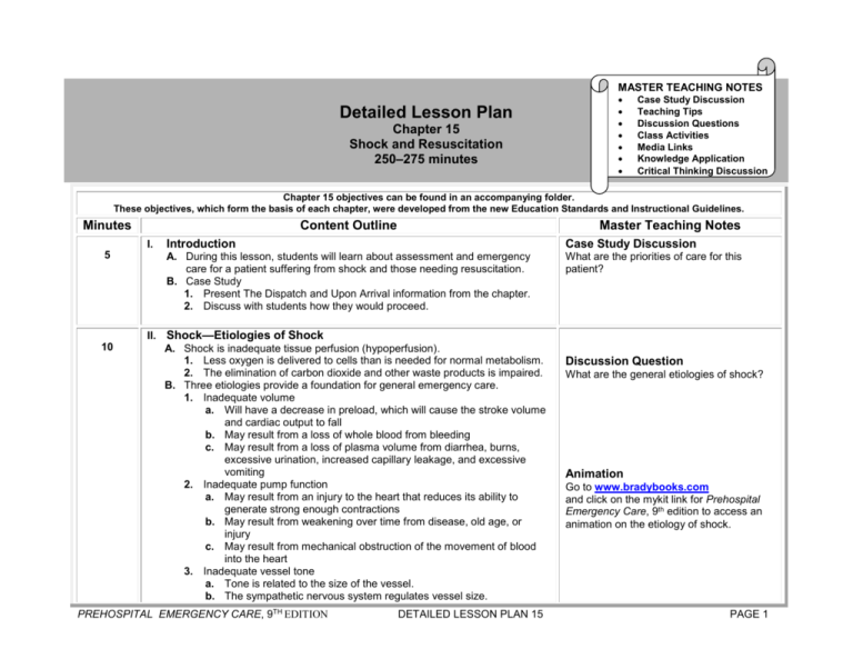
MASTER TEACHING NOTES Detailed Lesson Plan Chapter 15 Shock and Resuscitation 250–275 minutes Case Study Discussion Teaching Tips Discussion Questions Class Activities Media Links Knowledge Application Critical Thinking Discussion Chapter 15 objectives can be found in an accompanying folder. These objectives, which form the basis of each chapter, were developed from the new Education Standards and Instructional Guidelines. Minutes Content Outline I. 5 10 Master Teaching Notes Introduction Case Study Discussion A. During this lesson, students will learn about assessment and emergency care for a patient suffering from shock and those needing resuscitation. B. Case Study 1. Present The Dispatch and Upon Arrival information from the chapter. 2. Discuss with students how they would proceed. What are the priorities of care for this patient? II. Shock—Etiologies of Shock A. Shock is inadequate tissue perfusion (hypoperfusion). 1. Less oxygen is delivered to cells than is needed for normal metabolism. 2. The elimination of carbon dioxide and other waste products is impaired. B. Three etiologies provide a foundation for general emergency care. 1. Inadequate volume a. Will have a decrease in preload, which will cause the stroke volume and cardiac output to fall b. May result from a loss of whole blood from bleeding c. May result from a loss of plasma volume from diarrhea, burns, excessive urination, increased capillary leakage, and excessive vomiting 2. Inadequate pump function a. May result from an injury to the heart that reduces its ability to generate strong enough contractions b. May result from weakening over time from disease, old age, or injury c. May result from mechanical obstruction of the movement of blood into the heart 3. Inadequate vessel tone a. Tone is related to the size of the vessel. b. The sympathetic nervous system regulates vessel size. PREHOSPITAL EMERGENCY CARE, 9TH EDITION DETAILED LESSON PLAN 15 Discussion Question What are the general etiologies of shock? Animation Go to www.bradybooks.com and click on the mykit link for Prehospital Emergency Care, 9th edition to access an animation on the etiology of shock. PAGE 1 Chapter 15 objectives can be found in an accompanying folder. These objectives, which form the basis of each chapter, were developed from the new Education Standards and Instructional Guidelines. Minutes Content Outline Master Teaching Notes c. Inadequate vessel tone may result from an injury to the spinal cord or released chemical mediators that cause a systemic dilation of vessels. 10 10 III. Shock—Categories of Shock A. Hypovolemic shock 1. Caused by low blood volume 2. Most common type of shock 3. Generally caused by hemorrhage 4. Also be caused by burns and dehydration B. Distributive shock 1. Associated with a decrease in intravascular volume 2. Massive systemic vasodilation 3. Increase in capillary permeability 4. Reduction in systemic and peripheral vascular resistance 5. Reduction in systolic blood pressure C. Cardiogenic shock 1. Caused by ineffective pump function of the heart 2. Patient is prone to cardiogenic shock when more than 40 percent of the left ventricle is lost. D. Obstructive shock 1. Results from a condition that obstructs forward blood flow 2. Possible causes a. Blood clot b. Tension pneumothorax c. Pericardial tamponade E. Metabolic or respiratory shock 1. Described as a fifth type of shock in some sources 2. Dysfunction in the ability of oxygen to diffuse into the blood, be carried by hemoglobin, off-load at the cell, or be used by the cell for metabolism IV. Shock—Specific Types of Shock A. Hemorrhagic hypovolemic shock 1. Results from the loss of whole blood from the intravascular space. 2. Relates to whole blood loss that can occur from traumatic injury or medical illness. 3. Reduction in pressure and a decrease in oxygen-carrying capability. 4. Poor perfusion state from an inadequate intravascular volume. PREHOSPITAL EMERGENCY CARE, 9TH EDITION DETAILED LESSON PLAN 15 Discussion Question What are the categories of shock from each etiology? Discussion Question How can hypovolemia occur without hemorrhage? PAGE 2 Chapter 15 objectives can be found in an accompanying folder. These objectives, which form the basis of each chapter, were developed from the new Education Standards and Instructional Guidelines. Minutes Content Outline B. C. D. E. F. Master Teaching Notes 5. Bleeding must be stopped. 6. Administration of whole blood or blood components Nonhemorrhagic hypovolemic shock 1. Results from the loss of fluid from the intravascular space 2. Red blood cells and hemoglobin remain within the vessels. 3. Water, plasma proteins, and electrolytes are lost. 4. Blood volume, pressure and perfusion of cells are reduced. 5. Administration of intravenous fluids may be beneficial. Burn shock 1. Nonhemorrhagic hypovolemic shock resulting from a burn injury 2. Burns may interrupt the integrity of the capillaries and vessels. 3. “Pull” effect draws fluid into the interstitial space, causing edema. 4. Establish and maintain an adequate airway, ventilation, and oxygenation. 5. Prevent further contamination of the burn injury. Anaphylactic shock 1. This is a type of distributive shock. 2. Chemical mediators in the anaphylactic reaction cause massive and systemic vasodilation. 3. Capillaries become permeable and leak. 4. Fluid is forced out into the interstitial space. 5. Systemic vascular resistance is reduced. 6. Blood pressure and perfusion are decreased. 7. Epinephrine is the medication of choice. Septic shock 1. This is a type of distributive shock. 2. Results from an infection that releases bacteria or toxins in the blood. 3. Vessels dilate and become permeable. 4. Fluid leaks into the interstitial space. 5. Systemic vascular resistance, blood pressure, and perfusion are reduced. 6. Intravascular volume, preload, stroke volume, cardiac output, systolic blood pressure, and perfusion are decreased. 7. Manage the airway, ventilation, and oxygenation. 8. Administer intravenous fluids and medication to constrict the vessels. Neurogenic shock 1. This is a type of distributive shock, also known as vasogenic shock. 2. May be caused by spinal cord injury PREHOSPITAL EMERGENCY CARE, 9TH EDITION DETAILED LESSON PLAN 15 Animation Go to www.bradybooks.com and click on the mykit link for Prehospital Emergency Care, 9th edition to access an animation on types of shock. Weblink Go to www.bradybooks.com and click on the mykit link for Prehospital Emergency Care, 9th edition to access a web resource on toxic shock syndrome. PAGE 3 Chapter 15 objectives can be found in an accompanying folder. These objectives, which form the basis of each chapter, were developed from the new Education Standards and Instructional Guidelines. Minutes Content Outline Master Teaching Notes a. May damage the sympathetic nerve fibers that control vessel tone b. Vessels dilate. c. Systemic vascular resistance, blood pressure, and perfusion may drop. d. Blood will pool in the peripheral vessels. e. Preload, stroke volume, cardiac output, and systolic blood pressure will decrease. 3. Emergency care focuses on spinel immobilization and management of the airway, ventilation, and oxygenation. 4. Patient may also benefit from intravenous fluids and medication to constrict the vessels. G. Cardiogenic shock 1. Most common causes a. Myocardial infarction b. Congestive heart failure c. Abnormal cardiac rhythm d. Overdose on drugs that depress the pumping function of the heart 2. Emergency care focuses on management of the airway, ventilation, and oxygenation. 10 V. Shock—The Body’s Response to Shock A. The body attempts to compensate for a disturbance and returns perfusion and tissue function to a normal state. B. Compensatory mechanisms 1. Direct nerve stimulation a. Increase in heart rate b. Increase in force of ventricular contraction c. Vasoconstriction d. Stimulation of the release of epinephrine and norepinephrine 2. Release of hormones a. Epinephrine stimulates alpha and beta receptors. b. Norepinephrine stimulates alpha receptors. c. Other hormones decrease urine output, cause further vasoconstriction, cause an increase in heart rate and contractility, and cause an increase in glucose in the blood. PREHOSPITAL EMERGENCY CARE, 9TH EDITION DETAILED LESSON PLAN 15 Discussion Question What are the general signs and symptoms of shock? Video Clip Go to www.bradybooks.com and click on the mykit link for Prehospital Emergency Care, 9th edition to access a video clip on bleeding control in shock management. PAGE 4 Chapter 15 objectives can be found in an accompanying folder. These objectives, which form the basis of each chapter, were developed from the new Education Standards and Instructional Guidelines. Minutes 10 10 Content Outline Master Teaching Notes VI. Shock—Stages of Shock A. Compensatory shock 1. A near normal blood pressure and perfusion of the vital organs is maintained. 2. If the etiology of shock is reversed at this stage, the compensatory mechanisms will continue to maintain the blood pressure and perfusion. 3. A narrow pulse pressure should be noted as an early sign of shock. B. Decompensatory shock 1. An advanced stage of shock in which the compensatory mechanisms are no longer able to maintain a blood pressure and perfusion to vital organs. 2. If the shock state continues, the compensatory mechanisms will become exhausted. 3. Cells, tissues, and organs become ischemic. 4. Heart function is depressed. 5. Blood in the capillaries begins to sludge and form microemboli. 6. Blood leaks out of the vessels into the interstitial space. 7. When the vasomotor in the medulla becomes hypoxic, sympathetic nervous system stimulation is reduced. 8. Aggressive shock management may or may not reverse the process. C. Irreversible shock 1. A stage where the patient outcome is death 2. Cell, tissue, and organ failure is so severe that it cannot be reversed. 3. Microemboli block capillaries throughout the body. 4. Fibinolysis leads to widespread uncontrolled bleeding. VII. Shock—Shock Assessment A. History 1. Pay attention to chief complaint. 2. Identify signs or symptoms that might provide clues to the etiology of the shock. 3. Gather information about allergies, medications, past medical history, last oral intake, and events prior to the incident. B. Physical exam 1. Assess for physical signs of shock. 2. Obtain vital signs. a. Blood pressure b. Heart rate PREHOSPITAL EMERGENCY CARE, 9TH EDITION DETAILED LESSON PLAN 15 Weblink Go to www.bradybooks.com and click on the mykit link for Prehospital Emergency Care, 9th edition to access a web resource on stages of shock. Knowledge Application Given several descriptions of patient problems, students should be able to match the description to the patient’s type of shock. PAGE 5 Chapter 15 objectives can be found in an accompanying folder. These objectives, which form the basis of each chapter, were developed from the new Education Standards and Instructional Guidelines. Minutes Content Outline Master Teaching Notes c. Pulse character d. Respiratory rate and tidal volume e. Skin color, temperature, and condition f. Pulse oximeter reading 3. Note signs of poor perfusion. a. Altered mental status b. Pale, cool, clammy skin c. Delayed capillary refill d. Decreased urine output e. Weak or absent peripheral pulses 5 10 VIII. Shock—Age Considerations in Shock A. Elderly persons and infants deteriorate rapidly. B. Children and young adults exhibit minor signs over a long period of time and then decompensate suddenly. C. Medications in the elderly patient may prevent some signs and symptoms from appearing. D. An altered mental status and tachypnea may be most profound signs of shock in the elderly. Teaching Tip IX. Shock—General Goals of Prehospital Management of Shock Discussion Question A. Management of shock is geared to improving oxygenation of the blood and delivery of oxygen and glucose to the cells. B. General goals 1. Secure and maintain a patent airway. 2. Establish and maintain adequate ventilation. 3. Establish and maintain adequate oxygenation. 4. Do not hyperventilate. 5. Stop the bleeding using direct pressure. 6. Splint fractures. 7. Do not remove impaled objects. 8. Maintain the body temperature. 9. Keep the patient in a supine position. 10. Apply the pneumatic antishock garment (PASG). a. A pelvic fracture is suspected. b. Systolic blood pressure is less than 90 mmHg. c. Profound hypertension is present. d. Intra-abdominal hemorrhage is suspected with sever hypotension. e. Retroperitoneal hemorrhage is suspected with hypotension. PREHOSPITAL EMERGENCY CARE, 9TH EDITION DETAILED LESSON PLAN 15 Use questioning to assess students’ retention of material from pathophysiology. What are the general management goals for patients in shock? Critical Thinking Discussion Why is maintaining the body temperature so critical in patients in shock? PAGE 6 Chapter 15 objectives can be found in an accompanying folder. These objectives, which form the basis of each chapter, were developed from the new Education Standards and Instructional Guidelines. Minutes Content Outline Master Teaching Notes 11. Rapidly transport patient. 12. Consider ALS intercept. 15 X. Resuscitation in Cardiac Arrest— Pathophysiology of Cardiac Arrest A. Resuscitation is bringing a patient back from a potential or apparent death. B. Cardiac arrest occurs when the ventricles of the heart are not contracting or when the cardiac output is completely ineffective. C. Sudden death occurs when the patient dies within one hour of the onset of the signs and symptoms. D. Three phases the patient goes through following cardiac arrest that lead to biological death 1. Electrical phase a. Begins immediately upon cardiac arrest and ends four minutes afterward. b. Heart is in good condition for resuscitation. c. Restore an effective electrical rhythm. 2. Circulatory phase a. Begins at four minutes and last through ten minutes following a cardiac arrest. b. Myocardial cells shift from aerobic to anaerobic metabolism. c. Myocardial cells become ischemic. d. Heart is not prepared for defibrillation and is not prone to restarting. e. CPR will provide oxygen and glucose to the heart, improving chances for defibrillation. 3. Metabolic phase a. Begins ten minutes after a cardiac arrest. b. Heart is starved of oxygen and glucose. c. Acid has built up in the heart. d. Tissues are very ischemic and may begin to die. e. The sodium/potassium pump fails. f. The sodium that stays in the cells attracts water. g. Cells swell, rupture, and die. h. Resuscitation during this phase is typically unsuccessful. XI. 10 Resuscitation in Cardiac Arrest—Terms Related to Resuscitation A. Downtime—The time the patient goes into cardiac arrest until CPR is effectively being performed B. Total downtime—The total time from when the patient goes into cardiac PREHOSPITAL EMERGENCY CARE, 9TH EDITION DETAILED LESSON PLAN 15 Discussion Questions Why is defibrillation in the first four minutes of cardiac arrest more likely to be successful than delayed defibrillation? How can cardiac arrest patients benefit from CPR prior to defibrillation in the circulatory phase of cardiac arrest? Critical Thinking Discussion If you were a researcher trying to improve survival from cardiac arrest, what kind of study would you design? Weblink Go to www.bradybooks.com and click on the mykit link for Prehospital Emergency Care, 9th edition to access the PAGE 7 Chapter 15 objectives can be found in an accompanying folder. These objectives, which form the basis of each chapter, were developed from the new Education Standards and Instructional Guidelines. Minutes Content Outline Master Teaching Notes arrest until the patient is delivered to the emergency department C. Return of spontaneous circulation (ROSC)—The patient regains a spontaneous pulse during resuscitation. D. Survival—A patient who survives to be discharged from the hospital XII. Resuscitation in Cardiac Arrest—Withholding a Resuscitation Attempt 10 A. B. C. D. E. XIII. 10 DNR—Patient’s do not resuscitate order POLST—Physician’s orders for life-sustaining treatment MOLST—Medical orders for life-sustaining treatment A patient with injuries that are not compatible with life Obvious death in patients who are beyond the point of resuscitation Resuscitation in Cardiac Arrest—The Chain of Survival A. Early access 1. Early recognition of cardiac event 2. Easy access to the EMS system B. Early CPR 1. Immediate CPR can double or even triple a patient’s chance of survival from ventricular-fibrillation-induced sudden cardiac arrest (VF SCA). 2. It is important to begin CPR within two minutes of the cardiac arrest. C. Early defibrillation 1. Survival of VF SCA patients decreases approximately seven to ten percent for every minute that defibrillation is delayed. 2. Defibrillation is the procedure of sending an electrical current through the chest. D. Early advanced life support 1. Advanced life support (ALS) is delivered most often by paramedics who can provide advanced cardiac life support (ACLS). 2. Advanced EMTs may be able to provide either all or certain components of ALS interventions. XIV. 15 American Heart Association’s site on therapeutic hypothermia after cardiac arrest. Automated External Defibrillation and Cardiopulmonary Resuscitation—Types of Defibrillators A. External defibrillators are applied to the outside of the chest. 1. Manual 2. Automated (AED) B. Advantages of AEDs PREHOSPITAL EMERGENCY CARE, 9TH EDITION DETAILED LESSON PLAN 15 Discussion Question What are the links in the Chain of Survival? Teaching Tip Motivate students by emphasizing the importance of the basic skills they can perform in resuscitation from cardiac arrest: high quality CPR and early defibrillation. Class Activity Have small groups of students brainstorm ways to improve each of the links in the Chain of Survival in the local community. Teaching Tip Have at least one AED visible to students while teaching this section. Discussion Questions PAGE 8 Chapter 15 objectives can be found in an accompanying folder. These objectives, which form the basis of each chapter, were developed from the new Education Standards and Instructional Guidelines. Minutes Content Outline Master Teaching Notes 1. Initial training and education 2. Speed of operation 3. Safer, more effective delivery 4. More efficient monitoring C. Types of AEDs 1. Fully automated AEDs 2. Semiautomated AEDs XV. 15 What is the rationale for early defibrillation? What is the rationale for the “push hard and push fast” approach to chest compressions in CPR? What is meant by monophasic and biphasic defibrillation? What is the difference between a fully automatic AED and a semi-automatic AED? Automated External Defibrillation and Cardiopulmonary Resuscitation—Analysis of Cardiac Rhythms A. Ventricular fibrillation (VF or V-Fib) 1. Disorganized cardiac rhythm 2. No pulse or cardiac output 3. Commonly associated with advanced coronary disease B. Ventricular tachycardia (V-Tach) 1. Described as a very fast heart rhythm 2. Generated in the ventricle instead of the sinoatrial node in the atrium 3. Cardiac output is sharply reduced. 4. Be aware that some V-Tach patients are not appropriate candidates for defibrillation. 5. AED should only be used on patients who are pulseless, not breathing, and unresponsive. C. AED will detect rhythms for which no shock is indicated. 1. Asystole—Electrical activity and pumping action in the heart is absent. 2. Pulseless electrical activity (PEA)—The heart has an organized rhythm but does not pump. D. The AED is very sensitive. 1. No one should be touching the patient. 2. The ambulance should be stopped with the motor turned off. XVI. 15 Knowledge Application List each of the cardiac rhythms described in this section and have students indicate if it can be successfully treated with an AED or not. Critical Thinking Discussion How does defibrillation work? Automated External Defibrillation and Cardiopulmonary Resuscitation—When and When Not to Use the AED A. Infants—Do not apply the AED to infants. B. Patients between one and eight years of age—Use an AED preferably with a dose attenuating system to reduce defibrillation energy. C. Patients over eight years of age PREHOSPITAL EMERGENCY CARE, 9TH EDITION DETAILED LESSON PLAN 15 Teaching Tip Discuss specific protocols for your area. PAGE 9 Chapter 15 objectives can be found in an accompanying folder. These objectives, which form the basis of each chapter, were developed from the new Education Standards and Instructional Guidelines. Minutes Content Outline Master Teaching Notes 1. Within five minutes, immediately apply the AED. 2. If it has been more than five minutes, immediately perform five cycles of CPR at a ratio of 30 compressions to two ventilations and then apply the AED. D. The AED is not intended for trauma patients. E. Consult medication direction and local protocols if you are unsure about whether to use the AED. XVII. 15 Recognizing and Treating Cardiac Arrest—Assessment-Based Approach: Cardiac Arrest A. Scene size-up and primary assessment 1. Take appropriate Standard Precautions. 2. Ensure the scene is secure. 3. Form a general impression of the patient. 4. If a suspected cardiac patient is unresponsive, follow procedures for assessment and care for cardiac-related emergencies. B. With unresponsive patients 1. Open the airway. 2. Assess breathing and pulse. 3. If there is no breathing and pulse, the patient is in cardiac arrest. 4. One member of the team should deliver CPR. 5. Deliver emergency care appropriate for age. C. Secondary assessment 1. Gather the history from bystanders and relatives. 2. Identify signs and symptoms of cardiac arrest a. No breathing b. No pulse c. Unresponsiveness to stimuli D. Emergency medical care 1. Follow the proper steps to provide emergency care with an AED to cardiac arrest patients. 2. Use CPR and defibrillation as appropriate according to age and downtime. E. Reassessment 1. Once pulse is restored, continue to perform reassessments. 2. Monitor patient’s pulse, breathing, and mental status. 15 Discussion Question In what situations is CPR begun prior to applying the AED? Knowledge Application Describe several patient presentations and ask students to determine whether or not the AED should be applied. XVIII. Recognizing and Treating Cardiac Arrest—Performing Defibrillation PREHOSPITAL EMERGENCY CARE, 9TH EDITION DETAILED LESSON PLAN 15 PAGE 10 Chapter 15 objectives can be found in an accompanying folder. These objectives, which form the basis of each chapter, were developed from the new Education Standards and Instructional Guidelines. Minutes Content Outline Master Teaching Notes A. Using a semiautomated AED 1. Take Standard Precautions. 2. Perform a primary assessment of the patient. 3. Begin or resume CPR. 4. Attach the adhesive monitoring-defibrillation pads to the cables. 5. Turn on the power to the AED. 6. Apply the two defibrillation pads to the patient’s bared chest. 7. Stop ongoing CPR and clear anyone from touching the patient. 8. Begin analysis of the patient’s heart rhythms. 9. Deliver shock if indicated on the AED and then resume CPR. 10. Check for a pulse for no longer than ten seconds. 11. If a pulse is present, check for breathing to apply ventilation as needed. 12. If no pulse is present, deliver a second shock, and then resume CPR. 13. If ALS is not responding to the scene, transport after three shocks are delivered. B. Use of the AED by a single EMT 1. Only one EMT may be available for a cardiac arrest patient. 2. Adjust the procedure to the situation until help arrives. C. Using a fully automated AED 1. The fully automated AED will deliver the shock to the patient. 2. The device gives directions to the EMT throughout the defibrillation process. XIX. 10 Discussion Question Why is the pulse not checked immediately after the shock is delivered? Class Activity Ensure that students have ample opportunity for supervised practice of resuscitation scenarios. Recognizing and Treating Cardiac Arrest—Transporting the Cardiac Arrest Patient A. Transporting a patient with a pulse 1. Check airway and provide oxygen. 2. Provide pressure ventilation if breathing is inadequate. 3. Have suction ready for use. 4. Secure the patient to a stretcher or transfer to an ambulance. 5. Consult with dispatch to find out how to connect with an ALS unit. 6. Continue to keep the AED attached to the patient. 7. Perform the secondary assessment en route every five minutes. B. Patients brought out of a ventricular fibrillation through use of AED have a high likelihood of slipping back into that state. 1. Be alert if patient becomes unresponsive. 2. Check for breathing and pulse. 3. If the patient shows no breathing or pulse PREHOSPITAL EMERGENCY CARE, 9TH EDITION DETAILED LESSON PLAN 15 Critical Thinking Discussion Why is a patient who remains in cardiac arrest not transported until three shocks or three no-shock messages have been delivered? PAGE 11 Chapter 15 objectives can be found in an accompanying folder. These objectives, which form the basis of each chapter, were developed from the new Education Standards and Instructional Guidelines. Minutes Content Outline Master Teaching Notes a. Stop the vehicle. b. Start CPR. c. Stop CPR and initiate rhythm analysis. d. Deliver a shock if warranted. e. Resume CPR for two minutes. f. Deliver a second shock if warranted. g. Resume CPR for two minutes. h. Reanalyze until two or three “no shock” messages. i. Continue as per local protocol. j. Continue transport. C. Transporting a patient without a pulse 1. Provide CPR. 2. Contact medical direction. 3. Follow local protocol. XX. 10 Recognizing and Treating Cardiac Arrest—Providing for Advanced Cardiac Life Support A. Keep the AHA’s Chain of Survival in mind. B. Inform medical direction and request ACLS backup as soon as you can. C. Minimize the time from the delivery of CPR and defibrillatory shocks to the arrival of ACLS. XXI. 10 Recognizing and Treating Cardiac Arrest—Summary: Assessment and Care A. Review assessment findings associated with cardiac arrest. B. Review assessment findings associated emergency care for cardiac arrest. XXII. 5 Teaching Tip Demonstrate several cardiac arrest scenarios for students, explaining the steps as you go through the scenarios. Special Considerations for the AED—Safety Considerations A. The shock from an AED can travel through different substances. B. No one should be in contact with the patient during rhythm analysis or delivery of defibrillating shocks. C. The AED should not be operated if the machine or patient is in contact with water. D. Ensure that no one else is directly in contact with metal that is touching the patient before delivering a shock. E. Use gloves to remove any transversal medication patches and dry the area before delivering a shock. F. Do not put an AED adhesive pad on top of a surgically implanted pacemaker. PREHOSPITAL EMERGENCY CARE, 9TH EDITION DETAILED LESSON PLAN 15 Discussion Question What are the safety precautions that must be observed when using an AED? PAGE 12 Chapter 15 objectives can be found in an accompanying folder. These objectives, which form the basis of each chapter, were developed from the new Education Standards and Instructional Guidelines. Minutes Content Outline Master Teaching Notes G. Make sure that electrodes are adhering properly to a very hairy chest. XXIII. Special Considerations for the AED—AED Maintenance 4 3 A. B. C. D. Scheduled maintenance is crucial to ensuring proper functioning. Follow local protocols and manufacturer’s directions. Batteries should be replaced on a set schedule. Extra, fully charged batteries should always be available. XXIV. Special Considerations for the AED—Training and Skills Maintenance A. EMTs should be properly educated in operating the AED. B. Operators should review incidents of AED use, study new protocols, and practice working with the device. C. EMTs should check updated research on AED procedures at sources such as EMS journals, state EMS offices, and the AHA. 3 XXV. Special Considerations for the AED—Medical Direction and the AED A. Medical direction plays a significant role in providing AED services. B. Medical direction involvement 1. Make sure the EMS system has necessary links in the AHA Chain of Survival. 2. Oversee all levels of EMTs. 3. Review the continual competency skill review program. 4. Engage in audit and/or quality improvement. 3 Teaching Tip Show any mechanical CPR devices or circulation-enhancing devices used in your local EMS system. Discussion Question What is the role of a medical director with regard to AED use? XXVI. Special Considerations for the AED—Energy Levels of Defibrillators A. Electrical current for defibrillators is measured in joules. B. It is important that EMTs know how to deliver the appropriate amount of energy for their type of AED. XXVII. Special Considerations for the AED—Cardiac Pacemakers 3 A. A cardiac pacemaker is placed under the skin with electrodes connecting to the heart. B. Cardiac pacemakers are usually positioned beneath one of the clavicles. C. An AED can still be used in a patient with a cardiac pacemaker. D. The adhesive pad should not be placed directly over the pacemaker. XXVIII.Special Considerations for the AED—Automatic Implantable PREHOSPITAL EMERGENCY CARE, 9TH EDITION DETAILED LESSON PLAN 15 PAGE 13 Chapter 15 objectives can be found in an accompanying folder. These objectives, which form the basis of each chapter, were developed from the new Education Standards and Instructional Guidelines. Minutes 4 Content Outline Master Teaching Notes Cardioverter Defibrillators A. An automatic implantable cardioverter defibrillators (AICD) is used for ventricular heart rhythm disturbances that cannot be controlled by medication. B. For a responsive cardiac patient with an AICD, allow the device to operate, stabilize the patient, and prepare for transport. C. For an unresponsive cardiac patient, look for surgical scars or medical identification tags. 1. Treat as other unresponsive cardiac patients. 2. Do not apply the AED’s adhesive pads directly over the implanted AICD. 5 XXIX. Special Considerations for the AED—Automated Chest Compression Devices A. Mechanical piston device 1. Apparatus that depresses the sternum with a compressed-gas-powered plunger that has been affixed to a backboard 2. Can be configured to deliver a specific rate and depth of compressions 3. Delivers uniform compressions with no diminishment 4. Frees up an EMS provider 5. Can become useless if compressed gas runs out B. Load-distributing-band CPR or vest CPR 1. Composed of a wide band applied to the chest circumferentially 2. Is either pneumatically or electrically driven to provide an inward constrictive pressure on the thorax 3. Frees up an EMS provider 4. Has been shown to improve coronary and cerebral blood flow over traditional CPR C. Impedance threshold device 1. Piece of equipment with a valve that limits the air that enters the chest and lungs during the chest recoil phase of active compressions 2. Has been shown to improve blood flow through the heart during CPR 3. May be considered for use in a nonintubated patient 4. A tight mask seal must constantly be maintained. D. Other circulation-enhancing devices 1. Other devices have been developed to actively compress and decompress the thorax during resuscitation. 2. The compression phase generates the positive pressure to move blood out of the heart. PREHOSPITAL EMERGENCY CARE, 9TH EDITION DETAILED LESSON PLAN 15 Discussion Question What are the advantages and disadvantages of adjunctive equipment used for CPR? PAGE 14 Chapter 15 objectives can be found in an accompanying folder. These objectives, which form the basis of each chapter, were developed from the new Education Standards and Instructional Guidelines. Minutes Content Outline Master Teaching Notes 3. The decompression is designed to increase the negative pressure inside the thorax to improve blood return to the heart. XI. Follow-Up 10 Case Study Follow-Up Discussion B. Answer student questions. C. Case Study Follow-Up 1. Review the case study from the beginning of the chapter. 2. Remind students of some of the answers that were given to the discussion questions. 3. Ask students if they would respond the same way after discussing the chapter material. Follow up with questions to determine why students would or would not change their answers. D. Follow-Up Assignments 1. Review Chapter 15 Summary. 2. Complete Chapter 15 In Review questions. 3. Complete Chapter 15 Critical Thinking. E. Assessments 1. Handouts 2. Chapter 15 quiz PREHOSPITAL EMERGENCY CARE, 9TH EDITION DETAILED LESSON PLAN 15 What is your general impression of the patient’s condition? What priority will you assign this patient? Class Activity Alternatively, assign each question to a group of students and give them several minutes to generate answers to present to the rest of the class for discussion. Teaching Tips Answers to In Review and Critical Thinking questions are in the appendix to the Instructor’s Wraparound Edition. Advise students to review the questions again as they study the chapter. The Instructor’s Resource Package contains handouts that assess student learning and reinforce important information in each chapter. This can be found under mykit at www.bradybooks.com. PAGE 15
