Supplementary Table S3 (doc 89K)
advertisement
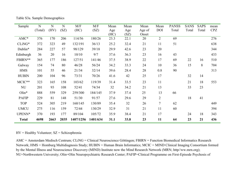
Table S3a. Sample Demographics Sample N (Total) N (HV) N (SZ) M/F (HC) M/F (SZ) Mean Age (SZ) 22.1 32.4 42.6 36.3 Mean Age of Onset 20 21 23 23 Mean DOI PANSS Total 180/26 36/13 39/18 9/7 Mean Age (HC) 23.3 25.2 29.9 37.6 AMC* CLiNG* Dublin* Edinburgh 376 372 284 36 170 323 227 20 206 49 57 16 114/56 132/191 98/129 10/10 2 11 20 16 69 51 FBIRN** Galway HMS HUBIN 365 154 101 200 177 74 55 104 186 80 46 96 127/51 46/28 21/34 73/31 141/46 56/24 32/14 70/26 37.5 34.2 39.6 41.6 38.9 33.3 28.4 42 22 24 28 25 17 10 0.8 17 69 36 90 MCIC** NU Olin* 323 201 888 165 93 559 158 108 329 103/62 52/41 259/300 119/39 74/34 184/145 31.4 32 37.9 33.5 34.2 37.4 23 21 25 11 13 13 PAFIP TOP 229 524 81 305 148 219 51/30 160/145 91/57 130/89 27.6 35.4 29.6 32 29 26 2 7 UMCU UPENN* 275 370 116 193 159 177 72/44 89/104 130/29 105/72 32.9 35.9 31 38.4 21 21 Total 4698 2663 2035 31.1 33.8 23 1407/1256 1401/634 SANS SAPS Total Total mean CPZ 276 638 344 433 43 22 15 16 8 32 14 21 33 18 23 18 41 510 704 313 553 66 62 449 11 17 60 11 64 24 18 394 343 23 21 436 HV = Healthy Volunteer; SZ = Schizophrenia AMC = Amsterdam Medisch Centrum; CLiNG = Clinical Neuroscience Göttingen; FBIRN = Function Biomedical Informatics Research Network; HMS = Homburg Multidiagnosis Study; HUBIN = Human Brain Informatics; MCIC = MIND Clinical Imaging Consortium formed by the Mental Illness and Neuroscience Discovery (MIND) Institute now the Mind Research Network (MRN; http//ww.mrn.org); NU=Northwestern University; Olin=Olin Neuropsychiatric Research Center; PAFIP=Clinical Programme on First-Episode Psychosis of Cantabria Psychiatry Research Unit; TOP= Tematisk Område Psykoser (Thematically Organized Psychosis Research); UMCU = Universitair Medisch Centrum Utrecht; UPENN=University of Pennsylvania. * 3 Tesla scanners; ** 1.5 and 3 Tesla scanners PAFIP sample SANS/SAPS Total converted from SANS(6)/SAPS(14) GLOBAL scores; Galway sample SANS/SAPS Total converted from PANNS POS(9.51)/NEG(11.12) Total scores using converteasy.org. CPZ = chlorpromazine dose equivalent based on Woods (2005; www.scottwilliamwoods.com/files/Equivtext.doc) Table S3b. Additional Medication Information and Imaging Parameters by Sample Sample % % % Atypical Typical Both Atypical & Typical % Scanner Imaging Protocols Vendor NotMedicated Slice Orientation FreeSurfer Version Operating System Number of subjects removed from analysis due to QC failure? v5.0.0 Linux centos4 x86_64 1 & Type AMC 0.86 0.11 0 0.03 Philips Intera 3T TR: 8-9.8, M=9,4 (0,41). TE:3,5-4,6, M=4,26 (0,46). Slice thickness: 1/1.2, flipangle: 8degr, rows/columns: 192-288, M=255,49 (7,00). Pixelspacing: 1mm CLiNG 0.76 0.06 0 0.18 3T Magnetom TIM Trio MRI scanning was performed on a 3.0-Tesla Magnetom TIM Trio (Siemens, Erlangen, Germany). A T1-weighted, 3D magnetization prepared rapid gradient echo sequence (MPRAGE) (TR/TE/TI/FA=2250 ms/3.26 ms/900 ms/9°; image matrix = 256 x 256; duration 8 min and 26 sec) was acquired generating 192 sagittal slices with a voxel size of 1 mm3.” sagittal v 5.1.0 Ubuntu 12.04 0 Dublin 0.82 0 0 0.03 3T Philips Intera Achieva 180 slice T1-weighted image using a TFE gradient echo pulse sequence (TR=8.4ms, TE=3.8ms, flip angle=8°, slice thickness=0.9mm, voxel size=0.9mm3, 180slices, duration=6min) axial v5.2.0 Linux 0 Edinburgh 0.25 0.69 0.06 0 1.5T Signa A coronal gradient echo sequence with magnetization preparation and produced 128 coronal high-resolution T1weighted images, which were used for structural image analysis (time of inversion [TI] 600 msec, echo time 3.4 msec, flip angle 15, field of view 22, slice thickness 1.7 mm, matrix 256 192). coronal v5.0.1 Linux 0 FBIRN (Phase3) 0.87 0.11 0.02 0 3T Siemens Tim Trio; 3T GE High-resolution structural imaging scans were acquired on six 3T Siemens Tim® Trio System and one 3T General Electric Discovery MR750 scanner. MP-RAGE scan parameters for the Siemens scanner were: scan plane=sagittal, TR/TE/TI=2300/2.94/1100ms, GRAPPA acceleration factor=2, flip angle=9°, resolution=256×256x160, FOV=220mm2, voxel size=0.86x0.86x1.2mm, and NEX=1. IR-SPGR scan parameters for the General Electric scanner were: scan plane=sagittal, TR/TE/TI=5.95/1.99/450ms, ASSET acceleration factor=2, a flip angle=12°, resolution=256×256x166, FOV=220mm2, voxel size=0.86x0.86x1.2mm, and NEX=1. All scans covered the entire brain. sagittal v5.1.0 Centos 64bit 2.6.18308.4.1.el5 0 Galway 0.95 0.02 0 0.04 1.5 Tesla Siemens Magnetom Symphony (Erlangen. Germany) A volumetric T1-weighted magnetization-prepared rapid acquisition of gradient echo (MPRAGE) sequence was acquired with the imaging parameters: Repetition time (TR): 1140ms, Echo time (TE): 4.38ms, flip angle 15; matrix size 256 x 256; an inplane pixel resolution of 0.9mm x 0.9mm and slice thickness 0.9mm axial v5.1.0 Linux 0 HMS 0.85 0 0.02 0.13 1.5 T Magnetom Sonata MRI scanning was performed on a 1.5-Tesla Magnetom Sonata (Siemens, Erlangen, Germany). A T1-weighted, magnetization prepared rapid gradient echo sequence (MPRAGE) (TR/TE/TI/FA=1900 ms/4.0 ms/700 ms/15°; image matrix = 256 x 256) was acquired generating 176 consecutive sagittal slices with a voxel size of 1 mm3. ~5 min sagittal v 5.1.0 centos6 x86_64 0 HUBIN 0.4 0.47 0.05 0.07 1.5 Tesla General Electronics Signa T1-weighted images, using a three-dimensional spoiled gradient recalled (SPGR) pulse sequence, were acquired with the following parameters; 1.5 mm coronal slices, no gap, 35° flip angle, repetition time (TR) = 24 ms, echo time (TE) = 6.0 ms, number of excitations (NEX) = 2, field of view (FOV) = 24 cm, acquisition matrix = 256 × 192. T2weighted images were acquired with the following coronal v5.3.0 RedHat 0 parameters; 2.0 mm coronal slices, no gap, TR = 6,000 ms, TE = 84 ms, NEX = 2, FOV = 24 cm, acquisition matrix = 256 × 192. MCIC 0.78 0.09 0.03 0.1 1.5, 3T Siemens and GE T1 scans: coronal v4.0.1 Linux of various flavors 5 subjects failed automated segmentation procedure due to excessive motion artifacts 2 participants’ MRI data failed the manual inspection axial v5.3.0 centos6 x86_64 1 subject had an oversaturated scan which caused thalamus to oversegment among other issues and we excluded this subject TR = 2530 ms for 3 T, TR = 12 ms for 1.5 T; TE = 3.79 ms for 3 T, TE = 4.76 ms for 1.5 T; FA = 7 for 3 T, FA = 20 for 1.5 T; TI = 1100 for 3 T; Bandwidth = 181 for 3 T, Bandwidth = 110 for 1.5 T; 0.625×0.625 mm voxel size; slice thickness 1.5 mm; FOV 256×256×128 cm matrix; FOV = 16 cm (could be increased to 18 cm when needed for full brain coverage). NU 0.75 0.16 0 0.09 1.5T Vision 1) 3D turbo-FLASH: TR=20 ms, TE=5.4 ms, flip=30˚, ACQ=1, 256x256 matrix, 1xl mm in-plane resolution, 180 slices, slice thickness 1 mm, 13:30 min scan time and 2) 3D MPRAGE (2-4 repeats): TR=9.7 ms, TE=4 ms, flip=10˚, ACQ=1, 256x256 matrix, 1xl mm in-plane resolution, 128 slices, slice thickness 1.25 mm, 5:36 min scan time each OLIN PAFIP TOP 0.83 0.71 0.17 0 0 0.05 0.15 0.1 completely; we excluded only Laccumb for 1 other subject and only Raccumb for 1 other subject 3T Alegra T1-weighted, 3D magnetization-prepared rapid gradient-echo (MPRAGE) sequence (TR/TE/TI=2200/4.13/766 ms, flip angle=13°, voxel size [isotropic]=0.8mm, image size=240 x 320 x 208 voxels), with axial slices parallel to the AC-PC line. axial v5.1.0 GE 1.5T Three-dimensional T1weighted images, using a spoiled grass (SPGR) sequence acquired in the coronal plane with: echo time (TE)=5 ms, repetition time (TR)=24 ms, numbers of excitations (NEX)=2, rotation angle=45°, field of view (FOV)=26×19.5 cm, slice thickness=1.5mm and a matrix of 256×192. coronal v5.0.0 Ubuntu 11,04 (x86_64) 1 subject was excluded because motion leading to very poor segmentation 1.5T Siemens Magnetom Sonata Two sagittal T1-weighted magnetization prepared rapid gradient echo (MPRAGE) volumes were acquired with the Siemens tfl3d1_ns pulse sequence (TE = 3.93 ms, TR = 2730 ms, TI = 1000 ms, flip angle = 7°; FOV = 24 cm, sagittal v4.5.0 Linux Centos or Ubuntu 0 voxel size= 1.33 x 0.94 x 1 mm3, number of partitions = 160) UMCU 0.58 0.41 0.01 0.01 UPENN 0.82 0.15 0.03 0 Philips 1.5T Intera and Achieva T1-weighted threedimensional fast-field echo (3D-FFE) scans with 160–180 contiguous coronal slices [256 3 256 matrix, echo time (TE)=4.6 ms, repetition time (TR)=30 ms, flip angle=30 degrees, 1x1x1.2 mm3 voxels, field of view [FOV] = 256 mm/ 70%] coronal v5.1.0 Siemens 3T MPRAGE, TR=1810 ms, TE= 3.51 ms, TI=1100 ms, flip angle 9, FOV= 240 x 180 mm, matrix= 256 × 192, resolution = 0.9 x 0.9 mm, slices = 160, slice/skip thickness = 1 mm/0 mm axial v5.3.0 0 Linux Redhat Enterprise 5 0
