The search for purely long-range molecules proved fruitful with
advertisement
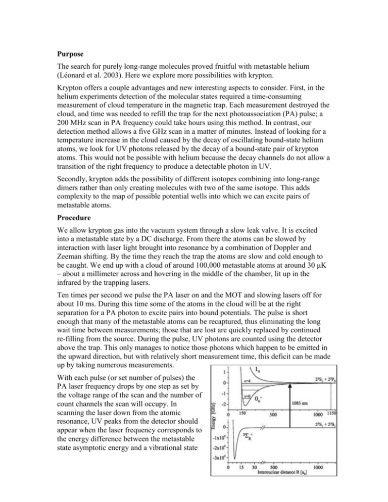
Purpose The search for purely long-range molecules proved fruitful with metastable helium (Léonard et al. 2003). Here we explore more possibilities with krypton. Krypton offers a couple advantages and new interesting aspects to consider. First, in the helium experiments detection of the molecular states required a time-consuming measurement of cloud temperature in the magnetic trap. Each measurement destroyed the cloud, and time was needed to refill the trap for the next photoassociation (PA) pulse; a 200 MHz scan in PA frequency could take hours using this method. In contrast, our detection method allows a five GHz scan in a matter of minutes. Instead of looking for a temperature increase in the cloud caused by the decay of oscillating bound-state helium atoms, we look for UV photons released by the decay of a bound-state pair of krypton atoms. This would not be possible with helium because the decay channels do not allow a transition of the right frequency to produce a detectable photon in UV. Secondly, krypton adds the possibility of different isotopes combining into long-range dimers rather than only creating molecules with two of the same isotope. This adds complexity to the map of possible potential wells into which we can excite pairs of metastable atoms. Procedure We allow krypton gas into the vacuum system through a slow leak valve. It is excited into a metastable state by a DC discharge. From there the atoms can be slowed by interaction with laser light brought into resonance by a combination of Doppler and Zeeman shifting. By the time they reach the trap the atoms are slow and cold enough to be caught. We end up with a cloud of around 100,000 metastable atoms at around 30 μK – about a millimeter across and hovering in the middle of the chamber, lit up in the infrared by the trapping lasers. Ten times per second we pulse the PA laser on and the MOT and slowing lasers off for about 10 ms. During this time some of the atoms in the cloud will be at the right separation for a PA photon to excite pairs into bound potentials. The pulse is short enough that many of the metastable atoms can be recaptured, thus eliminating the long wait time between measurements; those that are lost are quickly replaced by continued re-filling from the source. During the pulse, UV photons are counted using the detector above the trap. This only manages to notice those photons which happen to be emitted in the upward direction, but with relatively short measurement time, this deficit can be made up by taking numerous measurements. With each pulse (or set number of pulses) the PA laser frequency drops by one step as set by the voltage range of the scan and the number of count channels the scan will occupy. In scanning the laser down from the atomic resonance, UV peaks from the detector should appear when the laser frequency corresponds to the energy difference between the metastable state asymptotic energy and a vibrational state energy level in one of the accessible long-range bound state potentials. The figure on the right gives the schematic representation of a transition in helium like those we expect to observe in krypton. Generally the transition takes place with the internuclear distance near the outer classical turning point on the potential curve because of the relatively low cloud density. Once in this bound state the atoms oscillate, somewhat as though they are attached by a long, floppy spring. The inner turning point is what makes these molecules quite interesting: the inner classical turning point occurs at what is still a very large internuclear distance (no smaller than 40 Bohr radii according to Doery et al. 1998) for normally interacting atoms. This is why they are called “purely long-range” molecules. Because these potentials represent bound particle interactions, there are only discrete quantum energy levels allowed for the oscillation. Each of these levels should appear as a separate UV peak; however, with overlapping potentials involved (Doery et al. 1998), as well as the increasingly small spacing between levels near the top of the well, some peaks will likely appear in complex, multi-peaked features. The UV photon is emitted when an atom in the oscillating dimer molecule decays. Thus detection of a UV photon indicates the death of an exotic molecule, but it also assures that one did exist. Unfortunately, a single scan of the PA laser cannot go far enough below resonance to reach as many areas of interest as we would like to examine. The main reason for this is the laser is only stable (mode-hop free) over a range of up to about five GHz. It is also important to be sure the frequency step size is not larger than the typical linewidth of a molecular vibrational state. Therefore we do a series of overlapping scans and put them together to extend the overall scan. To accomplish this, we need a frequency scale corresponding to the channels in the data. This is found using a Fabry-Perot, which sends signals in channels coincident with the channels of the UV counts. Since its peaks are equidistant in frequency they can be used to set a relative frequency scale, assigning a nominal frequency to each peak separated by one free spectral range. FabryPerot peaks from consecutive scans are matched using overlapping features in the UV signal from those scans. 34000 Frequency (MHz) 32000 30000 28000 y = -0.098x 3 + 6.649x 2 + 565.68x + 26944 26000 24000 22000 0 2 4 6 Channel 8 10 12 Another difficulty arises in that the frequency scan is non-linear: early peaks from the Fabry-Perot are generally broader and further separated than later peaks in a single scan. Using the frequencies assigned to the peaks and the channels from which they come, a polynomial function can be found to relate frequency (y-axis) to channel number (x-axis) for each scan. The same set of frequencies then corresponds to the channels in the UV signal, thus yielding a counts-vs.-frequency table for the UV. The smaller the free-spectral range for the Fabry-Perot, the more points are available within a scan to fit the frequency-vs.-channel function, which makes the assignment of the frequency scale more accurate. However, it may also become more difficult with more closely-spaced Fabry-Perot peaks to decide which should align from scan to scan. Using this scaling method we were able to assign a rough frequency scale to a long series of scans. The resulting plot reveals some overlapping peaks as well as some that only appear once. Other peaks may show up in multiple scans, but the uncertainty in frequency scale offsets them a little on the plot. It seemed obvious that we needed an absolute reference point, so we decided to look also at the saturated absorption in the discharged Krypton. This gives us a curve, which we can run a fit to, and the minimum, which is the absorption of the largest abundance Krypton ion, Krypton 84, is then set to be zero. This gives us an absolute frequency scale, and makes it so that we do not need to try and align our data by hand. Both the initial frequency scale and the absolute zero are found in one Mathematica program, which makes it a fast and accurate process. Once we have an absolute frequency scale on our data, we then combine similar runs into one large excel file, and run them through a binning program in Mathematica, which in essence adds them together, so that if there is a peak in many of the runs, it shows up quite large. We are starting to see interesting peaks but we need to see them consistently to be convinced of anything. Below is a short movie that starts with the isotopes and hyperfine shifts of Krypton, and then broadens them with respect to the temperature they have. It broadens with temperature because the atoms are moving faster and therefore have a greater Doppler shift.
