Figure 5. Glo-Germ particles collected onto a glass slide after 10 min
advertisement
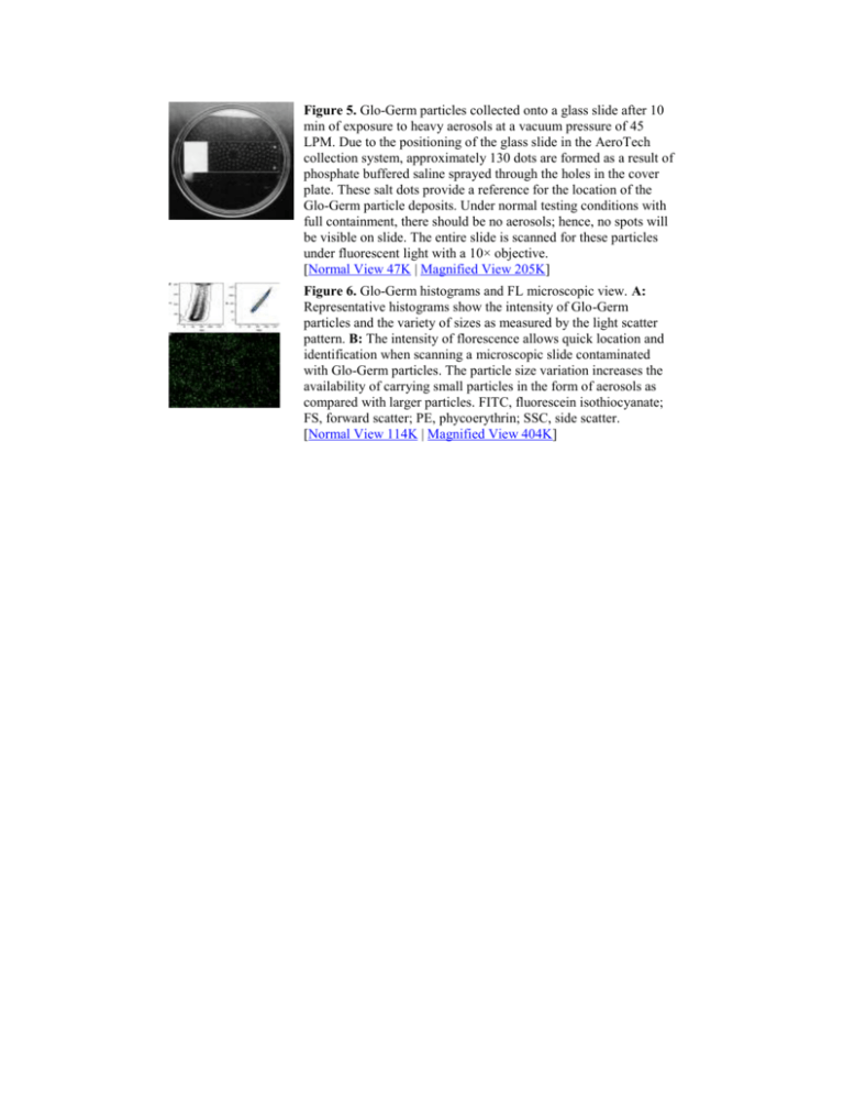
Figure 5. Glo-Germ particles collected onto a glass slide after 10 min of exposure to heavy aerosols at a vacuum pressure of 45 LPM. Due to the positioning of the glass slide in the AeroTech collection system, approximately 130 dots are formed as a result of phosphate buffered saline sprayed through the holes in the cover plate. These salt dots provide a reference for the location of the Glo-Germ particle deposits. Under normal testing conditions with full containment, there should be no aerosols; hence, no spots will be visible on slide. The entire slide is scanned for these particles under fluorescent light with a 10× objective. [Normal View 47K | Magnified View 205K] Figure 6. Glo-Germ histograms and FL microscopic view. A: Representative histograms show the intensity of Glo-Germ particles and the variety of sizes as measured by the light scatter pattern. B: The intensity of florescence allows quick location and identification when scanning a microscopic slide contaminated with Glo-Germ particles. The particle size variation increases the availability of carrying small particles in the form of aerosols as compared with larger particles. FITC, fluorescein isothiocyanate; FS, forward scatter; PE, phycoerythrin; SSC, side scatter. [Normal View 114K | Magnified View 404K]
