Comet Assay™ Protocol - ImmunoKontact, Immunok
advertisement
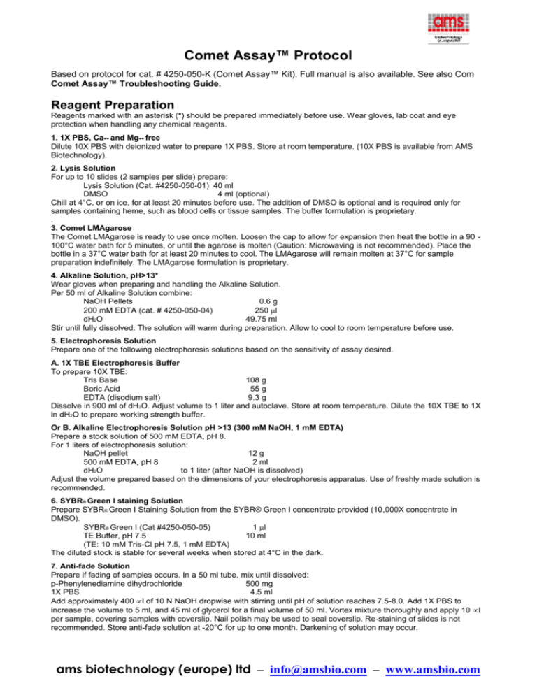
Comet Assay™ Protocol Based on protocol for cat. # 4250-050-K (Comet Assay™ Kit). Full manual is also available. See also Com Comet Assay™ Troubleshooting Guide. Reagent Preparation Reagents marked with an asterisk (*) should be prepared immediately before use. Wear gloves, lab coat and eye protection when handling any chemical reagents. 1. 1X PBS, Ca++ and Mg++ free Dilute 10X PBS with deionized water to prepare 1X PBS. Store at room temperature. (10X PBS is available from AMS Biotechnology). 2. Lysis Solution For up to 10 slides (2 samples per slide) prepare: Lysis Solution (Cat. #4250-050-01) 40 ml DMSO 4 ml (optional) Chill at 4°C, or on ice, for at least 20 minutes before use. The addition of DMSO is optional and is required only for samples containing heme, such as blood cells or tissue samples. The buffer formulation is proprietary. . 3. Comet LMAgarose The Comet LMAgarose is ready to use once molten. Loosen the cap to allow for expansion then heat the bottle in a 90 100°C water bath for 5 minutes, or until the agarose is molten (Caution: Microwaving is not recommended). Place the bottle in a 37°C water bath for at least 20 minutes to cool. The LMAgarose will remain molten at 37°C for sample preparation indefinitely. The LMAgarose formulation is proprietary. 4. Alkaline Solution, pH>13* Wear gloves when preparing and handling the Alkaline Solution. Per 50 ml of Alkaline Solution combine: NaOH Pellets 0.6 g 200 mM EDTA (cat. # 4250-050-04) 250 l dH2O 49.75 ml Stir until fully dissolved. The solution will warm during preparation. Allow to cool to room temperature before use. 5. Electrophoresis Solution Prepare one of the following electrophoresis solutions based on the sensitivity of assay desired. A. 1X TBE Electrophoresis Buffer To prepare 10X TBE: Tris Base 108 g Boric Acid 55 g EDTA (disodium salt) 9.3 g Dissolve in 900 ml of dH2O. Adjust volume to 1 liter and autoclave. Store at room temperature. Dilute the 10X TBE to 1X in dH2O to prepare working strength buffer. Or B. Alkaline Electrophoresis Solution pH >13 (300 mM NaOH, 1 mM EDTA) Prepare a stock solution of 500 mM EDTA, pH 8. For 1 liters of electrophoresis solution: NaOH pellet 12 g 500 mM EDTA, pH 8 2 ml dH2O to 1 liter (after NaOH is dissolved) Adjust the volume prepared based on the dimensions of your electrophoresis apparatus. Use of freshly made solution is recommended. 6. SYBR® Green I staining Solution Prepare SYBR® Green I Staining Solution from the SYBR® Green I concentrate provided (10,000X concentrate in DMSO). SYBR® Green I (Cat #4250-050-05) 1 l TE Buffer, pH 7.5 10 ml (TE: 10 mM Tris-Cl pH 7.5, 1 mM EDTA) The diluted stock is stable for several weeks when stored at 4°C in the dark. 7. Anti-fade Solution Prepare if fading of samples occurs. In a 50 ml tube, mix until dissolved: p-Phenylenediamine dihydrochloride 500 mg 1X PBS 4.5 ml Add approximately 400 l of 10 N NaOH dropwise with stirring until pH of solution reaches 7.5-8.0. Add 1X PBS to increase the volume to 5 ml, and 45 ml of glycerol for a final volume of 50 ml. Vortex mixture thoroughly and apply 10 l per sample, covering samples with coverslip. Nail polish may be used to seal coverslip. Re-staining of slides is not recommended. Store anti-fade solution at -20°C for up to one month. Darkening of solution may occur. ams biotechnology (europe) ltd – info@amsbio.com – www.amsbio.com Sample Preparation and Storage Cell samples should be prepared immediately before starting the assay, although success has been obtained using cryopreserved cells (see below). Cell samples should be handled under dimmed or yellow light to prevent DNA damage from ultraviolet light. Buffers should be chilled to 4°C or on ice to inhibit endogenous damage occurring during sample preparation and to inhibit repair in the unfixed cells. PBS must be calcium and magnesium free to inhibit endonuclease activities. The appropriate controls should also be included (see below). Optimal results in the CometAssay ™ are usually obtained with 500-1000 cells per CometSlide™ sample area. Using 50 l of a cell suspension at 1 x 10⁵ cells per ml combined with 500 l of LMAgarose will provide the correct agarose concentration and cell density for optimal results when plating 75 l per sample. Suspension Cells Cell suspensions are harvested by centrifugation. Resuspend cells at 1 x 10⁵ cells/ml in ice cold 1X PBS (Ca++ and Mg++ free). The media used for cell culture can reduce the adhesion of the agarose on the CometSlide ™. Adherent Cells Gently scrape cells using a rubber policeman. Transfer cells and medium to centrifuge tube, perform cell count, then pellet cells. Wash once in ice cold 1X PBS (Ca++ and Mg++ free). Resuspend cells at 1 x 10⁵ cells/ml in ice cold 1X PBS (Ca++ and Mg++ free). Tissue Preparation Place a small piece of tissue into 1-2 ml of ice cold 1X PBS (Ca++ and Mg++ free), 20 mM EDTA. Using small dissecting scissors, mince the tissue into very small pieces and let stand for 5 minutes. Recover the cell suspension, avoiding transfer of debris. Count cells, pellet by centrifugation, and resuspend at 1 x 10 5 cells/ml in ice cold 1X PBS (Ca++ and Mg++ free). For blood rich organs ( e.g., liver, spleen), chop tissue into large pieces (1-2 mm3), let settle for 5 minutes then aspirate and discard medium. Add 1-2 ml of ice cold 20 mM EDTA in 1X PBS (Ca++ and Mg++ free), mince the tissue into very small pieces and let stand for 5 minutes. Recover the cell suspension, avoiding transfer of debris. Count cells, pellet, and resuspend at 1 x 10⁵ cells/ml in ice cold 1X PBS (Ca++ and Mg++ free). Controls A sample of untreated cells should always be processed to control for endogenous levels of damage within cells, and for damage that may occur during sample preparation. Control cells and treated cells should be handled in an identical manner. If UV damage is being studied, the cells should be kept in low level yellow light during processing. If you require a sample that will be positive for comet tails, treat cells with 100 M hydrogen peroxide or 25 M KMnO4 for 20 minutes at 4°C. Treatment will generate significant oxidative damage in the majority of cells, thereby providing a positive control for each step in the comet assay. Note that the dimensions and characteristics of the comet tail, as a consequence of H2O2 or KMnO4 treatment, may be different to those induced by the damage under investigation. Method for Cryopreservation of Cells Prior to CometAssay™ Certain cells, e.g. lymphocytes, may be successfully cryopreserved prior to performing CometAssay™ (Visvardis et al.). A pilot study should be performed to determine if cryopreservation is appropriate for the cells in use. 1. Centrifuge cells at 200 x g for 5 minutes. 2. Resuspend cell pellet at 1 x 10⁷ cells/ml in 10% (v/v) dimethylsulfoxide, 40% (v/v) medium, 50% (v/v) fetal calf serum. 3. Transfer aliquots of 2 x 10⁶ cells into freezing vials. 4. Freeze at -70°C with -1°C per minute freezing rate. 5. Recover cells by submerging in 37°C water bath until the last trace of ice has melted. 6. Transfer to 15 ml of prechilled 40% (v/v) medium, 10% (w/v) dextrose, 50% (v/v) fetal calf serum. 7. Centrifuge at 200 x g for 10 minutes at 4°C. 8. Resuspend in ice cold 1X PBS (Ca++ and Mg++ free) and proceed with CometAssay™. Assay Protocol Both protocols provided are for alkali unwinding conditions. The electrophoresis conditions will determine the sensitivity of the assay. TBE electrophoresis or Neutral electrophoresis after alkali unwinding will detect single-stranded DNA breaks, double-stranded DNA breaks, and may detect a few apurinic sites, apyrimidinic sites. Alkaline electrophoresis will detect single-stranded DNA breaks, double-stranded DNA breaks, and the majority of apurinic sites, apyrimidinic sites as well as alkali labile DNA adducts ( e.g. phosphoglycols, phosphotriesters). The comet assay has been reported to detect DNA damage associated with low doses (0.6 cGy) of gamma irradiation, providing a simple technique for quantitation of low levels of DNA damage. Prior to performing the comet assay, a viability assay should be performed to determine the dose of the test substance that gives at least 75% viability. False positives may occur when high doses of cytotoxic agents are used. For information on performing the neutral comet assay that will predominantly detect doublestranded DNA breaks, see Section XII: Appendix in full manual of cat. # 4250-050-K. For cryopreservation of cells, fixing the CometSlide™ samples, and storage, refer to Sample Preparation and Storage in this protocol. The CometAssay™ requires approximately 2 - 3 hours to complete, including the incubations and electrophoresis. Once the cells or tissues have been prepared the procedure is not labor intensive. The Lysis Solution may be chilled and the LMAgarose melted while the cell and tissue samples are being prepared. All steps are performed at room temperature unless otherwise specified. Work under dimmed or yellow light to prevent damage from UV. ams biotechnology (europe) ltd – info@amsbio.com – www.amsbio.com 1. Prepare Lysis Solution (see Section V: Reagent Preparation) and chill at 4°C or on ice for at least 20 minutes before use. 2. Melt LMAgarose in a beaker of boiling water for 5 minutes, with the cap loosened. Place bottle in a 37°C water bath for at least 20 minutes to cool. The temperature of the agarose is critical or the cells may undergo heat shock. Heat blocks are not recommended for regulating the temperature of the agarose. 3. Combine cells at 1 x 10⁵/ml with molten LMAgarose (at 37°C) at a ratio of 1: 10 (v/v) and immediately pipette 75 l onto CometSlide™. If necessary, use side of pipette tip to spread agarose/cells over sample area to ensure complete coverage of the sample area. When working with many samples it may be convenient to place aliquots of the molten agarose into prewarmed microcentrifuge tubes and place the tubes at 37°C. Add cells to one tube, mix by gently pipetting once or twice, then transfer 75 l aliquots onto each sample area as required. Then proceed with the next sample of cells. Comet LMAgarose (molten and at 37°C from step 2) 500 l Cells in 1X PBS (Ca++and Mg++ free) at 1 x 10⁵/ml 50 l Note: If sample is not spreading evenly on the slide, warm the slide at 37 °C before application. 4. Place slide flat at 4°C in the dark ( e.g. place in refrigerator) for 10 minutes. A 0.5 mm clear ring appears at edge of CometSlide™ area. Increasing gelling time to 30 minutes improves adherence of samples in high humidity environments. 5. Immerse slide in prechilled Lysis Solution and leave on ice, or at 4°C, for 30 minutes to 60 minutes. 6. Tap off excess buffer from slide and immerse in freshly prepared Alkaline Solution, pH>13 (see Reagent Preparation). WEAR GLOVES WHEN PREPARING OR HANDLING THIS SOLUTION. 7. Leave CometSlide™ in Alkaline Solution for 20 to 60 minutes at room temperature, in the dark. To perform TBE Electrophoresis go to step 8 or for Alkaline go to step 13. 8. Remove slide from Alkaline Solution, gently tap excess buffer from slide and wash by immersing in 1X TBE buffer for 5 minutes, 2 times (see Reagent Preparation). 9. Transfer slide from 1X TBE buffer to an horizontal electrophoresis apparatus. Place slides flat onto a gel tray and align equidistant from the electrodes. Pour 1X TBE buffer until level just covers samples. Set power supply to 1 volt per cm (measured electrode to electrode). Apply voltage for 10 minutes. 10. Very gently tap off excess TBE, and dip slide in 70% ethanol for 5 minutes. 11. Air dry samples. Drying brings all the cells in a single plane to facilitate observation. At this stage, samples may be stored at room temperature, with desiccant. NOTE: AMS Biotechnology and Trevigen offer the CometAssay™ Silver Staining Kit designed for comet staining (Cat#4254-200-K). Silver staining allows visualization of comets on any transmission light microscope and permanently stains the samples for archiving and long term storage. It is recommended that samples be dried before silver staining. 12. Proceed to Step 16. For Alkaline Electrophoresis 13. Transfer slide from Alkaline Solution to a horizontal electrophoresis apparatus. Place slides flat onto a gel tray and align equidistant from the electrodes. Carefully pour the Alkaline Solution until level just covers samples. Set the voltage to about 1 Volt/cm. Add or remove buffer until the current is approximately 300 mA and perform electrophoresis for 20-40 minutes. Tips: Since the Alkaline Electrophoresis Solution is a non-buffered system, temperature control is highly recommended. Inhouse testing has shown great temperature fluctuations when conducting the alkaline electrophoresis at ambient temperature. To improve temperature control, the use of a large electrophoresis apparatus (25-30 cm between electrodes) is recommended along with recirculation of the electrophoresis solution. Alternatively, performing the electrophoresis at cooler temperatures ( e.g. 16°C or 4°C) will diminish background damage, increase sample adherence at high pHs and significantly improves reproducibility. Choose the method that is most convenient for your laboratory and always use the same conditions, power supplies and electrophoresis chambers for comparative analysis. 14. Gently tap off excess electrophoresis solution, rinse by dipping several times in dH 2O, then immerse slide in 70% ethanol for 5 minutes. 15. Air dry samples. Drying brings all the cells in a single plane to facilitate observation. Samples may be stored at room temperature, with desiccant prior to scoring at this stage. NOTE: Trevigen offers the CometAssay™ Silver Staining Kit designed for comet staining (Cat#4254-200-K). Silver staining allows visualization of comets on any transmission light microscope and permanently stains the samples for archiving and long term storage. It is recommended that samples be dried before silver staining. 16. Place 50 l of diluted SYBR® Green I (See Section V: Reagent Preparation) onto each circle of dried agarose. 17. View slide by epifluorescence microscopy. (SYBR® Green I’s maximum excitation and emission are respectively 494nm/521nm. Fluorescein filter is adequate). ams biotechnology (europe) ltd – info@amsbio.com – www.amsbio.com Data Analysis When excited (425 - 500 nm) the DNA-bound SYBR® Green I emits green light. In healthy cells the fluorescence is confined to the nucleoid: undamaged DNA is supercoiled and thus does not migrate very far of the nucleoid under the influence of an electric current. In cells that have accrued damage to the DNA, the alkali treatment unwinds the DNA, releasing fragments that migrate from the cell when subjected to an electric field. The negatively charged DNA migrates toward the anode and the extrusion length reflects increasing relaxation of supercoiling, which is indicative of damage. When using TBE as the electrophoresis buffer, the length of the comet tail may be correlated with DNA damage. When using alkaline electrophoresis conditions, the distribution of DNA between the tail and the head of the comet should be used to evaluate the degree of DNA damage. The characteristics of the comet tail including length, width, and DNA content may also be useful in assessing qualitative differences in the type of DNA damage. Qualitative Analysis The comet tail can be scored according to DNA content (intensity). The control (untreated cells) should be used to determine the characteristics of data for a healthy cell. Scoring can then be made according to nominal, medium or high intensity tail DNA content. At least 75 cells should be scored per sample. Quantitative Analysis There are several image analysis systems that are suitable for quantitation of CometAssay data. The more sophisticated systems include the microscope, camera and computer analysis package. These systems can be set up to establish the length of DNA migration, image length, nuclear size, and calculate the tail moment. At least 75 randomly selected cells should be analyzed per sample. A list of commercially available software package is available on request. ams biotechnology (europe) ltd – info@amsbio.com – www.amsbio.com

![Student Objectives [PA Standards]](http://s3.studylib.net/store/data/006630549_1-750e3ff6182968404793bd7a6bb8de86-300x300.png)
