TOPIC 2
OVERHEAD MASTER
BLM 2-2
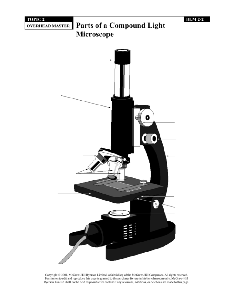
Parts of a Compound Light
Microscope
Copyright © 2001, McGraw-Hill Ryerson Limited, a Subsidiary of the McGraw-Hill Companies. All rights reserved.
Permission to edit and reproduce this page is granted to the purchaser for use in his/her classroom only. McGraw-Hill
Ryerson Limited shall not be held responsible for content if any revisions, additions, or deletions are made to this page.
DATE:
NAME:
TOPIC 2
REINFORCEMENT
CLASS:
BLM 2-3
The Compound Light
Microscope
Goal • Use this page to review the function of each part of the compound light
microscope.
What to Do
Each part of the compound light microscope is listed in the left column of the table below.
In the right column, describe the function of each microscope part. For assistance, refer to
pages 106-107 SCIENCEFOCUS™ 8.
Microscope part
Function
eyepiece
tube
revolving nosepiece
objective lens
fine-adjustment knob
coarse-adjustment knob
stage
condenser lens
diaphragm
light source
Copyright © 2001, McGraw-Hill Ryerson Limited, a Subsidiary of the McGraw-Hill Companies. All rights reserved.
Permission to edit and reproduce this page is granted to the purchaser for use in his/her classroom only. McGraw-Hill
Ryerson Limited shall not be held responsible for content if any revisions, additions, or deletions are made to this page.
DATE:
NAME:
TOPIC 2
INFORMATION
HANDOUT
CLASS:
BLM 2-4
Use and Care of a
Microscope
Goal • This page tells you how to use and care for a microscope.
Introduction
Using microscopes can be exciting.
Microscopes show you a whole new
world of micro-organisms to explore!
Sometimes our excitement can make us
careless. Keep this page in your science
notebook as a quick reference for the
proper use and care of your microscope.
Proper care will keep the microscope
working well. Proper use of the microscope will allow you to gather accurate
scientific data.
Setting Up the Microscope
Use both hands to carry the microscope.
One hand should be on the base and
the other hand should be on the arm.
Place the microscope carefully on the
desk, keeping it away from the edge.
If the lenses are dirty, clean them using
lens paper only. Do not use tissue or
paper towels since they may scratch the
delicate lens surfaces.
Plug in the microscope light so that the
cord is not in your way or dangling over
the edge of the desk.
Low Power-Coarse Focus
Always start with the low-power
objective lens. This is the shortest lens
on the revolving nosepiece.
Carefully place the slide on the stage and
centre it over the light source.
Use the stage clips to hold the slide in
place once it is centred.
While viewing the slide, slowly adjust
the focus with the coarse-adjustment
knob.
If necessary, re-centre your specimen
and follow the procedure below for fine
focus.
Medium Power-Fine Focus
Once you have focussed under low
power, carefully turn the revolving
nosepiece until the medium-power lens
“clicks” into place.
Centre your object in the field of view.
If necessary, use the fine-adjustment
knob to focus.
High Power-Fine Focus
Once you have focussed under medium
power, carefully turn the revolving
nosepiece until the high-power lens
“clicks” into place.
Centre your object in the field of view.
If necessary, use the fine-adjustment
knob to focus.
Clean-Up
Remove the slide from the stage.
Turn the revolving nosepiece to the
low-power lens.
If the stage is wet, wipe it off with a
paper towel.
Wrap the electric cord around the
microscope base and carry
the microscope back to its proper place
using both hands.
Copyright © 2001, McGraw-Hill Ryerson Limited, a Subsidiary of the McGraw-Hill Companies. All rights reserved.
Permission to edit and reproduce this page is granted to the purchaser for use in his/her classroom only. McGraw-Hill
Ryerson Limited shall not be held responsible for content if any revisions, additions, or deletions are made to this page.
DATE:
NAME:
TOPIC 2
ASSESSMENT
CLASS:
BLM 2-6
Using Microscopes:
A Partner Checklist
Goal • You and your partner will use this page to assess each other’s use and knowledge
of the microscope.
What to Do
You will need a partner. You may not use your textbook or notebook. Follow the
instructions for Parts A and B.
Part A: Identifying the Parts of the Microscope
Use the following two-question format when asking your partner to identify the parts of
the microscope and their functions. Ask your partner: On the microscope, point to the
_______________. What is the function of this microscope part? Place a check mark beside each
part that your partner identifies and describes correctly. (12 marks)
eyepiece
nosepiece
fine-adjustment knob
diaphragm
condenser lens
arm
objective lens
light source
stage
stage clips
tube
coarse-adjustment knob
Part B: Focussing the Microscope
Observe your partner’s ability to set up and properly focus the microscope to view a slide. Your
partner must do each of these steps correctly and in sequence. For every step performed
correctly, place a check mark in the blank. If something is performed incorrectly or if a step has
been skipped, then place an “X” in the blank. (8 marks)
Carry the microscope using both hands. Place one hand securely under the base and use the
other to hold the arm of the microscope.
Place the microscope on the centre of the lab bench. Carefully plug it in.
Turn on the microscope using the on/off switch.
Make sure the low-power objective lens is in position.
While looking from the side, lower the objective lens to about 1 cm above the stage using the
coarse-adjustment knob.
Place a sample on the stage under the stage clips.
While looking through the eyepiece, use the coarse-adjustment knob to get the sample
into focus.
Use the fine-adjustment knob to obtain the best image.
Give your partner a score:
Part A:
/12
Part B:
/8
Total:
/20
Copyright © 2001, McGraw-Hill Ryerson Limited, a Subsidiary of the McGraw-Hill Companies. All rights reserved.
Permission to edit and reproduce this page is granted to the purchaser for use in his/her classroom only. McGraw-Hill
Ryerson Limited shall not be held responsible for content if any revisions, additions, or deletions are made to this page.
DATE:
NAME:
TOPIC 2
PROBLEM SOLVING
CLASS:
BLM 2-5
Calculate Magnification
Goal • Practise calculating different magnifications of a microscope.
Think About It
A magnifying lens that magnifies the size of an image by ten times has a magnification of
10x. A compound microscope, like the ones in your classroom, uses two lenses – an
ocular lens and an objective lens. Combining two lenses creates higher magnifications.
What to Do
On this page, or on a separate sheet, answer questions, 1-4 in full sentence form. You must also
show your mathematical calculations for each question.
To calculate the total magnification of a compound microscope, you must multiply the
magnification of the ocular lens by the magnification of the objective lens.
1. What would be the magnification of a microscope with two lenses that each enlarges
an image by 10x?
2. An ocular lens on a microscope has a magnification of 10x. The objective lenses on
the microscope have magnifications of 4x at low power, 10x at medium power, and
40x at high power.
(a) Using the information how would you combine lenses on a microscope if
you wanted to magnify an object 40x?
(b) How would you combine lenses if you wanted to magnify an object 100x?
(c) How would you combine lenses if you wanted to magnify an object 400x?
3. If a compound microscope has an ocular lens of 15x magnification and a scientist
selects an objective lens with a power of 40x, what is the total magnification of the object
in view?
4. Fill in the blanks within the brackets to express total magnification as a word equation.
Total magnification = (_______________) x (_______________)
Copyright © 2001, McGraw-Hill Ryerson Limited, a Subsidiary of the McGraw-Hill Companies. All rights reserved.
Permission to edit and reproduce this page is granted to the purchaser for use in his/her classroom only. McGraw-Hill
Ryerson Limited shall not be held responsible for content if any revisions, additions, or deletions are made to this page.
DATE:
NAME:
TOPIC 2
SKILL BUILDER
CLASS:
BLM 2-7
Estimating the Size of
Microscopic Objects
Goal • This page helps you develop your skills at estimating the size of objects
under the microscope.
Think About It
Once you know the diameter of a microscope’s field of view, how do you estimate
the size of the object you are viewing?
What to Do
Read the information below and answer the questions. You may also refer to pages
110-111 of SCIENCEFOCUS™ 8 for additional information.
Part A: Estimating Object Size
1. Look at the four circles below. Assume that each circle has a diameter of 2.5 cm. (Diameter
is the distance across a circle.) You do not know the size of the happy faces within the
circles. Try to estimate the size of one happy face inside each of the four circles. Write
your answer in the blank space under each circle. Leave some space to write the answers
that you will calculate in question 2.
cm
cm
cm
cm
2. Use the following formula to calculate the exact size of one happy face in each of the
circles:
Size of one happy face = Diameter of circle ÷ Number of happy faces
Write your answers in the blank space beside your estimates from question 1.
Compare the exact size of the smiling faces with your original estimates.
Copyright © 2001, McGraw-Hill Ryerson Limited, a Subsidiary of the McGraw-Hill Companies. All rights reserved.
Permission to edit and reproduce this page is granted to the purchaser for use in his/her classroom only. McGraw-Hill
Ryerson Limited shall not be held responsible for content if any revisions, additions, or deletions are made to this page.
DATE:
NAME:
TOPIC 2
SKILL BUILDER
CLASS:
BLM 2-7
Estimating the Size of
Microscopic Objects (continued)
Part B: Estimating Size Under the Microscope
Once you know the diameter of the field of view of a microscope, you can estimate the
size of the object you are viewing. The field of view is what you see when you look
through the microscope. To find the diameter of the field of view, use a ruler to measure
the distance across its centre. The diagram below represents a field of view when looking
at millimetre markings on a ruler.
The diameter of the field of view
represented on the left is 2.5mm.
Most objects under the microscope are much smaller than a millimetre. Try using a
smaller unit, the micrometre (µm). Multiply the field diameter by 1000 to
convert it from millimetres (mm) to micrometres (µm).
Convert the field of view represented above (2.5 mm) to micrometres:
The diameter of the field of view is __________ µm.
Copyright © 2001, McGraw-Hill Ryerson Limited, a Subsidiary of the McGraw-Hill Companies. All rights reserved.
Permission to edit and reproduce this page is granted to the purchaser for use in his/her classroom only. McGraw-Hill
Ryerson Limited shall not be held responsible for content if any revisions, additions, or deletions are made to this page.
DATE:
NAME:
TOPIC 2
ASSESSMENT
CLASS:
BLM 2-8
Cell Size
Goal • This page tests your ability to estimate the size of cells in a field of view.
What to Do
Read the information given for each question. Answer the questions in the space provided.
1. As scientists, we must determine how small cells really are. To do this, we need to measure
the diameter of the field of view.
(a) What is a field of view? (1 mark)
(b) What is a diameter? (1 mark)
2.
When Molly looks under a microscope, before placing her specimen on the stage, she
observes an empty field of view.
(a) Use your ruler to draw in the diameter of the field of view;
that is, draw a line that cuts the circle exactly in half. (1 mark)
(b) What is the measurement of the circle’s diameter? (2 marks)
in centimeters _____
in millimeters _____
3.
Imagine that ten cells of equal size fit across the diameter of the circle below.
(a) Measure the diameter of the circle. __________ (1 mark)
(b) What is the span of the ten cells? __________ (1 mark)
(c) What is the span of one cell? __________ (1 mark)
(d) Explain how you arrived at your answer for question (c).
(1 mark)
4.
If ten cells fit across a field diameter of 40mm, what is the length of one cell? Show your
work. (3 marks)
Total:
/12 marks
Copyright © 2001, McGraw-Hill Ryerson Limited, a Subsidiary of the McGraw-Hill Companies. All rights reserved.
Permission to edit and reproduce this page is granted to the purchaser for use in his/her classroom only. McGraw-Hill
Ryerson Limited shall not be held responsible for content if any revisions, additions, or deletions are made to this page.


