Mitochondria, Chloroplasts, and Peroxisomes
advertisement

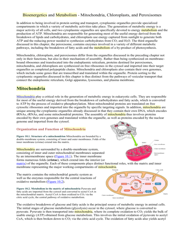
Bioenergetics and Metabolism - Mitochondria, Chloroplasts, and Peroxisomes In addition to being involved in protein sorting and transport, cytoplasmic organelles provide specialized compartments in which a variety of metabolic activities take place. The generation of metabolic energy is a major activity of all cells, and two cytoplasmic organelles are specifically devoted to energy metabolism and the production of ATP. Mitochondria are responsible for generating most of the useful energy derived from the breakdown of lipids and carbohydrates, and chloroplasts use energy captured from sunlight to generate both ATP and the reducing power needed to synthesize carbohydrates from CO2 and H2O. The third organelle discussed in this chapter, the peroxisome, contains enzymes involved in a variety of different metabolic pathways, including the breakdown of fatty acids and the metabolism of a by-product of photosynthesis. Mitochondria, chloroplasts, and peroxisomes differ from the organelles discussed in the preceding chapter not only in their functions, but also in their mechanism of assembly. Rather than being synthesized on membranebound ribosomes and translocated into the endoplasmic reticulum, proteins destined for peroxisomes, mitochondria, and chloroplasts are synthesized on free ribosomes in the cytosol and imported into their target organelles as completed polypeptide chains. Mitochondria and chloroplasts also contain their own genomes, which include some genes that are transcribed and translated within the organelle. Protein sorting to the cytoplasmic organelles discussed in this chapter is thus distinct from the pathways of vesicular transport that connect the endoplasmic reticulum, Golgi apparatus, lysosomes, and plasma membrane. Mitochondria Mitochondria play a critical role in the generation of metabolic energy in eukaryotic cells. They are responsible for most of the useful energy derived from the breakdown of carbohydrates and fatty acids, which is converted to ATP by the process of oxidative phosphorylation. Most mitochondrial proteins are translated on free cytosolic ribosomes and imported into the organelle by specific targeting signals. In addition, mitochondria are unique among the cytoplasmic organelles already discussed in that they contain their own DNA, which encodes tRNAs, rRNAs, and some mitochondrial proteins. The assembly of mitochondria thus involves proteins encoded by their own genomes and translated within the organelle, as well as proteins encoded by the nuclear genome and imported from the cytosol. Organization and Function of Mitochondria Figure 10.1. Structure of a mitochondrion Mitochondria are bounded by a double-membrane system, consisting of inner and outer membranes. Folds of the inner membrane (cristae) extend into the matrix. Mitochondria are surrounded by a double-membrane system, consisting of inner and outer mitochondrial membranes separated by an intermembrane space (Figure 10.1). The inner membrane forms numerous folds (cristae), which extend into the interior (or matrix) of the organelle. Each of these components plays distinct functional roles, with the matrix and inner membrane representing the major working compartments of mitochondria. The matrix contains the mitochondrial genetic system as well as the enzymes responsible for the central reactions of oxidative metabolism (Figure 10.2). Figure 10.2. Metabolism in the matrix of mitochondria Pyruvate and fatty acids are imported from the cytosol and converted to acetyl CoA in the mitochondrial matrix. Acetyl CoA is then oxidized to CO2 via the citric acid cycle, the central pathway of oxidative metabolism. The oxidative breakdown of glucose and fatty acids is the principal source of metabolic energy in animal cells. The initial stages of glucose metabolism (glycolysis) occur in the cytosol, where glucose is converted to pyruvate. Pyruvate is then transported into mitochondria, where its complete oxidation to CO2 yields the bulk of usable energy (ATP) obtained from glucose metabolism. This involves the initial oxidation of pyruvate to acetyl CoA, which is then broken down to CO2 via the citric acid cycle. The oxidation of fatty acids also yields acetyl CoA, which is similarly metabolized by the citric acid cycle in mitochondria. The enzymes of the citric acid cycle (located in the matrix of mitochondria) thus are central players in the oxidative breakdown of both carbohydrates and fatty acids. The oxidation of acetyl CoA to CO2 is coupled to the reduction of NAD+ and FAD to NADH and FADH2, respectively. Most of the energy derived from oxidative metabolism is then produced by the process of oxidative phosphorylation (discussed in detail in the next section), which takes place in the inner mitochondrial membrane. The high-energy electrons from NADH and FADH2 are transferred through a series of carriers in the membrane to molecular oxygen. The energy derived from these electron transfer reactions is converted to potential energy stored in a proton gradient across the membrane, which is then used to drive ATP synthesis. The inner mitochondrial membrane thus represents the principal site of ATP generation, and this critical role is reflected in its structure. First, its surface area is substantially increased by its folding into cristae. In addition, the inner mitochondrial membrane contains an unusually high percentage (greater than 70%) of proteins, which are involved in oxidative phosphorylation as well as in the transport of metabolites (e.g., pyruvate and fatty acids) between the cytosol and mitochondria. Otherwise, the inner membrane is impermeable to most ions and small molecules a property critical to maintaining the proton gradient that drives oxidative phosphorylation. In contrast to the inner membrane, the outer mitochondrial membrane is freely permeable to small molecules. This is because it contains proteins called porins, which form channels that allow the free diffusion of molecules smaller than about 6000 daltons. The composition of the intermembrane space is therefore similar to the cytosol with respect to ions and small molecules. Consequently, the inner mitochondrial membrane is the functional barrier to the passage of small molecules between the cytosol and the matrix and maintains the proton gradient that drives oxidative phosphorylation. The Genetic System of Mitochondria Mitochondria contain their own genetic system, which is separate and distinct from the nuclear genome of the cell. Mitochondria are thought to have evolved from bacteria that developed a symbiotic relationship in which they lived within larger cells (endosymbiosis). This hypothesis has recently been substantiated by the results of DNA sequence analysis, which revealed striking similarities between the genomes of mitochondria and of the bacterium Rickettsia prowazekii. Rickettsia are intracellular parasites which, like mitochondria, are only able to reproduce within eukaryotic cells. Consistent with their similar symbiotic lifestyles, the genomic DNA sequences of Rickettsia and mitochondria suggest that they share a common ancestor, from which the genetic system of present-day mitochondria evolved. Mitochondrial genomes are usually circular DNA molecules, like those of bacteria, which are present in multiple copies per organelle. They vary considerably in size between different species. The genomes of human and most other animal mitochondria are only about 16 kb, but substantially larger mitochondrial genomes are found in yeasts (approximately 80 kb) and plants (more than 200 kb). However, these larger mitochondrial genomes are composed predominantly of noncoding sequences and do not appear to contain significantly more genetic information. For example, the largest sequenced mitochondrial genome is that of the plant Arabidopsis thaliana. Although Arabidopsis mitochondrial DNA is approximately 367 kb, it encodes only 32 proteins: just more than twice the number encoded by human mitochondrial DNA. The largest number of mitochondrial genes has been found in mitochondrial DNA of the protozoan Reclinomonas americana, which is 69 kb and contains 97 genes. The mitochondrial genome of Reclinomonas appears to more closely resemble the bacterial genome from which mitochondria evolved than most present-day mitochondrial genomes, which encode only a small number of proteins that are essential components of the oxidative phosphorylation system. In addition, mitochondrial genomes encode all of the ribosomal RNAs and most of the transfer RNAs needed for translation of these protein-coding sequences within mitochondria. Other mitochondrial proteins are encoded by nuclear genes, which are thought to have been transferred to the nucleus from the ancestral mitochondrial genome.The human mitochondrial genome encodes 13 proteins involved in electron transport and oxidative phosphorylation (Figure 10.3). Figure 10.3. The human mitochondrial genome The genome contains 13 proteincoding sequences, which are designated as components of respiratory complexes I, III, IV, or V. In addition, the genome contains genes for 12S and 16S rRNAs and for 22 tRNAs, which are designated by the one-letter code for the corresponding amino acid. The region of the genome designated "D loop" contains an origin of DNA replication and transcriptional promoter sequences. In addition, human mitochondrial DNA encodes 16S and 12S rRNAs and 22 tRNAs, which are required for translation of the proteins encoded by the organelle genome. The two rRNAs are the only RNA components of animal and yeast mitochondrial ribosomes, in contrast to the three rRNAs of bacterial ribosomes (23S, 16S, and 5S). Plant mitochondrial DNAs, however, also encode a third rRNA of 5S. The mitochondria of plants and protozoans also differ in importing and utilizing tRNAs encoded by the nuclear as well as the mitochondrial genome, whereas in animal mitochondria, all the tRNAs are encoded by the organelle. The small number of tRNAs encoded by the mitochondrial genome highlights an important feature of the mitochondrial genetic system the use of a slightly different genetic code, which is distinct from the "universal" genetic code used by both prokaryotic and eukaryotic cells.There are 64 possible triplet codons, of which 61 encode the 20 different amino acids incorporated into proteins. Many tRNAs in both prokaryotic and eukaryotic cells are able to recognize more than a single codon in mRNA because of "wobble," which allows some mispairing between the tRNA anticodon and the third position of certain complementary codons. However, at least 30 different tRNAs are required to translate the universal code according to the wobble rules. Yet human mitochondrial DNA encodes only 22 tRNA species, and these are the only tRNAs used for translation of mitochondrial mRNAs. This is accomplished by an extreme form of wobble in which U in the anticodon of the tRNA can pair with any of the four bases in the third codon position of mRNA, allowing four codons to be recognized by a single tRNA. In addition, some codons specify different amino acids in mitochondria than in the universal code. Like the DNA of nuclear genomes, mitochondrial DNA can be altered by mutations, which are frequently deleterious to the organelle. Since almost all the mitochondria of fertilized eggs are contributed by the oocyte rather than by the sperm, germ-line mutations in mitochondrial DNA are transmitted to the next generation by the mother. Such mutations have been associated with a number of diseases. For example, Leber's hereditary optic neuropathy, a disease that leads to blindness, can be caused by mutations in mitochondrial genes that encode components of the electron transport chain. In addition, the progressive accumulation of mutations in mitochondrial DNA during the lifetime of individuals has been suggested to contribute to the process of aging. Protein Import and Mitochondrial Assembly In contrast to the RNA components of the mitochondrial translation apparatus (rRNAs and tRNAs), most mitochondrial genomes do not encode the proteins required for DNA replication, transcription, or translation. Instead, the genes that encode proteins required for the replication and expression of mitochondrial DNA are contained in the nucleus. In addition, the nucleus contains the genes that encode most of the mitochondrial proteins required for oxidative phosphorylation and all of the enzymes involved in mitochondrial metabolism (e.g., enzymes of the citric acid cycle). The proteins encoded by these genes (more than 95% of mitochondrial proteins) are synthesized on free cytosolic ribosomes and imported into mitochondria as completed polypeptide chains. Because of the double-membrane structure of mitochondria, the import of proteins is considerably more complicated than the transfer of a polypeptide across a single phospholipid bilayer. Proteins targeted to the matrix have to cross both the inner and outer mitochondrial membranes, while other proteins need to be sorted to distinct compartments within the organelle (e.g., the intermembrane space). The import of proteins to the matrix is the best-understood aspect of mitochondrial protein sorting (Figure 10.4). Figure 10.4. Import of proteins into mitochondria Proteins are targeted for mitochondria by an amino-terminal presequence containing positively charged amino acids. Proteins are maintained in a partially unfolded state by association with a cytosolic Hsp70 and are recognized by a receptor on the surface of mitochondria. The unfolded polypeptide chains are then translocated through the Tom complex in the outer membrane and transferred to the Tim complex in the inner membrane. The voltage component of the electrochemical gradient is required for translocation across the inner membrane. The presequence is cleaved by a matrix protease, and a mitochondrial Hsp70 binds the polypeptide chain as it crosses the inner membrane, driving further protein translocation. A mitochondrial Hsp60 then facilitates folding of the imported polypeptide within the matrix Most proteins are targeted to mitochondria by amino-terminal sequences of 20 to 35 amino acids (called presequences) that are removed by proteolytic cleavage following their import into the organelle. The presequences of mitochondrial proteins, first characterized by Gottfried Schatz, contain multiple positively charged amino acid residues, usually in an amphipathic helix. The first step in protein import is the binding of these presequences to receptors on the surface of mitochondria. The polypeptide chains are then inserted into a protein complex that directs translocation across the outer membrane (the translocase of the outer membrane or Tom complex). The proteins are then transferred to a second protein complex in the inner membrane (the translocase of the inner membrane or Tim complex). Continuing protein translocation requires the electrochemical potential established across the inner mitochondrial membrane during electron transport. As discussed in the next section of this chapter, the transfer of high-energy electrons from NADH and FADH2 to molecular oxygen is coupled to the transfer of protons from the mitochondrial matrix to the intermembrane space. Since protons are charged particles, this transfer establishes an electric potential across the inner membrane, with the matrix being negative. During protein import, this electric potential drives translocation of the positively charged presequence. To be translocated across the mitochondrial membrane, proteins must be at least partially unfolded. Consequently, protein import into mitochondria requires molecular chaperones in addition to the membrane proteins involved in translocation (see Figure 10.4). On the cytosolic side, members of the Hsp70 family of chaperones maintain proteins in a partially unfolded state so that they can be inserted into the mitochondrial membrane. As they cross the inner membrane, the unfolded polypeptide chains bind to another member of the Hsp70 family, which is associated with the Tim complex and acts as a motor that drives protein import. The polypeptide is then transferred to a chaperone of the Hsp60 family (a chaperonin), within which protein folding takes place. Since these interactions of polypeptide chains with molecular chaperones depend on ATP, protein import requires ATP both outside and inside the mitochondria, in addition to the electric potential across the inner membrane. As noted above, some mitochondrial proteins are targeted to the outer membrane, inner membrane, or intermembrane space rather than to the matrix, so additional mechanisms are needed to direct these proteins to the correct submitochondrial compartment. These proteins are targeted to their destinations by a second sorting signal following the positively charged presequence that directs mitochondrial import. The targeting of proteins to the mitochondrial membranes appears to be mediated by hydrophobic stop-transfer sequences that halt translocation of the polypeptide chains through the Tim or Tom complexes, leading to their insertion into the inner or outer mitochondrial membranes, respectively (Figure 10.5). Figure 10.5. Insertion of mitochondrial membrane proteins Proteins targeted for the mitochondrial membranes contain hydrophobic stop-transfer sequences that halt their translocation through the Tom or Tim complexes and lead to their incorporation into the outer or inner membranes, respectively. Proteins may be targeted to the intermembrane space by several different mechanisms (Figure 10.6). Figure 10.6. Sorting proteins to the intermembrane space Proteins can be targeted to the intermembrane space by several mechanisms. Some proteins (I) are translocated through the Tom complex and released into the intermembrane space. Other proteins (II) are transferred from the Tom complex to the Tim complex, but they contain hydrophobic stoptransfer sequences that halt translocation through the Tim complex. These stop-transfer sequences are then cleaved to release the proteins into the intermembrane space. Still other proteins (III) are imported to the matrix, as depicted in Figure 10.4. Removal of the presequence within the matrix then exposes a hydrophobic signal sequence, which targets the protein back across the inner membrane to the intermembrane space. Some proteins are transferred across the outer membrane through the Tom complex but are then released within the intermembrane space instead of being transferred to the Tim complex. Other proteins are transferred to the Tim complex but are then released into the intermembrane space as a result of cleavage of hydrophobic stoptransfer sequences. Still other proteins may be completely imported into the mitochondrial matrix and then exported back across the inner membrane to the intermembrane space. Not only the proteins, but also the phospholipids of mitochondrial membranes are imported from the cytosol. In animal cells, phosphatidylcholine and phosphatidylethanolamine are synthesized in the ER and carried to mitochondria by phospholipid transfer proteins, which extract single phospholipid molecules from the membrane of the ER. The lipid can then be transported through the aqueous environment of the cytosol, buried in a hydrophobic binding site of the protein, and released when the complex reaches a new membrane, such as that of mitochondria. The mitochondria then synthesize phosphatidylserine from phosphatidylethanolamine, in addition to catalyzing the synthesis of the unusual phospholipid cardiolipin, which contains four fatty acid chains (Figure 10.7). Figure 10.7. Structure of cardiolipin Cardiolipin is an unusual "double" phospholipid, containing four fatty acid chains, that is found primarily in the inner mitochondrial membrane. The Mechanism of Oxidative Phosphorylation Most of the usable energy obtained from the breakdown of carbohydrates or fats is derived by oxidative phosphorylation, which takes place within mitochondria. For example, the breakdown of glucose by glycolysis and the citric acid cycle yields a total of four molecules of ATP, ten molecules of NADH, and two molecules of FADH2. Electrons from NADH and FADH2 are then transferred to molecular oxygen, coupled to the formation of an additional 32 to 34 ATP molecules by oxidative phosphorylation. Electron transport and oxidative phosphorylation are critical activities of protein complexes in the inner mitochondrial membrane, which ultimately serve as the major source of cellular energy. The Electron Transport Chain During oxidative phosphorylation, electrons derived from NADH and FADH2 combine with O2, and the energy released from these oxidation/ reduction reactions is used to drive the synthesis of ATP from ADP. The transfer of electrons from NADH to O2 is a very energy-yielding reaction, with G°´ = -52.5 kcal/mol for each pair of electrons transferred. To be harvested in usable form, this energy must be produced gradually, by the passage of electrons through a series of carriers, which constitute the electron transport chain. These carriers are organized into four complexes in the inner mitochondrial membrane. A fifth protein complex then serves to couple the energyyielding reactions of electron transport to ATP synthesis. Electrons from NADH enter the electron transport chain in complex I, which consists of nearly 40 polypeptide chains (Figure 10.8). Figure 10.8. Transport of electrons from NADH These electrons are initially transferred from NADH to flavin mononucleotide and then, through an iron-sulfur carrier, to coenzyme Q an energy-yielding process with G°´ = -16.6 kcal/mol. Coenzyme Q (also called ubiquinone) is a small, lipid-soluble molecule that carries electrons from complex I through the membrane to complex III, which consists of about ten polypeptides. In complex III, electrons are transferred from cytochrome b to cytochrome c an energy-yielding reaction with G°´ = -10.1 kcal/mol. Cytochromec, a peripheral membrane protein bound to the outer face of the inner membrane, then carries electrons to complex IV (cytochrome oxidase), where they are finally transferred to O2 (G°´ = -25.8 kcal/mol). A distinct protein complex (complex II), which consists of four polypeptides, receives electrons from the citric acid cycle intermediate, succinate (Figure 10.9). Figure 10.9. Transport of electrons from FADH2 Electrons from succinate enter the electron transport chain via FADH2 in complex II. They are then transferred to coenzyme Q and carried through the rest of the electron transport chain as described in Figure 10.8. The transfer of electrons from FADH2 to coenzyme Q is not associated with a significant decrease in free energy, so protons are not pumped across the membrane at complex II. These electrons are transferred to FADH2, rather than to NADH, and then to coenzyme Q. From coenzyme Q, electrons are transferred to complex III and then to complex IV as already described. In contrast to the transfer of electrons from NADH to coenzyme Q at complex I, the transfer of electrons from FADH2 to coenzyme Q is not associated with a significant decrease in free energy and, therefore, is not coupled to ATP synthesis. Consequently, the passage of electrons derived from FADH2 through the electron transport chain yields free energy only at complexes III and IV. The free energy derived from the passage of electrons through complexes I, III, and IV is harvested by being coupled to the synthesis of ATP. Importantly, the mechanism by which the energy derived from these electron transport reactions is coupled to ATP synthesis is fundamentally different from the synthesis of ATP during glycolysis or the citric acid cycle. In the latter cases, a high-energy phosphate is transferred directly to ADP from the other substrate of an energy-yielding reaction. For example, in the final reaction of glycolysis, the high-energy phosphate of phosphoenolpyruvate is transferred to ADP, yielding pyruvate plus ATP. Such direct transfer of high-energy phosphate groups does not occur during electron transport. Instead, the energy derived from electron transport is coupled to the generation of a proton gradient across the inner mitochondrial membrane. The potential energy stored in this gradient is then harvested by a fifth protein complex, which couples the energetically favorable flow of protons back across the membrane to the synthesis of ATP. Chemiosmotic Coupling The mechanism of coupling electron transport to ATP generation, chemiosmotic coupling, is a striking example of the relationship between structure and function in cell biology. The hypothesis of chemiosmotic coupling was first proposed in 1961 by Peter Mitchell, who suggested that ATP is generated by the use of energy stored in the form of proton gradients across biological membranes, rather than by direct chemical transfer of high-energy groups. Biochemists were initially highly skeptical of this proposal, and the chemiosmotic hypothesis took more than a decade to win general acceptance in the scientific community. Overwhelming evidence eventually accumulated in its favor, however, and chemiosmotic coupling is now recognized as a general mechanism of ATP generation, operating not only in mitochondria but also in chloroplasts and in bacteria, where ATP is generated via a proton gradient across the plasma membrane. Electron transport through complexes I, III, and IV is coupled to the transport of protons out of the interior of the mitochondrion (see Figure 10.8). Thus, the energy-yielding reactions of electron transport are coupled to the transfer of protons from the matrix to the intermembrane space, which establishes a proton gradient across the inner membrane. Complexes I and IV appear to act as proton pumps that transfer protons across the membrane as a result of conformational changes induced by electron transport. In complex III, protons are carried across the membrane by coenzyme Q, which accepts protons from the matrix at complexes I or II and releases them into the intermembrane space at complex III. Complexes I and III each transfer four protons across the membrane per pair of electrons. In complex IV, two protons per pair of electrons are pumped across the membrane and another two protons per pair of electrons are combined with O2 to form H2O within the matrix. Thus, the equivalent of four protons per pair of electrons are transported out of the mitochondrial matrix at each of these three complexes. This transfer of protons from the matrix to the intermembrane space plays the critical role of converting the energy derived from the oxidation/reduction reactions of electron transport to the potential energy stored in a proton gradient. Because protons are electrically charged particles, the potential energy stored in the proton gradient is electric as well as chemical in nature. The electric component corresponds to the voltage difference across the inner mitochondrial membrane, with the matrix of the mitochondrion negative and the intermembrane space positive. The corresponding free energy is given by the equation where F is the Faraday constant and V is the membrane potential. The additional free energy corresponding to the difference in proton concentration across the membrane is given by the equation where [H+]i and [H+]o refer, respectively, to the proton concentrations inside and outside the mitochondria. In metabolically active cells, protons are typically pumped out of the matrix such that the proton gradient across the inner membrane corresponds to about one pH unit, or a tenfold lower concentration of protons within mitochondria (Figure 10.10). Figure 10.10. The electrochemical nature of the proton gradient Since protons are positively charged, the proton gradient established across the inner mitochondrial membrane has both chemical and electric components. The chemical component is the proton concentration, or pH, gradient, which corresponds to about a tenfold higher concentration of protons on the cytosolic side of the inner mitochondrial membrane (a difference of one pH unit). In addition, there is an electric potential across the membrane, resulting from the net increase in positive charge on the cytosolic side. The pH of the mitochondrial matrix is therefore about 8, compared to the neutral pH (approximately 7) of the cytosol and intermembrane space. This gradient also generates an electric potential of approximately 0.14 V across the membrane, with the matrix negative. Both the pH gradient and the electric potential drive protons back into the matrix from the cytosol, so they combine to form an electrochemical gradient across the inner mitochondrial membrane, corresponding to a G of approximately -5 kcal/mol per proton. Because the phospholipid bilayer is impermeable to ions, protons are able to cross the membrane only through a protein channel. This restriction allows the energy in the electrochemical gradient to be harnessed and converted to ATP as a result of the action of the fifth complex involved in oxidative phosphorylation, complex V, or ATP synthase (see Figure 10.8). ATP synthase is organized into two structurally distinct components, F0 and F1, which are linked by a slender stalk (Figure 10.11). Figure 10.11. Structure of ATP synthase The mitochondrial ATP synthase (complex V) consists of two multisubunit components, F 0 and F1, which are linked by a slender stalk. F0 spans the lipid bilayer, forming a channel through which protons can cross the membrane. F1 harvests the free energy derived from proton movement down the electrochemical gradient by catalyzing the synthesis of ATP. The F0 portion spans the inner membrane and provides a channel through which protons are able to flow back from the intermembrane space to the matrix. The energetically favorable return of protons to the matrix is coupled to ATP synthesis by the F1 subunit, which catalyzes the synthesis of ATP from ADP and phosphate ions (Pi). Detailed structural studies have established the mechanism of ATP synthase action, which involves mechanical coupling between the F0 and F1 subunits. In particular, the flow of protons through F0 drives the rotation of F1, which acts as a rotary motor to drive ATP synthesis. It appears that the flow of four protons back across the membrane through F0 is required to drive the synthesis of one molecule of ATP by F1, consistent with the proton transfers at complexes I, III, and IV each contributing sufficient free energy to the proton gradient to drive the synthesis of one ATP molecule. The oxidation of one molecule of NADH thus leads to the synthesis of three molecules of ATP, whereas the oxidation of FADH2, which enters the electron transport chain at complex II, yields only two ATP molecules. Transport of Metabolites across the Inner Membrane In addition to driving the synthesis of ATP, the potential energy stored in the electrochemical gradient drives the transport of small molecules into and out of mitochondria. For example, the ATP synthesized within mitochondria has to be exported to the cytosol, while ADP and Pi need to be imported from the cytosol for ATP synthesis to continue. The electrochemical gradient generated by proton pumping provides energy required for the transport of these molecules and other metabolites that need to be concentrated within mitochondria (Figure 10.12). Figure 10.12. Transport of metabolites across the mitochondrial inner membrane The transport of small molecules across the inner membrane is mediated by membrane-spanning transport proteins and driven by the electrochemical gradient. For example, ATP is exported from mitochondria to the cytosol by a transporter that exchanges it for ADP. The voltage component of the electrochemical gradient drives this exchange: ATP carries a greater negative charge (-4) than ADP (-3), so ATP is exported from the mitochondrial matrix to the cytosol while ADP is imported to mitochondria. In contrast, the transport of phosphate (P i) and pyruvate is coupled to an exchange for hydroxyl ions (OH-); in this case, the pH component of the electrochemical gradient drives the export of hydroxyl ions, coupled to the transport of Pi and pyruvate into mitochondria. The transport of ATP and ADP across the inner membrane is mediated by an integral membrane protein, the adenine nucleotide translocator, which transports one molecule of ADP into the mitochondrion in exchange for one molecule of ATP transferred from the mitochondrion to the cytosol. Because ATP carries more negative charge than ADP (-4 compared to -3), this exchange is driven by the voltage component of the electrochemical gradient. Since the proton gradient establishes a positive charge on the cytosolic side of the membrane, the export of ATP in exchange for ADP is energetically favorable. The synthesis of ATP within the mitochondrion requires phosphate ions (Pi) as well as ADP, so Pi must also be imported from the cytosol. This is mediated by another membrane transport protein, which imports phosphate (H2PO4-) and exports hydroxyl ions (OH-). This exchange is electrically neutral because both phosphate and hydroxyl ions have a charge of -1. However, the exchange is driven by the proton concentration gradient; the higher pH within mitochondria corresponds to a higher concentration of hydroxyl ions, favoring their translocation to the cytosolic side of the membrane. Energy from the electrochemical gradient is similarly used to drive the transport of other metabolites into mitochondria. For example, the import of pyruvate from the cytosol (where it is produced by glycolysis) is mediated by a transporter that exchanges pyruvate for hydroxyl ions. Other intermediates of the citric acid cycle are able to shuttle between mitochondria and the cytosol by similar exchange mechanisms. Chloroplasts and Other Plastids Chloroplasts, the organelles responsible for photosynthesis, are in many respects similar to mitochondria. Both chloroplasts and mitochondria function to generate metabolic energy, evolved by endosymbiosis, contain their own genetic systems, and replicate by division. However, chloroplasts are larger and more complex than mitochondria, and they perform several critical tasks in addition to the generation of ATP. Most importantly, chloroplasts are responsible for the photosynthetic conversion of CO2 to carbohydrates. In addition, chloroplasts synthesize amino acids, fatty acids, and the lipid components of their own membranes. The reduction of nitrite (NO2-) to ammonia (NH3), an essential step in the incorporation of nitrogen into organic compounds, also occurs in chloroplasts. Moreover, chloroplasts are only one of several types of related organelles (plastids) that play a variety of roles in plant cells. The Structure and Function of Chloroplasts Plant chloroplasts are large organelles (5 to 10 m long) that, like mitochondria, are bounded by a double membrane called the chloroplast envelope (Figure 10.13). Figure 10.13. Structure of a chloroplast In addition to the inner and outer membranes of the envelope, chloroplasts contain a third internal membrane system: the thylakoid membrane. These membranes divide chloroplasts into three internal compartments In addition to the inner and outer membranes of the envelope, chloroplasts have a third internal membrane system, called the thylakoid membrane. The thylakoid membrane forms a network of flattened discs called thylakoids, which are frequently arranged in stacks called grana. Because of this three-membrane structure, the internal organization of chloroplasts is more complex than that of mitochondria. In particular, their three membranes divide chloroplasts into three distinct internal compartments: (1) the intermembrane space between the two membranes of the chloroplast envelope; (2) the stroma, which lies inside the envelope but outside the thylakoid membrane; and (3) the thylakoid lumen. Despite this greater complexity, the membranes of chloroplasts have clear functional similarities with those of mitochondria as expected, given the role of both organelles in the chemiosmotic generation of ATP. The outer membrane of the chloroplast envelope, like that of mitochondria, contains porins and is therefore freely permeable to small molecules. In contrast, the inner membrane is impermeable to ions and metabolites, which are therefore able to enter chloroplasts only via specific membrane transporters. These properties of the inner and outer membranes of the chloroplast envelope are similar to the inner and outer membranes of mitochondria: In both cases the inner membrane restricts the passage of molecules between the cytosol and the interior of the organelle. The chloroplast stroma is also equivalent in function to the mitochondrial matrix: It contains the chloroplast genetic system and a variety of metabolic enzymes, including those responsible for the critical conversion of CO2 to carbohydrates during photosynthesis. The major difference between chloroplasts and mitochondria, in terms of both structure and function, is the thylakoid membrane. This membrane is of central importance in chloroplasts, where it fills the role of the inner mitochondrial membrane in electron transport and the chemiosmotic generation of ATP (Figure 10.14). Figure 10.14. Chemiosmotic generation of ATP in chloroplasts and mitochondria In mitochondria, electron transport generates a proton gradient across the inner membrane, which is then used to drive ATP synthesis in the matrix. In chloroplasts, the proton gradient is generated across the thylakoid membrane and used to drive ATP synthesis in the stroma. The inner membrane of the chloroplast envelope (which is not folded into cristae) does not function in photosynthesis. Instead, the chloroplast electron transport system is located in the thylakoid membrane, and protons are pumped across this membrane from the stroma to the thylakoid lumen. The resulting electrochemical gradient then drives ATP synthesis as protons cross back into the stroma. In terms of its role in generation of metabolic energy, the thylakoid membrane of chloroplasts is thus equivalent to the inner membrane of mitochondria. The Chloroplast Genome Like mitochondria, chloroplasts contain their own genetic system, reflecting their evolutionary origins from photosynthetic bacteria. The genomes of chloroplasts are similar to those of mitochondria in that they consist of circular DNA molecules present in multiple copies per organelle. However, chloroplast genomes are larger and more complex than those of mitochondria, ranging from 120 to 160 kb and containing approximately 120 genes. The chloroplast genomes of several plants have been completely sequenced, leading to the identification of many of the genes contained in the organelle DNAs. These chloroplast genes encode both RNAs and proteins involved in gene expression, as well as a variety of proteins that function in photosynthesis. Both the ribosomal and transfer RNAs used for translation of chloroplast mRNAs are encoded by the organelle genome. These include four rRNAs (23S, 16S, 5S, and 4.5S) and 30 tRNA species. In contrast to the smaller number of tRNAs encoded by the mitochondrial genome, the chloroplast tRNAs are sufficient to translate all the mRNA codons according to the universal genetic code. In addition to these RNA components of the translation system, the chloroplast genome encodes about 20 ribosomal proteins, which represent approximately a third of the proteins of chloroplast ribosomes. Some subunits of RNA polymerase are also encoded by chloroplasts, although additional RNA polymerase subunits and other factors needed for chloroplast gene expression are encoded in the nucleus. The chloroplast genome also encodes approximately 30 proteins that are involved in photosynthesis, including components of photosystems I and II, of the cytochrome bf complex, and of ATP synthase. In addition, one of the subunits of ribulose bisphosphate carboxylase (rubisco) is encoded by chloroplast DNA. Rubisco is the critical enzyme that catalyzes the addition of CO2 to ribulose-1,5-bisphosphate during the Calvin cycle. Not only is it the major protein component of the chloroplast stroma, but it is also thought to be the single most abundant protein on Earth, so it is noteworthy that one of its subunits is encoded by the chloroplast genome. Import and Sorting of Chloroplast Proteins Although chloroplasts encode more of their own proteins than mitochondria, about 90% of chloroplast proteins are still encoded by nuclear genes. As with mitochondria, these proteins are synthesized on cytosolic ribosomes and then imported into chloroplasts as completed polypeptide chains. They must then be sorted to their appropriate location within chloroplasts an even more complicated task than protein sorting in mitochondria, since chloroplasts contain three separate membranes that divide them into three distinct internal compartments. Protein import into chloroplasts generally resembles mitochondrial protein import (Figure 10.15). Figure 10.15. Protein import into the chloroplast stroma Proteins are targeted for import into chloroplasts by a transit peptide at their amino terminus. The transit peptide directs polypeptide translocation through the Toc complex in the chloroplast outer membrane and the Tic complex in the chloroplast inner membrane. This peptide is then removed by proteolytic cleavage within the stroma. Both cytosolic and chloroplast chaperones (Hsp60 and Hsp70) are required for protein import. Proteins are targeted for import into chloroplasts by N-terminal sequences of 30 to 100 amino acids, called transit peptides, which direct protein translocation across the two membranes of the chloroplast envelope and are then removed by proteolytic cleavage. The transit peptides are recognized by the translocation complex of the chloroplast outer member (the Toc complex), and proteins are transported through this complex across the membrane. They are then transferred to the translocation complex of the inner membrane (the Tic complex) and transported across the inner membrane to the stroma. As in mitochondria, molecular chaperones on both the cytosolic and stromal sides of the envelope are required for protein import, which requires energy in the form of ATP. In contrast to the presequences of mitochondrial import, however, transit peptides are not positively charged and the translocation of polypeptide chains into chloroplasts does not require an electric potential across the membrane. Proteins incorporated into the thylakoid lumen are transported to their destination in two steps (Figure 10.16). Figure 10.16. Import of proteins into the thylakoid lumen Proteins are imported into the thylakoid lumen in two steps. The first step is import into the chloroplast stroma, as illustrated in Figure 10.15. Cleavage of the transit peptide then exposes a second hydrophobic signal sequence, which directs protein translocation across the thylakoid membrane. They are first imported into the stroma, as already described, and are then targeted for translocation across the thylakoid membrane by a second hydrophobic signal sequence, which is exposed following cleavage of the transit peptide. The hydrophobic signal sequence directs translocation of the polypeptide across the thylakoid membrane and is finally removed by a second proteolytic cleavage within the lumen. The pathways of protein sorting to the other four compartments of chloroplasts the inner and outer membranes, thylakoid membrane, and intermembrane space are less well established. As with mitochondria, proteins appear to be inserted directly into the outer membrane of the chloroplast envelope. In contrast, proteins destined for either the thylakoid membrane or the inner membrane of the chloroplast envelope are initially targeted for import into the stroma by N-terminal transit peptides. Following cleavage of the transit peptides, these proteins are then targeted for insertion into the appropriate membrane by other sequences, which are not yet well characterized. Finally, neither the sequences that target proteins to the intermembrane space nor the pathways by which they travel to that destination have been identified. Other Plastids Chloroplasts are only one, albeit the most prominent, member of a larger family of plant organelles called plastids. All plastids contain the same genome as chloroplasts, but they differ in both structure and function. Chloroplasts are specialized for photosynthesis and are unique in that they contain the internal thylakoid membrane system. Other plastids, which are involved in different aspects of plant cell metabolism, are bounded by the two membranes of the plastid envelope but lack both the thylakoid membranes and other components of the photosynthetic apparatus. The different types of plastids are frequently classified according to the kinds of pigments they contain. Chloroplasts are so named because they contain chlorophyll. Chromoplasts lack chlorophyll but contain carotenoids; they are responsible for the yellow, orange, and red colors of some flowers and fruits, although their precise function in cell metabolism is not clear. Leucoplasts are nonpigmented plastids, which store a variety of energy sources in nonphotosynthetic tissues. Amyloplasts and elaioplasts are examples of leucoplasts that store starch and lipids, respectively. All plastids, including chloroplasts, develop from proplastids, small (0.5 to 1 m in diameter) undifferentiated organelles present in the rapidly dividing cells of plant roots and shoots. Proplastids then develop into the various types of mature plastids according to the needs of differentiated cells. In addition, mature plastids are able to change from one type to another. Chromoplasts develop from chloroplasts, for example, during the ripening of fruit (e.g., tomatoes). During this process, chlorophyll and the thylakoid membranes break down, while new types of carotenoids are synthesized. An interesting feature of plastids is that their development is controlled both by environmental signals and by intrinsic programs of cell differentiation. In the photosynthetic cells of leaves, for example, proplastids develop into chloroplasts (Figure 10.18). Figure 10.18. Development of chloroplasts Chloroplasts develop from proplastids in the photosynthetic cells of leaves. Proplastids contain only the inner and outer envelope membranes; the thylakoid membrane is formed by vesicle budding from the inner membrane during chloroplast development. If the plant is kept in the dark, chloroplast development is arrested at an intermediate stage (etioplasts). Etioplasts lack chlorophyll and contain semicrystalline arrays of membrane tubules. In the presence of light, they continue their development to chloroplasts. During this process, the thylakoid membrane is formed by vesicles budding from the inner membrane of the plastid envelope and the various components of the photosynthetic apparatus are synthesized and assembled. However, chloroplasts develop only in the presence of light. If plants are kept in the dark, the development of proplastids in leaves is arrested at an intermediate stage (called etioplasts), in which a semicrystalline array of tubular internal membranes has formed but chlorophyll has not been synthesized. If dark-grown plants are then exposed to light, the etioplasts continue their development to chloroplasts. It is noteworthy that this dual control of plastid development involves the coordinated expression of genes within both the plastid and nuclear genomes. The mechanisms responsible for such coordinated gene expression are largely unknown, and their elucidation represents a challenging problem in plant molecular biology Photosynthesis During photosynthesis, energy from sunlight is harvested and used to drive the synthesis of glucose from CO2 and H2O. By converting the energy of sunlight to a usable form of potential chemical energy, photosynthesis is the ultimate source of metabolic energy for all biological systems. Photosynthesis takes place in two distinct stages. In the light reactions, energy from sunlight drives the synthesis of ATP and NADPH, coupled to the formation of O2 from H2O. In the dark reactions, so named because they do not require sunlight, the ATP and NADPH produced by the light reactions drive glucose synthesis. In eukaryotic cells, both the light and dark reactions of photosynthesis occur within chloroplasts the light reactions in the thylakoid membrane and the dark reactions within the stroma. This section discusses the light reactions of photosynthesis, which are related to oxidative phosphorylation in mitochondria. Electron Flow through Photosystems I and II Sunlight is absorbed by photosynthetic pigments, the most abundant of which in plants are the chlorophylls. Absorption of light excites an electron to a higher energy state, thus converting the energy of sunlight to potential chemical energy. The photosynthetic pigments are organized into photocenters in the thylakoid membrane, each of which contains hundreds of pigment molecules (Figure 10.20). Figure 10.20. Organization of a photocenter Each photocenter consists of hundreds of antenna pigment molecules, which absorb photons and transfer energy to a reaction center chlorophyll. The reaction center chlorophyll then transfers its excited electron to an acceptor in the electron transport chain. The reaction center illustrated is that of photosystem II, in which electrons are transferred from the reaction center chlorophyll to pheophytin and then to quinones (QA, QB, and QH2). The many pigment molecules in each photocenter act as antennae to absorb light and transfer the energy of their excited electrons to a chlorophyll molecule that serves as a reaction center. The reaction center chlorophyll then transfers its high-energy electron to an acceptor molecule in an electron transport chain. High-energy electrons are then transferred through a series of membrane carriers, coupled to the synthesis of ATP and NADPH. The best characterized photosynthetic reaction center is that of the bacterium Rhodopseudomonas viridis, the structure of which was determined by Johann Deisenhofer, Hartmut Michel, Robert Huber, and their colleagues in 1985. The reaction center consists of three transmembrane polypeptides, bound to a c-type cytochrome on the exterior side of the membrane. Energy from sunlight is captured by a pair of chlorophyll molecules known as the special pair. Electrons are then transferred from the special pair to another pair of chlorophylls and from there to other prosthetic groups (pheophytins and quinones). From there the electrons are transferred to a cytochrome bc complex in which electron transport is coupled to the generation of a proton gradient. The electrons are then transferred to the reaction center cytochrome and finally returned to the chlorophyll special pair. The reaction center thus converts the energy of sunlight to high-energy electrons, the potential energy of which is converted to a proton gradient by the cytochrome bc complex. The proteins involved in the light reactions of photosynthesis in plants are organized into five complexes in the thylakoid membrane (Figure 10.22). Figure 10.22. Electron transport and ATP synthesis during photosynthesis Five protein complexes in the thylakoid membrane function in electron transport and the synthesis of ATP and NADPH. Photons are absorbed by complexes of pigment molecules associated with photosystems I and II (PS I and PS II). At photosystem II, energy derived from photon absorption is used to split a water molecule within the thylakoid lumen. Electrons are then carried by plastoquinone (PQ) to the cytochrome bf complex, where they are transferred to a lower energy state and protons are pumped into the thylakoid lumen. Electrons are then transferred to photosystem I by plastocyanin (PC). At photosystem I, energy derived from light absorption again generates high-energy electrons, which are transferred to ferrodoxin (Fd) and used to reduce NADP + to NADPH in the stroma. ATP synthase then uses the energy stored in the proton gradient to convert ADP to ATP. Two of these complexes are photosystems (photosystems I and II), in which light is absorbed and transferred to reaction center chlorophylls. High-energy electrons are then transferred through a series of carriers in both photosystems and in a third protein complex, the cytochromebfcomplex. As in mitochondria, these electron transfers are coupled to the transfer of protons into the thylakoid lumen, thereby establishing a proton gradient across the thylakoid membrane. The energy stored in this proton gradient is then harvested by a fourth protein complex in the thylakoid membrane, ATP synthase, which (like the mitochondrial enzyme) couples proton flow back across the membrane to the synthesis of ATP. One important difference between electron transport in chloroplasts and that in mitochondria is that the energy derived from sunlight during photosynthesis not only is converted to ATP but also is used to generate the NADPH required for subsequent conversion of CO2 to carbohydrates. This is accomplished by the use of two different photosystems in the light reactions of photosynthesis, one to generate ATP and the other to generate NADPH. Electrons are transferred sequentially between the two photosystems, with photosystem I acting to generate NADPH and photosystem II acting to generate ATP. The pathway of electron flow starts at photosystem II, which is homologous to the photosynthetic reaction center of R. viridis already described. However, at photosystem II the energy derived from absorption of photons is used to split water molecules to molecular oxygen and protons (see Figure 10.22). This reaction takes place within the thylakoid lumen, so the release of protons from H2O establishes a proton gradient across the thylakoid membrane. The high-energy electrons derived from this process are transferred through a series of carriers to plastoquinone, a lipid-soluble carrier similar to coenzyme Q (ubiquinone) of mitochondria. Plastoquinone carries electrons from photosystem II to the cytochrome bf complex, within which electrons are transferred to plastocyanin and additional protons are pumped into the thylakoid lumen. Electron transport through photosystem II is thus coupled to establishment of a proton gradient, which drives the chemiosmotic synthesis of ATP. From photosystem II, electrons are carried by plastocyanin (a peripheral membrane protein) to photosystem I, where the absorption of additional photons again generates high-energy electrons. Photosystem I, however, does not act as a proton pump; instead, it uses these high-energy electrons to reduce NADP+ to NADPH. The reaction center chlorophyll of photosystem I transfers its excited electrons through a series of carriers to ferrodoxin, a small protein on the stromal side of the thylakoid membrane. The enzyme NADP reductase then transfers electrons from ferrodoxin to NADP+, generating NADPH. The passage of electrons through photosystems I and II thus generates both ATP and NADPH, which are used by the Calvin cycle enzymes in the chloroplast stroma to convert CO2 to carbohydrates. Cyclic Electron Flow A second electron transport pathway, called cyclic electron flow, produces ATP without the synthesis of NADPH, thereby supplying additional ATP for other metabolic processes. In cyclic electron flow, light energy harvested at photosystem I is used for ATP synthesis rather than NADPH synthesis (Figure 10.23). Figure 10.23. The pathway of cyclic electron flow Light energy absorbed at photosystem I (PS I) is used for ATP synthesis rather than NADPH synthesis. High-energy electrons generated by photon absorption are transferred to the cytochrome bf complex rather than to NADP+. At the cytochrome bf complex, electrons are transferred to a lower energy state and protons are pumped into the thylakoid lumen. The electrons are then returned to photosystem I by plastocyanin (PC). Instead of being transferred to NADP+, high-energy electrons from photosystem I are transferred to the cytochrome bf complex. Electron transfer through the cytochrome bf complex is then coupled, as in photosystem II, to the establishment of a proton gradient across the thylakoid membrane. Plastocyanin then returns these electrons to photosystem I in a lower energy state, completing a cycle of electron transport in which light energy harvested at photosystem I is used to pump protons at the cytochrome bf complex. Electron transfer from photosystem I can thus generate either ATP or NADPH, depending on the metabolic needs of the cell. ATP Synthesis The ATP synthase of the thylakoid membrane is similar to the mitochondrial enzyme. However, the energy stored in the proton gradient across the thylakoid membrane, in contrast to the inner mitochondrial membrane, is almost entirely chemical in nature. This is because the thylakoid membrane, although impermeable to protons, differs from the inner mitochondrial membrane in being permeable to other ions, particularly Mg2+ and Cl-. The free passage of these ions neutralizes the voltage component of the proton gradient, so the energy derived from photosynthesis is conserved mainly as the difference in proton concentration (pH) across the thylakoid membrane. However, because the thylakoid lumen is a closed compartment, this difference in proton concentration can be quite large, corresponding to a differential of more than three pH units between the stroma and the thylakoid lumen. Because of the magnitude of this pH differential, the total free energy stored across the thylakoid membrane is similar to that stored across the inner mitochondrial membrane. For each pair of electrons transported, two protons are transferred across the thylakoid membrane at photosystem II and two to four protons at the cytochrome bf complex. Since four protons are needed to drive the synthesis of one molecule of ATP, passage of each pair of electrons through photosystems I and II by noncyclic electron flow yields between 1 and 1.5 ATP molecules. Cyclic electron flow has a lower yield, corresponding to between 0.5 and 1 ATP molecules per pair of electrons. Peroxisomes Peroxisomes are small, membrane-enclosed organelles (Figure 10.24) that contain enzymes involved in a variety of metabolic reactions, including several aspects of energy metabolism. Although peroxisomes are morphologically similar to lysosomes, they are assembled, like mitochondria and chloroplasts, from proteins that are synthesized on free ribosomes and then imported into peroxisomes as completed polypeptide chains. Although peroxisomes do not contain their own genomes, they are similar to mitochondria and chloroplasts in that they replicate by division. Functions of Peroxisomes Peroxisomes contain at least 50 different enzymes, which are involved in a variety of biochemical pathways in different types of cells. Peroxisomes originally were defined as organelles that carry out oxidation reactions leading to the production of hydrogen peroxide. Because hydrogen peroxide is harmful to the cell, peroxisomes also contain the enzyme catalase, which decomposes hydrogen peroxide either by converting it to water or by using it to oxidize another organic compound. A variety of substrates are broken down by such oxidative reactions in peroxisomes, including uric acid, amino acids, and fatty acids. The oxidation of fatty acids (Figure 10.25) is a particularly important example, since it provides a major source of metabolic energy. In animal cells, fatty acids are oxidized in both peroxisomes and mitochondria, but in yeasts and plants fatty acid oxidation is restricted to peroxisomes. Figure 10.25. Fatty acid oxidation in peroxisomes The oxidation of a fatty acid is accompanied by the production of hydrogen peroxide (H2O2) from oxygen. The hydrogen peroxide is decomposed by catalase, either by conversion to water or by oxidation of another organic compound (designated AH2). In addition to providing a compartment for oxidation reactions, peroxisomes are involved in lipid biosynthesis. In animal cells, cholesterol and dolichol are synthesized in peroxisomes as well as in the ER. In the liver, peroxisomes are also involved in the synthesis of bile acids, which are derived from cholesterol. In addition, peroxisomes contain enzymes required for the synthesis of plasmalogens a family of phospholipids in which one of the hydrocarbon chains is joined to glycerol by an ether bond rather than an ester bond (Figure 10.26). Plasmalogens are important membrane components in some tissues, particularly heart and brain, although they are absent in others. Figure 10.26. Structure of a plasmalogen The plasmalogen shown is analogous to phosphatidylcholine. However, one of the fatty acid chains is joined to glycerol by an ether, rather than an ester, bond. Peroxisomes play two particularly important roles in plants. First, peroxisomes in seeds are responsible for the conversion of stored fatty acids to carbohydrates, which is critical to providing energy and raw materials for growth of the germinating plant. This occurs via a series of reactions termed the glyoxylate cycle, which is a variant of the citric acid cycle(Figure 10.27). The peroxisomes in which this takes place are sometimes called glyoxysomes. Figure 10.27. The glyoxylate cycle Plants are capable of synthesizing carbohydrates from fatty acids via the glyoxylate cycle, which is a variant of the citric acid cycle (see Figure 2.34). As in the citric acid cycle, acetyl CoA combines with oxaloacetate to form citrate, which is converted to isocitrate. However, instead of being degraded to CO2 and -ketoglutarate, isocitrate is converted to succinate and glyoxylate. Glyoxylate then reacts with another molecule of acetyl CoA to yield malate, which is converted to oxaloacetate and used for glucose synthesis. Second, peroxisomes in leaves are involved in photorespiration, which serves to metabolize a side product formed during photosynthesis (Figure 10.28). CO2 is converted to carbohydrates during photosynthesis via a series of reactions called the Calvin cycle. The first step is the addition of CO2 to the five-carbon sugar ribulose1,5-bisphosphate, yielding two molecules of 3-phosphoglycerate (three carbons each). However, the enzyme involved (ribulose bisphosphate carboxylase or rubisco) sometimes catalyzes the addition of O2 instead of CO2, producing one molecule of 3phosphoglycerate and one molecule of phosphoglycolate (two carbons). Figure 10.28. Role of peroxisomes in photorespiration During photosynthesis, CO2 is converted to carbohydrates by the Calvin cycle, which initiates with the addition of CO2 to the five-carbon sugar ribulose-1,5-bisphosphate. However, the enzyme involved sometimes catalyzes the addition of O2 instead, resulting in production of the two-carbon compound phosphoglycolate. Phosphoglycolate is converted to glycolate, which is then transferred to peroxisomes, where it is oxidized and converted to glycine. Glycine is then transferred to mitochondria and converted to serine. The serine is returned to peroxisomes and converted to glycerate, which is transferred back to chloroplasts. This is a side reaction, and phosphoglycolate is not a useful metabolite. It is first converted to glycolate and then transferred to peroxisomes, where it is oxidized and converted to glycine. Glycine is then transferred to mitochondria, where two molecules of glycine are converted to one molecule of serine, with the loss of CO2 and NH3. The serine is then returned to peroxisomes, where it is converted to glycerate. Finally, the glycerate is transferred back to chloroplasts, where it reenters the Calvin cycle. Photorespiration does not appear to be beneficial for the plant, since it is essentially the reverse of photosynthesis O2 is consumed and CO2 is released without any gain of ATP. However, the occasional utilization of O2 in place of CO2 appears to be an inherent property of rubisco, so photorespiration is a general accompaniment of photosynthesis. Peroxisomes thus play an important role by allowing most of the carbon in glycolate to be recovered and utilized. Peroxisome Assembly As already noted, the assembly of peroxisomes is fundamentally similar to that of mitochondria and chloroplasts, rather than to that of the endoplasmic reticulum, Golgi apparatus, and lysosomes. Proteins destined for peroxisomes are translated on free cytosolic ribosomes and then transported into peroxisomes as completed polypeptide chains (Figure 10.29). Figure 10.29. Assembly of peroxisomes Proteins destined for peroxisomes are synthesized on free ribosomes and imported into preexisting peroxisomes as completed polypeptide chains. Protein import results in peroxisome growth and the formation of new peroxisomes by division of old ones. Phospholipids are also imported to peroxisomes, via phospholipid transfer proteins, from their major site of synthesis in the ER. The import of proteins and phospholipids results in peroxisome growth, and new peroxisomes are then formed by division of old ones. Proteins are targeted to the interior of peroxisomes by at least two pathways, which are conserved from yeasts to humans. Most proteins are targeted to peroxisomes by the simple amino acid sequence Ser-Lys-Leu at their carboxy terminus (peroxisome targeting signal 1, or PTS1). Other proteins are targeted by a sequence of nine amino acids (PTS2) at their amino terminus, and some proteins may be targeted by alternative signals that have not yet been well defined. PTS1 and PTS2 are recognized by distinct receptors and then transferred to a translocation complex that mediates their transport across the peroxisome membrane. However, the mechanism of protein import into peroxisomes is less well characterized than the mechanisms of protein translocation across the membranes of other subcellular organelles. In contrast to the translocation of polypeptide chains across the membranes of the endoplasmic reticulum, mitochondria, and chloroplasts, targeting signals are usually not cleaved during the import of proteins into peroxisomes. Cytosolic Hsp70 has been implicated in protein import to peroxisomes, but the possible role of molecular chaperones within peroxisomes is unclear. Moreover, it appears that proteins can be transported into peroxisomes in at least partially folded conformations, rather than as extended polypeptide chains. Some peroxisome membrane proteins are similarly synthesized on cytosolic ribosomes and targeted to the peroxisome membrane by distinct internal signals. However, other experiments suggest that some peroxisomal membrane proteins may be synthesized on membrane-bound polysomes of the endoplasmic reticulum and then transported to peroxisomes, suggesting a role for the endoplasmic reticulum in peroxisome maintenance. The import of proteins into peroxisomes thus appears to have several novel features, making it an active area of investigation. Interestingly, some components of peroxisome import pathways have been identified not only as mutants of yeasts but also as mutations associated with serious human diseases involving disorders of peroxisomes. In some such diseases, only a single peroxisomal enzyme is deficient. However, in other diseases resulting from defects in peroxisome function, multiple peroxisomal enzymes fail to be imported to peroxisomes, instead being localized in the cytosol. The latter group of diseases results from deficiencies in the PTS1 or PTS2 pathways responsible for peroxisomal protein import. The prototypical example is Zellweger syndrome, which is lethal within the first ten years of life. Zellweger syndrome can result from mutations in at least ten different genes affecting peroxisomal protein import, one of which has been identified as the gene encoding the receptor for the peroxisome targeting signal PTS1.


