Propagation of Polymer Nanosheets from Silica Opal Membrane
advertisement
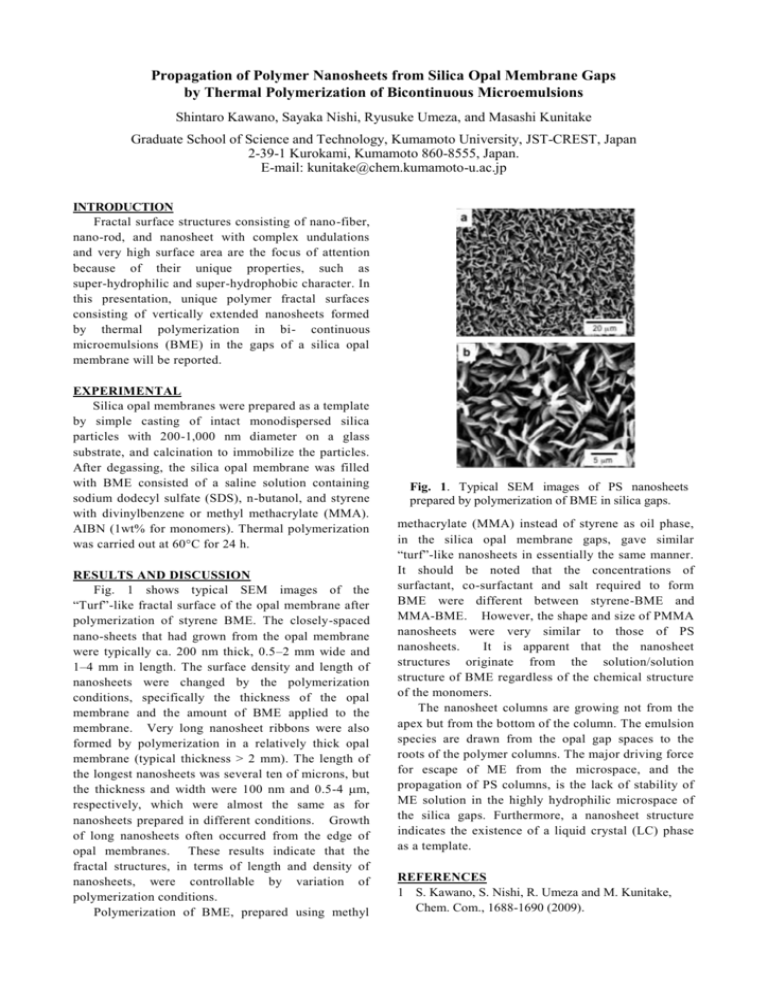
Propagation of Polymer Nanosheets from Silica Opal Membrane Gaps by Thermal Polymerization of Bicontinuous Microemulsions Shintaro Kawano, Sayaka Nishi, Ryusuke Umeza, and Masashi Kunitake Graduate School of Science and Technology, Kumamoto University, JST-CREST, Japan 2-39-1 Kurokami, Kumamoto 860-8555, Japan. E-mail: kunitake@chem.kumamoto-u.ac.jp INTRODUCTION Fractal surface structures consisting of nano-fiber, nano-rod, and nanosheet with complex undulations and very high surface area are the focus of attention because of their unique properties, such as super-hydrophilic and super-hydrophobic character. In this presentation, unique polymer fractal surfaces consisting of vertically extended nanosheets formed by thermal polymerization in bi- continuous microemulsions (BME) in the gaps of a silica opal membrane will be reported. EXPERIMENTAL Silica opal membranes were prepared as a template by simple casting of intact monodispersed silica particles with 200-1,000 nm diameter on a glass substrate, and calcination to immobilize the particles. After degassing, the silica opal membrane was filled with BME consisted of a saline solution containing sodium dodecyl sulfate (SDS), n-butanol, and styrene with divinylbenzene or methyl methacrylate (MMA). AIBN (1wt% for monomers). Thermal polymerization was carried out at 60°C for 24 h. RESULTS AND DISCUSSION Fig. 1 shows typical SEM images of the “Turf”-like fractal surface of the opal membrane after polymerization of styrene BME. The closely-spaced nano-sheets that had grown from the opal membrane were typically ca. 200 nm thick, 0.5–2 mm wide and 1–4 mm in length. The surface density and length of nanosheets were changed by the polymerization conditions, specifically the thickness of the opal membrane and the amount of BME applied to the membrane. Very long nanosheet ribbons were also formed by polymerization in a relatively thick opal membrane (typical thickness > 2 mm). The length of the longest nanosheets was several ten of microns, but the thickness and width were 100 nm and 0.5-4 m, respectively, which were almost the same as for nanosheets prepared in different conditions. Growth of long nanosheets often occurred from the edge of opal membranes. These results indicate that the fractal structures, in terms of length and density of nanosheets, were controllable by variation of polymerization conditions. Polymerization of BME, prepared using methyl Fig. 1. Typical SEM images of PS nanosheets prepared by polymerization of BME in silica gaps. methacrylate (MMA) instead of styrene as oil phase, in the silica opal membrane gaps, gave similar “turf”-like nanosheets in essentially the same manner. It should be noted that the concentrations of surfactant, co-surfactant and salt required to form BME were different between styrene-BME and MMA-BME. However, the shape and size of PMMA nanosheets were very similar to those of PS nanosheets. It is apparent that the nanosheet structures originate from the solution/solution structure of BME regardless of the chemical structure of the monomers. The nanosheet columns are growing not from the apex but from the bottom of the column. The emulsion species are drawn from the opal gap spaces to the roots of the polymer columns. The major driving force for escape of ME from the microspace, and the propagation of PS columns, is the lack of stability of ME solution in the highly hydrophilic microspace of the silica gaps. Furthermore, a nanosheet structure indicates the existence of a liquid crystal (LC) phase as a template. REFERENCES 1 S. Kawano, S. Nishi, R. Umeza and M. Kunitake, Chem. Com., 1688-1690 (2009).
