Approach to Wide Complex Tachycardia
advertisement
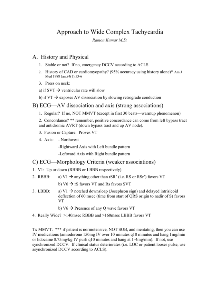
Approach to Wide Complex Tachycardia Ramon Kumar M.D. A. History and Physical 1. Stable or not? If no, emergency DCCV according to ACLS 2. History of CAD or cardiomyopathy? (95% accuracy using history alone)* Am J Med 1988 Jan;84(1):53-6 3. Press on neck: a) if SVT ventricular rate will slow b) if VT exposes AV dissociation by slowing retrograde conduction B) ECG—AV dissociation and axis (strong associations) 1. Regular? If no, NOT MMVT (except in first 30 beats—warmup phenomenon) 2. Concordance? ** remember, positive concordance can come from left bypass tract and antidromic AVRT (down bypass tract and up AV node). 3. Fusion or Capture: Proves VT 4. Axis: - Northwest -Rightward Axis with Left bundle pattern -Leftward Axis with Right bundle pattern C) ECG—Morphology Criteria (weaker associations) 1. V1: Up or down (RBBB or LBBB respectively) 2. RBBB: a) V1 anything other than rSR’ (i.e. RS or RSr’) favors VT b) V6 rS favors VT and Rs favors SVT 3. LBBB: a) V1 notched downsloap (Josephson sign) and delayed intrisicoid deflection of 60 msec (time from start of QRS origin to nadir of S) favors VT b) V6 Presence of any Q wave favors VT 4. Really Wide? >140msec RBBB and >160msec LBBB favors VT Tx MMVT: *** if patient is normotensive, NOT SOB, and mentating, then you can use IV medications (amiodorone 150mg IV over 10 minutes q10 minutes and hang 1mg/min or lidocaine 0.75mg/kg IV push q10 minutes and hang at 1-4mg/min). If not, use synchronized DCCV. If clinical status deteriorates (i.e. LOC or patient looses pulse, use asynchronized DCCV according to ACLS).

