Cytogenetic and FISH analysis of cell lines
advertisement
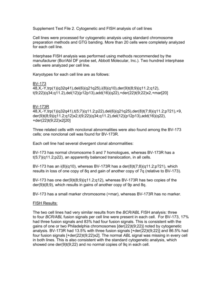
Supplement Text File 2. Cytogenetic and FISH analysis of cell lines Cell lines were processed for cytogenetic analysis using standard chromosome preparation methods and GTG banding. More than 20 cells were completely analyzed for each cell line. Interphase FISH analysis was performed using methods recommended by the manufacturer (Bcr/Abl DF probe set, Abbott Molecular, Inc.). Two hundred interphase cells were analyzed per cell line. Karyotypes for each cell line are as follows: BV-173 48,X,-Y,trp(1)(q32q41),del(6)(q21q25),i(8)(q10),der(9)t(8;9)(q11.2;q12), t(9;22)(q34;q11.2),del(12)(p12p13),add(16)(q22),+der(22)t(9;22)x2,+mar[20] BV-173R 48,X,-Y,trp(1)(q32q41),t(5;7)(q11.2;p22),del(6)(q21q25),der(8)t(7;8)(q11.2;p?21),+9, der(9)t(8;9)(q11.2;q12)x2,t(9;22)(q34;q11.2),del(12)(p12p13),add(16)(q22), +der(22)t(9;22)x2[20] Three related cells with nonclonal abnormalities were also found among the BV-173 cells; one nonclonal cell was found for BV-173R. Each cell line had several divergent clonal abnormalities: BV-173 has normal chromosome 5 and 7 homologues, whereas BV-173R has a t(5;7)(q11.2;p22), an apparently balanced translocation, in all cells. BV-173 has an i(8)(q10), whereas BV-173R has a der(8)t(7;8)(q11.2;p?21), which results in loss of one copy of 8q and gain of another copy of 7q (relative to BV-173). BV-173 has one der(9)t(8;9)(q11.2;q12), whereas BV-173R has two copies of the der(9)t(8;9), which results in gains of another copy of 9p and 8q. BV-173 has a small marker chromosome (+mar), whereas BV-173R has no marker. FISH Results: The two cell lines had very similar results from the BCR/ABL FISH analysis: three to four BCR/ABL fusion signals per cell line were present in each cell. For BV-173, 17% had three fusion signals and 83% had four fusion signals. This is consistent with the gains of one or two Philadelphia chromosomes [der(22)t(9;22)] noted by cytogenetic analysis. BV-173R had 13.5% with three fusion signals [+der(22)t(9;22)] and 86.5% had four fusion signals [+der(22)t(9;22)x2]. The normal ABL signal was missing in every cell in both lines. This is also consistent with the standard cytogenetic analysis, which showed one der(9)t(9;22) and no normal copies of 9q in each cell.
