(Undergraduate Medical Education, Microbiology: Student Study Module:
Geriatrics Integration Case)
CASE 1: ACUTE ABDOMINAL PAIN (POLYMICROBIAL COLON FLORA)
A 35-year-old woman presents to the ER (emergency room) with acute abdominal
pain that began 17 hours ago She describes the pain as being colicky in the
beginning, with an intensity of 4/10 and located in the center of her abdomen. The
pain has gotten progressively worse, and now she describes it as constant and sharp,
with a level of 7/10. Her pain was worsened by the bumps on the road while being
brought to the ER by her husband. The pain moved to her right lower quadrant just
before she arrived at the hospital. The patient reports a recent loss of appetite and
two episodes of clear vomiting. She has not had a bowel movement for three days.
She denies diarrhea, or weight loss. She has not taken her temperature, but she
admits to feeling “a little warm” with a few chills.
Physical examination reveals the following: oral temperature=38.4C, blood
pressure=125/70 mmHg, pulse rate=100 beats/min, and respiratory rate=16
breaths/min. HEENT: Flushed face and dry mucosa. Abdominal exam: Slightly
distended, almost no bowel sounds. Right lower quadrant tenderness and rebound
pain present with a positive Rovsing sign. Iliopsoas sign on the right is positive.
Rectal exam shows no masses and diffuse mild tenderness bilaterally, with stool
guaiac negative for blood. Other physical findings, including pelvic, pulmonary, and
cardiac examinations, were normal.
The patient’s ER lab tests are as follows: white blood cell count, 13,900 per mm3
(13.9 × 10⁹ per L); hemoglobin, 12.7 g per dL (127 g per L); neutrophils, 85 percent;
sodium, 138 mEq per L (138 mmol per L); potassium, 4.1 mEq per L (4.1 mmol per
L); blood urea nitrogen, 12 mg per dL (4.5 mmol per L); creatinine, 0.7 mg per dL
(60 µmol per L); aspartate transaminase, 15 U per L; and alanine transaminase, 25
U per L. Amylase and lipase are elevated to double normal values.
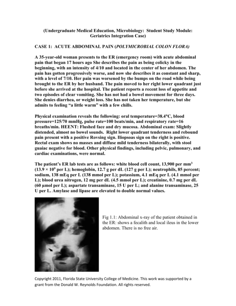
Fig 1.1: Abdominal x-ray of the patient obtained in
the ER: shows a fecalith and local ileus in the lower
abdomen. There is no free air.
Copyright 2011, Florida State University College of Medicine. This work was supported by a
grant from the Donald W. Reynolds Foundation. All rights reserved.
Question 1.1: What is your preliminary diagnosis?
This patient has the typical presentation of acute appendicitis, the single most common
acute abdominal emergency. It occurs most often between the ages of 10 and 20 years,
but no age is exempt. A male preponderance exists, with a male to female ratio of 1.4:1;
the overall lifetime risk is 8.6% for males and 6.7% for females in the United States.
Question 1.2: How do you diagnose it?
The diagnosis of acute appendicitis relies on a thorough history which elicits the
symptom sequence of colicky periumbilical abdominal pain, followed by vomiting, with
migration of the pain to the right iliac fossa and physical examination which establishes
classic tenderness and muscular rigidity after localization of the pain to the right iliac
fossa. The classic history above was first described by Murphy but may only be present
in 50% of patients. Loss of appetite is often a predominant feature; constipation and
nausea are also often present. Physical exam most commonly shows abdominal rigidity,
McBurney’s tenderness, Rovsing’s sign, guarding and rebound. Rectal tenderness, a
positive psoas sign and a positive obturator sign are not always present. The patient is
often flushed, with dry mucosa and an associated fetor oris. Pyrexia up to 38°C with
tachycardia is common.
Question 1.3: What are the top differential diagnoses?
There are multiple potential involved organ systems due to the location of abdominal
pain, which will vary by age and gender. Potential gastrointestinal causes include
intestinal obstruction, intussusception, acute cholecystitis, gastroenteritis and terminal
ileitis; genitourinary causes may include ureteral colic, pyelonephritis, urinary tract
infection, ovarian cyst torsion or follicle rupture, pelvic inflammatory disease, and
ectopic pregnancy. Pneumonia may also present with referred pain to the abdomen due to
diaphragmatic irritation.
Questions 1.4: What studies will you request now to confirm your clinical suspicion?
The patient has a blood count with predominant leucocytosis and a neutrophil shift,
findings which are present in 80-90% of cases. Urinalysis may be abnormal in up to 40%
of patients. Females of reproductive age should also be evaluated for pregnancy. The
amylase and lipase are typically elevated due to inflammation but not specific to
appendicitis. For imaging studies: plain radiography does not have any role in diagnosing
acute appendicitis, although it may be useful when evaluating potential complications
such as perforation showing free air, or as in our patient’s case, an ileus may be shown
(Fig 1)
Copyright 2011, Florida State University College of Medicine. This work was supported by a
grant from the Donald W. Reynolds Foundation. All rights reserved.
Computed tomography is the most accurate diagnostic test, with a sensitivity and
specificity of 95%. It can show an abnormal appendix, calcified appendicolith seen in
association with periappendiceal inflammation, or diameter >6 mm. In this patient’s case,
it shows a thick-walled structure with surrounding inflammation in the right lower
abdomen (Fig 2, red arrow)
Fig 1.2: Computed tomography of the patient.
Question 1.5: What are the risk factors for this disease?
The cause of acute appendicitis is unknown but is probably multifactorial; luminal
obstruction, dietary and familial factors have all been suggested. An increased incidence
in adolescent males has been noted. The obstruction of the appendicular lumen can be
caused by fecaliths, lymphoid tissue hypertrophy, strictures, intra-abdominal tumors, and
foreign bodies. Foreign bodies may include vegetable and fruit seeds, impacted barium,
and intestinal worms (ascaris).
Question 1.6: What are the complications associated with this condition?
Perforation: Profuse vomiting may indicate development of generalized peritonitis after
perforation but is rarely a major feature in simple appendicitis. Delayed diagnosis
increases the risk of perforation. After the first 36 hours from the onset of symptoms the
average rate of perforation is between 16% and 36%, and the risk of perforation is 5% for
every subsequent 12 hour period.
Abscess: Patients with an appendix abscess have a tender mass with a swinging pyrexia,
tachycardia, and leucocytosis. It is most often located in the lateral aspect of the right iliac
fossa but may be pelvic; a rectal examination is useful to identify a pelvic collection. The
abscess can be shown by ultrasonography or computed tomography scanning, and
percutaneous radiological drainage can be done. However, open drainage has the added
advantage of allowing an appendectomy to be done.
Question 1.7: What is the treatment of this condition?
Copyright 2011, Florida State University College of Medicine. This work was supported by a
grant from the Donald W. Reynolds Foundation. All rights reserved.
Expedient appendectomy is the treatment of choice. All patients should receive broad
spectrum perioperative antibiotics to decrease the incidence of postoperative wound
infection and intra-abdominal abscess formation.
Appendectomy is a relatively safe procedure with a mortality rate for non-perforated
appendicitis of 0.8 per 1000.
Open appendectomy is done through an incision over McBurney's point. Laparoscopic
appendectomy in adults is also possible and reduces wound infections, postoperative pain,
length of hospital stay, and time taken to return to work. Initial laparoscopy for diagnosis
may show alternative pathology as the cause of the presentation.
Fig 1.3: Laparoscopic appendectomy
Question 1.8: What are potential infectious surgical complications?
Wound infection and intra-abdominal wound abscess are the most serious potential
complications. The rate of postoperative wound infection is determined by intraoperative
wound contamination. Rates of infection vary from < 5% in simple appendicitis to 20%
in cases with perforation and gangrene. Intra-abdominal or pelvic abscesses may form in
the postoperative period after gross contamination of the peritoneal cavity. Diagnosis can
be confirmed by ultrasonography or computed tomography scanning. Abscesses can be
treated radiologically with a drain, although open or rectal drainage may be needed for a
pelvic abscess. Organisms commonly cultured include Escherichia coli, Viridans
streptococci, Pseudomonas aeruginosa, Bacteroides fragilis, and Peptostreptococcus
micros.
Question 1.9: Are there any differences in the clinical presentation and course of
this condition in an older adult?
The main three causes of acute abdominal pain in the geriatric population in order of
frequency are cholecystitis, bowel obstruction and appendicitis. The oldest old (age 85
years and up) patients with acute appendicitis tend to present with fewer classic
symptoms and signs than younger patients, notably migrating pain and right lower
quadrant tenderness. Instead, abdominal pain tends to be vague and diffuse. Because
vague abdominal pain is a common complaint in older persons, its significance can be
frequently overlooked. Many patients will present with the acute onset of delirium and
little pain. In one study, only 55% of patients older than age 80 had right lower quadrant
pain and 18% had no abdominal pain at all. Other signs of acute appendicitis are also
Copyright 2011, Florida State University College of Medicine. This work was supported by a
grant from the Donald W. Reynolds Foundation. All rights reserved.
unreliable in the elderly. White blood cells counts are less than 10,000/mm3 in 20% to
50% of older patients with simple appendicitis. Temperature is less than 37.6°C in up to
half of cases, and bacteremic elders may even be hypothermic. Nausea, vomiting, and
anorexia are also found less frequently in older patients.
The indolent nature of the initial symptoms of appendicitis in the older adult usually leads
to delays of 48 to 72 hours before medical attention is sought. In only 30% to 70% of
cases is the correct diagnosis made on admission. Delays to operation of greater than 24
hours are three times as likely to occur in older than in younger patients. The rate of
perforation is significantly increased in older adults, reaching up to 97%, with a mortality
rate of over 30%, usually because of a delay in diagnosis.
Specific physiological alterations, comorbidities and psychosocial factors may also
contribute to a late diagnosis. The perception of pain and its localization is altered due to
modifications of neural mechanisms and diminished immune function, but the traditional
stereotype that older adults have a higher pain threshold has been questioned. T cell
function is decreased, the bone marrow capacity is depressed and the inflammatory
response is dampened. Multiple co morbidities and polypharmacy may mask the acute
presentation of appendicitis. Psychosocial aspects that may delay presentation to
healthcare include living alone, and fear of the medical care system. Careful examination
and, maintaining a high index of suspicion for the diagnosis are extremely important in
avoiding a delayed diagnosis and its consequences.
References
Andrassay R. Appendix. Ch 47 in: Townsend C, Beauchamp E, Evers B, Mattox
K, eds. Sabiston Textbook of Surgery, 17th ed. Elsevier. Philadelphia: 2004.
Fenton, A. Appendicitis, in InfoRetriever: 5 minute clinical consult. Accessed
December 07, 2007 (PDA and online).
Ferri F. Appendicitis, in FirstConsult. Accessed on December 12, 2007.
Hardin, D. Acute appendicitis. Am Fam Phys. 1999: 60; 2027-34
Humes D, Simpson, J. Acute appendicitis. BMJ 2006:333;530-4.
Langer J. Gastroenterology and Hepatology: Edited by Mark Feldman (series
editor), Paul E. Hyman. ©1997 Current Medicine Group LLC. Accessed at
imagesmd.com on December 12, 1007
Lunca S, Buras G, Romedea N. Acute appendicitis in the elderly patient:
diagnosis problems, prognostic factors and outcomes. Rom J Gastroenterology.
2004 :13;299-303.
Copyright 2011, Florida State University College of Medicine. This work was supported by a
grant from the Donald W. Reynolds Foundation. All rights reserved.

