URINARY SYSTEM II
advertisement
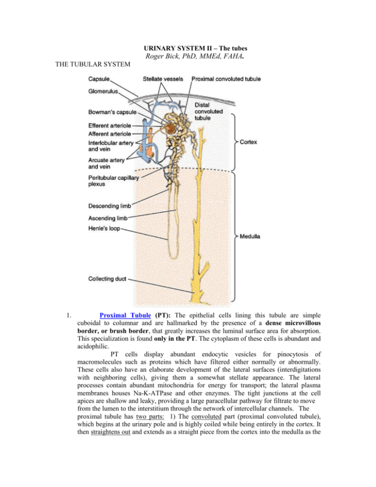
URINARY SYSTEM II – The tubes Roger Bick, PhD, MMEd, FAHA. THE TUBULAR SYSTEM 1. Proximal Tubule (PT): The epithelial cells lining this tubule are simple cuboidal to columnar and are hallmarked by the presence of a dense microvillous border, or brush border, that greatly increases the luminal surface area for absorption. This specialization is found only in the PT. The cytoplasm of these cells is abundant and acidophilic. PT cells display abundant endocytic vesicles for pinocytosis of macromolecules such as proteins which have filtered either normally or abnormally. These cells also have an elaborate development of the lateral surfaces (interdigitations with neighboring cells), giving them a somewhat stellate appearance. The lateral processes contain abundant mitochondria for energy for transport; the lateral plasma membranes houses Na-K-ATPase and other enzymes. The tight junctions at the cell apices are shallow and leaky, providing a large paracellular pathway for filtrate to move from the lumen to the interstitium through the network of intercellular channels. The proximal tubule has two parts: 1) The convoluted part (proximal convoluted tubule), which begins at the urinary pole and is highly coiled while being entirely in the cortex. It then straightens out and extends as a straight piece from the cortex into the medulla as the thick descending limb. Its function is to reabsorb about 85% of the filtrate isosmotically via active and passive transport processes. It is thus the major site of reabsorption of glucose, amino acids, electrolytes, water, and due to its pinocytotic activity, proteins. Thin Limbs - Structure The tall columnar-cuboidal cells of the thick descending limb abruptly change to the simple squamous lining cells of the thin descending limb. These cells have scanty cytoplasm with few mitochondria. All nephrons have thin descending limbs that travel different depths into the medulla. In the long-looped nephrons the thin limbs go all the way down to the tip of the papilla, turn, and go back up to the border of the inner and outer medulla where the thick ascending limb (TAL) begins. In short-looped nephrons, the thin descending limb curves back up at the border of the inner and outer medulla and immediately become TAL’s. In these nephrons, therefore, there is no thin ascending limb at all. Function - Studies indicate that the thin limbs participate in urine concentration. The long looped nephrons, Juxtamedullary nephrons, are of prime importance in establishing the gradient of hypertonicity in the medullary interstitium. Distal Tubule: Thick Ascending Limb (TAL) to Distal Convoluted (DCT), Structure - The beginning of the TAL defines the border between inner and outer medulla, with an abrupt transition in this nephron segment from squamous to low columnar/high cuboidal. It courses back to the parent glomerulus in the cortex, and as it does its epithelium gradually becomes lower. At the glomerulus it becomes juxtaposed between the afferent and efferent arterioles, with its epithelium is almost squamous at this point, except for a specialized area called the macula densa (dark spot), at the beginning of the DCT. Cells here are very tightly packed and the relative density of the nuclei, make this area appear darker than in the rest of the epithelial lining cells. The macula densa is in intimate contact with Extraglomerular mesangial cells and renin-secreting juxtaglomerular cells. Juxtaglomerular Apparatus (JGA) is composed of: Macula densa Extraglomerular mesangial cells Juxtaglomerular cells (afferent + efferent arterioles) Function - The TAL actively removes salt from the filtrate while being impermeable to water. Since water does not cross its epithelium, filtrate osmolality declines (diluting segment). It is postulated that filtrate composition (NA concentration? Osmolality ?) is sensed by the macula densa, and a signal is transmitted to the JG cells which results in renin release. Systemic renin is necessary for generation of the powerful vasopressor, angiotensin II, which stimulates the adrenal cortex to secrete aldosterone. Aldosterone feeds back to the collecting duct for absorption of sodium and water and together these mechanisms help maintain blood pressure. Distal Convoluted Tubule (DCT)-Macula densa found here Structure - After passing the glomerular hilus, the nephron abruptly displays uniformly cuboidal cells, and convolutes tortuously in the. Several distal convoluted tubules merge into a single cortical collecting duct. Function - Active absorption of salt and concomitant movement of water occur in this segment. Collecting Duct/Tubule Structure - Collecting ducts are straight, and lined with an initially low cuboidal epithelium that becomes columnar in the papilla. The cell population of the epithelium is heterogeneous and contains two morphologically different cell types: Principle Cell: is the most abundant throughout the collecting duct. It is also called a light cell because of its light cytoplasmic appearance. Intercalated Cell: less numerous and is interposed between groups of principal cells. Abundant mitochondria and an electron-dense cytoplasm give it a dark appearance when viewed with TEM, a dark cell. It has more subapical vesicles, and surface specializations,microplicae, that further distinguish it from the principal cell. Intercalated cells account for up to 40% of the total cell population of the cortical CD, 18% of the outer medullary CD, and less than 1% of the inner medullary CD. Function - The initial portion of the CD is sensitive to aldosterone, a hormone that stimulates sodium absorption and potassium secretion. The entire CD is sensitive to antidiuretic hormone (ADH; vasopressin). With ADH stimulation, water leaves the filtrate and goes into the interstitium without parallel movement of salt. The CD is the major site for concentration of filtrate. INTERSTITIUM While tubules occupy much of the total renal volume, connective tissue surrounds each tubule and is collectively called the interstitium. Fibroblast-like cells are the predominant cell type of this tissue, and are more numerous and prominent in the medulla. Much of the interstitium is occupied by capillaries, not surprising since both kidneys receive a quarter of the total cardiac output. Although a subtle morphologic feature in paraffin sections, the capillary plexuses and interstitium are as critical in renal physiology as the tubules. Interstitial cells are also the site of prostaglandin production. TYPES OF NEPHRONS Cortical, and juxtamedullary. Location of Glomerulus Length of Henle’s Loop Cortical (Upper cortex) Juxtamedullary (Deep cortex) Short Long Efferent gives rise to arteriole Cortical peritubular capillaries Medullary vasa rectae POST-GLOMERULAR CIRCULATION Blood leaving the glomerulus via efferent arterioles has a variable course, depending on the location of the parent glomerulus. 1. Cortical Nephrons - Blood leaving the efferent arteriole breaks upon into capillaries that surround the portions of the tubules lying in the cortex, peritubular capillaries. The passage of blood through these nephrons is: Efferent arteriole to peritubular capillaries to venule to interlobular vein to arcuate vein to interlobar vein to renal vein The significance of this unique system, with two capillary beds in series separated by an arteriole, is that hydrostatic pressure in the glomerular capillary bed will normally be very high relative to other capillaries in the body, facilitating large amounts of filtration. On the distal side of the arteriole, pressure in the peritubular capillary system will be inordinately low, favoring reabsorption of fluid from the interstitium. In this way, the large amount of fluid passing from the tubules, particularly the proximals, will be quickly taken back into the systemic circulation. 2. Juxtamedullary Nephrons - Efferent arterioles of juxtamedullary glomeruli pass down into the medulla as vasa rectae, which make a "hair-pin" turn, and leave the medulla in a parallel course as ascending vasa recta. Descending vasa recta give off branches that supply the capillary plexuses of the medulla, which then drain back into the ascending vasa rectae. Blood flow then is as follows: a. b. c. d. Efferent arteriole Descending vasa recta Capillaries Ascending vasa recta e. Arcuate vein f. Interlobar vein g. Renal vein Blood flow in the medulla is completely separate from that of the cortex and serves a different purpose. Efferent arterioles insure high juxtamedullary glomerular capillary filtration, but then the distal capillary network, connecting descending and ascending vasa rectae, are designed as hairpin loops to facilitate concentration of the filtrate. ZONES OF THE KIDNEY The zones are presented here in outline form and diagrammatically. It is strongly suggested that when you are finished studying this lecture you practice roughly drawing juxtaglomerular and cortical nephrons as they course through the different zones of the kidney to be sure you understand the topography of the nephron and that of the lobe. 1. Cortex 2. Medulla a. Outer zone (Outer medulla) (1) Outer stripe (2) Inner stripe b. Inner zone (Inner medulla) LABORATORY GUIDE FOR URINARY SYSTEM - PART II I. NEPHRON SEGMENTS: Use your basic diagram for orientation. Slides 74 and 76 Begin in the inner medulla (papilla), and work your way out to the cortex. The medulla has the simplest structure and fewer tube types, whereas the cortex is more complex. Even to the experienced, not all tubule types are identifiable in paraffin sections! If some tubules are indeterminate, as many will be, don't concern yourself, but concentrate on being able to identify good examples of each structure underlined. A. B. 1. Inner Medulla (Inner Zone of the Medulla) The largest circular profiles with large lumens are collecting ducts (usually), with a simple tall columnar lining epithelium. At the tip of the papilla, these ducts can be seen in longitudinal section opening into a minor calyx. Other small circular profiles represent thin limbs and vasa recta, which cannot be distinguished with certainty unless you see erythrocytes in the lumen of the latter. These are both lined by a simple squamous epithelium. These thin-walled structures are both descending and ascending here. The interstitium fills in between these structures. Outer Medulla (Outer Zone of the Medulla) Inner Stripe The border between inner and outer medulla is sharply divided by the sudden presence in the inner stripe of the thick ascending limbs, which have cuboidal cells with apical nuclei. So to positively identify these positively, go to this border and note tubules lined by a cuboidal epithelium which is slightly more eosinophilic than that of the collecting ducts. Thin limbs and capillaries are again difficult to distinguish from each other, but tend to aggregate together in bundles, separate from the thick ascending limbs. As usual, they are lined by a squamous epithelium. 2. Outer Stripe In the outer stripe of the outer medulla, the straight part of the proximal tubule appears abruptly. These are very eosinophilic in the rat (less so in human) and have a brush border. They are cuboidal to low columnar. Look carefully for a profile showing the transition from a proximal tubular epithelium to the squamous epithelium of the thin descending limb. This is the only site you will definitely be able to identify a thin limb and state with certainty that it is descending. Thick ascending limbs are still coursing out to the cortex, but are being lined by a progressively lower epithelium than was found in the inner stripe. Collecting ducts continue here, but are lined by a lower epithelium than in the inner medulla. C. Cortex This region of the kidney is defined by the presence of renal corpuscles, which you studied in Part I. Look for proximal tubules in the rat kidney first, focusing up and down to resolve the brush border. These tubules may appear collapsed and a brush border may not even be resolvable in some (it may look fluffy). Convoluted and straight segments appear identical, but the straight sections will be seen coursing in long rays parallel to thick ascending limbs and collecting ducts. Cells of proximal tubules are low columnar to cuboidal; their nuclei are located in a central position in each cell. Whereas the thick ascending limb displays a tall cuboidal epithelium with central to apical nuclei in the outer medulla, as it approaches the glomerulus it becomes flatter and nuclei are mostly apical. Identify by the presence of extraglomerular mesangial cells, perhaps one or both arterioles, and a macula densa in the thick ascending limb. You will know the macula densa when you see nuclei clustered closely together in this tubule as it abuts the parietal epithelium. In the rat the thick ascending limb is quite squamous at this point, and the nuclei of the macula densa are very dark. In the human, this tubule is cuboidal and the nuclei of the macula densa are not dark, but they are much more closely spaced than those of the other parts of the thick ascending. After leaving the JGA, the thick ascending limb is called the distal convoluted tubule that is cuboidal in both species. Distal convoluted tubules lack a brush border and have apically-located, oblong nuclei. Although we will discuss it and try to find it, you will not be tested on it since it can look a lot like thick ascendings and collecting ducts. To find cortical collecting ducts, look for longitudinally-oriented tubules running in rays perpendicular to the surface of the kidney. In humans, they are lined by a cuboidal epithelium and tend to have wide open lumens. Both here and in the inner part of the kidney, another trick to identifying them is that only in this part of the nephron can you see the lateral plasma membrane separating each cell. Thus the cell borders are easily visible. There may also be halo effect around the nuclei. While dark or intercalated cells occur frequently in the cortex of both species and are identified by their eosinophilia and large size. The space between the tubules is the interstitium. It is quite sparse and occupied primarily by peritubular capillaries.
