OMB No. 0925-0046, Biographical Sketch Format Page
advertisement
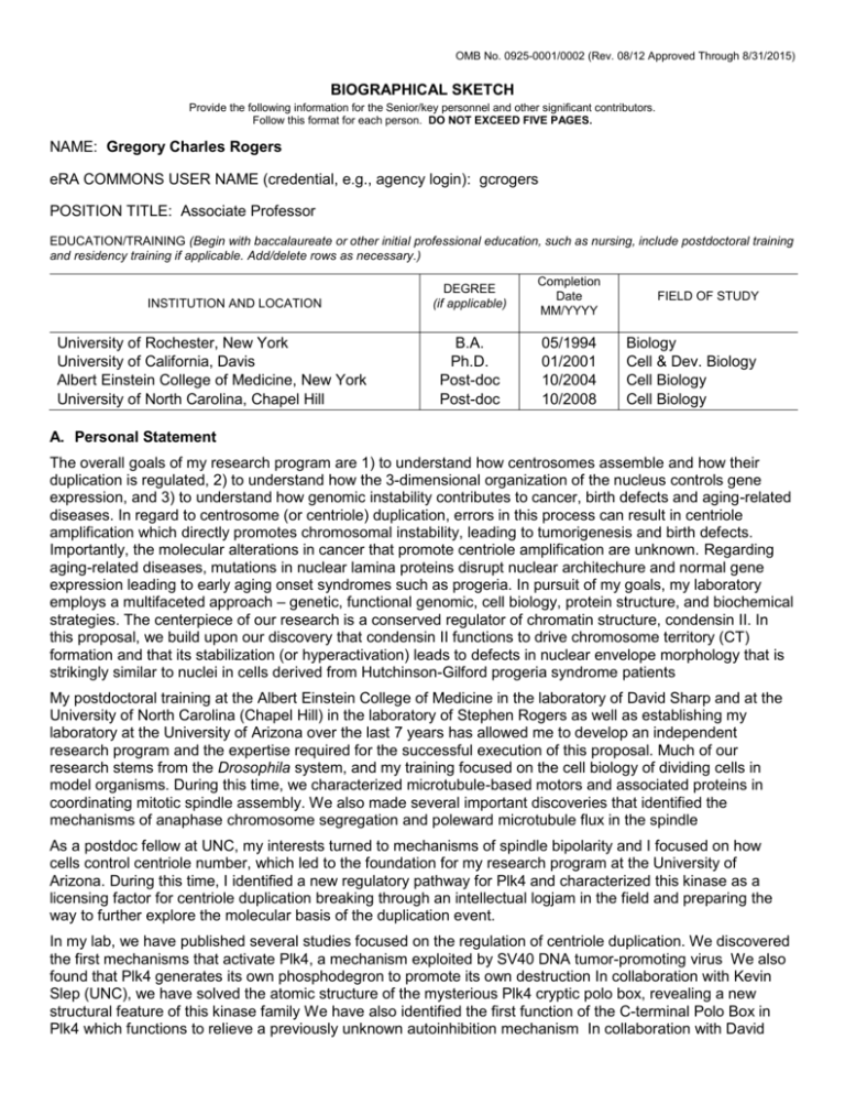
OMB No. 0925-0001/0002 (Rev. 08/12 Approved Through 8/31/2015) BIOGRAPHICAL SKETCH Provide the following information for the Senior/key personnel and other significant contributors. Follow this format for each person. DO NOT EXCEED FIVE PAGES. NAME: Gregory Charles Rogers eRA COMMONS USER NAME (credential, e.g., agency login): gcrogers POSITION TITLE: Associate Professor EDUCATION/TRAINING (Begin with baccalaureate or other initial professional education, such as nursing, include postdoctoral training and residency training if applicable. Add/delete rows as necessary.) INSTITUTION AND LOCATION University of Rochester, New York University of California, Davis Albert Einstein College of Medicine, New York University of North Carolina, Chapel Hill DEGREE (if applicable) Completion Date MM/YYYY B.A. Ph.D. Post-doc Post-doc 05/1994 01/2001 10/2004 10/2008 FIELD OF STUDY Biology Cell & Dev. Biology Cell Biology Cell Biology A. Personal Statement The overall goals of my research program are 1) to understand how centrosomes assemble and how their duplication is regulated, 2) to understand how the 3-dimensional organization of the nucleus controls gene expression, and 3) to understand how genomic instability contributes to cancer, birth defects and aging-related diseases. In regard to centrosome (or centriole) duplication, errors in this process can result in centriole amplification which directly promotes chromosomal instability, leading to tumorigenesis and birth defects. Importantly, the molecular alterations in cancer that promote centriole amplification are unknown. Regarding aging-related diseases, mutations in nuclear lamina proteins disrupt nuclear architechure and normal gene expression leading to early aging onset syndromes such as progeria. In pursuit of my goals, my laboratory employs a multifaceted approach – genetic, functional genomic, cell biology, protein structure, and biochemical strategies. The centerpiece of our research is a conserved regulator of chromatin structure, condensin II. In this proposal, we build upon our discovery that condensin II functions to drive chromosome territory (CT) formation and that its stabilization (or hyperactivation) leads to defects in nuclear envelope morphology that is strikingly similar to nuclei in cells derived from Hutchinson-Gilford progeria syndrome patients My postdoctoral training at the Albert Einstein College of Medicine in the laboratory of David Sharp and at the University of North Carolina (Chapel Hill) in the laboratory of Stephen Rogers as well as establishing my laboratory at the University of Arizona over the last 7 years has allowed me to develop an independent research program and the expertise required for the successful execution of this proposal. Much of our research stems from the Drosophila system, and my training focused on the cell biology of dividing cells in model organisms. During this time, we characterized microtubule-based motors and associated proteins in coordinating mitotic spindle assembly. We also made several important discoveries that identified the mechanisms of anaphase chromosome segregation and poleward microtubule flux in the spindle As a postdoc fellow at UNC, my interests turned to mechanisms of spindle bipolarity and I focused on how cells control centriole number, which led to the foundation for my research program at the University of Arizona. During this time, I identified a new regulatory pathway for Plk4 and characterized this kinase as a licensing factor for centriole duplication breaking through an intellectual logjam in the field and preparing the way to further explore the molecular basis of the duplication event. In my lab, we have published several studies focused on the regulation of centriole duplication. We discovered the first mechanisms that activate Plk4, a mechanism exploited by SV40 DNA tumor-promoting virus We also found that Plk4 generates its own phosphodegron to promote its own destruction In collaboration with Kevin Slep (UNC), we have solved the atomic structure of the mysterious Plk4 cryptic polo box, revealing a new structural feature of this kinase family We have also identified the first function of the C-terminal Polo Box in Plk4 which functions to relieve a previously unknown autoinhibition mechanism In collaboration with David Bilder (UCB) and Giovanni Bosco (Dartmouth College), we have expanded our studies of ubiquitin-mediated regulation to nuclear organization and we recently discovered a new pathway for condensin II regulation that may contribute to a host of laminopathies in humans. 1. Rogers GC, Rogers SL, Schwimmer TA, Ems-McClung SC, Walczak CE, Vale RD, Scholey JM, Sharp DJ. Two mitotic kinesins cooperate to drive sister chromatid separation during anaphase Nature. 2004 Jan 22;427(6972):364-70. Epub 2003 Dec 14. PMID: 14681690 2. Rogers GC, Rusan NM, Roberts DM, Peifer M, Rogers SL. The SCF Slimb ubiquitin ligase regulates Plk4/Sak levels to block centriole reduplication. J Cell Biol. 2009 Jan 26;184(2):225-39. doi: 10.1083/jcb.200808049. PMCID: PMC2654306 3. Brownlee CW, Klebba JE, Buster DW, Rogers GC. The Protein Phosphatase 2A regulatory subunit Twins stabilizes Plk4 to induce centriole amplification. J Cell Biol. 2011 Oct 17;195(2):231-43. doi: 10.1083/jcb.201107086. Epub 2011 Oct 10. PMCID: PMC3198173 4. Klebba JE, Buster DW, Nguyen AL, Swatkoski S, Gucek M, Rusan NM, Rogers GC. Polo-like kinase 4 autodestructs by generating its Slimb-binding phosphodegron. Curr Biol. 2013 Nov 18;23(22):2255-61. doi: 10.1016/j.cub.2013.09.019. Epub 2013 Oct 31. PMCID: PMC3844517 5. Klebba JE, Buster DW, McLamarrah TA, Rusan NM, Rogers GC. Autoinhibition and relief mechanism for Polo-like kinase 4. Proc Natl Acad Sci U S A. 2015 Feb 17;112(7):E657-66. Doi 10.1073/pnas.1417967112. Epub 2015 Feb 2 PMCID: PMC4343128 B. Positions and Honors Positions 2014-present University of Arizona, Associate Professor, Department of Cellular and Molecular Medicine 2008-2014 University of Arizona, Assistant Professor, Department of Cellular and Molecular Medicine, Joint Appointment, Department of Molecular and Cellular Biology Honors 1991-1994 University of Rochester Undergraduate Scholarship 1993 Die Kiewet Undergraduate Summer Research Fellowship 1994 Cum Laude, General Honors, University of Rochester 1997-1998 Jastro-Shields Graduate Research Award 1996-1998 Molecular and Cellular Biology NIH Graduate Training Grant 2009-2010 American Cancer Society Institutional Research Grant Award 2009-2010 Arizona Cancer Center SPORE in GI Cancer Career Development Award 2010 Joseph S. Pagano Postdoctoral Award – UNC Lineberger Cancer Center 2010 March of Dimes Basil O’Connor Award C. Contribution to Science When I began my first post-doctoral stint, a fairly elaborate picture of the nature and structure of mitotic spindles had been developed, but a large question remained: how are mitotic chromosomes segregated? For over 100 years, the fundamental qualities of the question have kept it intriguing. My publication explained the mechanisms of anaphase A chromosome segregation, when the sister chromatids disjoin, thus ensuring genomic inheritance to each daughter cell. Using Drosophila cells as my model system, I showed that chromosome-to-pole motion during anaphase A are driven by kinetochore microtubules, which are actively depolymerized at both their ends simultaneously to move chromatids poleward. Specifically, I characterized two microtubule-depolymerizing kinesins, KLP10A and KLP59C, whose complementary activities drive anaphase A. I showed that KLP10A depolymerizes spindle pole-associated microtubule minus-ends, causing microtubules to ‘flux’ and chromosomes to be ‘reeled-in’ toward spindle poles. My analyses revealed KLP10A as the first molecular component of poleward microtubule flux and, in doing so, revealed a function for flux. At the opposite ends of spindle microtubules, I showed that KLP59C depolymerizes kinetochore-associated microtubule plus-ends, participating in the ‘Pacman’ mechanism of anaphase A (KLP59C was the first molecular component of ‘Pacman’ motility to be identified). Lastly, I showed that poleward flux constrains mitotic spindle lengths and that inactivation of flux might trigger the spindle elongation during anaphase B – a conclusion verified 9 years later. a. Buster, D.W., Daniel, S.G., Nguyen, H.Q., Windler, S.L., Skwarek, L.C., Peterson, M., Roberts, M., Meserve, J.H., Hartl, T., Klebba, J.E., Bilder, D., Bosco, G. and Rogers, G.C. (2013) SCFSlimb ubiquitin- ligase suppresses condensin II-mediated nuclear reorganization by degrading Cap-H2. Journal of Cell Biology. 201, 49-63. PMID: 23530065 b. Rogers, G.C., Rogers, S.L., Schwimmer, T.A., Ems-McClung, S.C., Walczak, C.E., Vale, R.D., Scholey, J.M. and Sharp, D.J. (2004) Two mitotic kinesins cooperate to drive sister chromatid separation during anaphase. Nature. 427, 364-370. Received News & Views article in same issue. This paper is cited in two textbooks: Lodish et al., Mol. Cell. Biol., 6th edition and Berg et al., Biochemistry, 7th edition. PMID: 14681690 This study lays the foundation for the current application. Eukaryotic genomes are spatially organized in a nonrandom manner, and this 3-dimensional genomic structure is likely functionally important for control of gene expression, DNA replication and repair. Chromosome-conformation capture techniques have revealed that interphase chromosomes exist as globule-like structures, called chromosome territories (CTs), capable of longrange chromatin interactions. Notably, these studies provide little or no data that speaks to the mechanism of biological regulation of chromosome folding and functional organization of genomes. Even though such chromosome organization states (i.e., Rabl and CTs) were described ~100 years ago, essentially nothing was known of the molecules regulating any type of 3-dimensional genome organization. In this study, we provide the first mechanistic explanation of interphase genome remodeling. Specifically, we found that the SCFSlimb ubiquitinligase controls condensin II activity during interphase by targeting the condensin II regulatory subunit, Cap-H2, for degradation, thereby diminishing condensin II-mediated compaction of chromatin. Thus, ubiquitin-mediated suppression of condensin II activity blocks chromosome compaction, chromosome individualization, and promotes homolog pairing. Our study was significant as it (1) identified the first molecular mechanism for chromosome individualization and CT formation, (2) was the first to demonstrate that condensins are regulated by ubiquitin-mediated proteolysis, (3) was the first to show how condensin II is regulated during interphase, and (4) demonstrated a new role for the SCFSlimb ubiquitin-ligase in nuclear reorganization. Lastly, we discovered that hyperactivation of condensin II also deforms nuclear envelopes in a way strikingly similar in appearance to those in cells from Hutchinson-Gilford progeria syndrome patients. Our work revealing the mechanism controlling interphase chromosome organization provides the basis to functionally link chromosome organization, topological regulation of genes, and a variety of laminopathies. a. Rogers, G.C., Rusan, N.M., Roberts, D.M., Peifer, M. and Rogers, S.L. (2009) The SCFSlimb ubiquitinligase regulates Plk4/Sak levels to block centriole reduplication. Journal of Cell Biology. 184, 225-239. Received cover illustration and an In This Issue commentary. PMID: 19171756 The idea that centrosome number requires close regulation is an old one, dating back more than 100 years ago to Theodor Boveri who hypothesized that centrosome overduplication (or amplification) causes aneuploidy and, consequently, tumorigenesis. But even beyond the possible medical consequences, understanding the schemes used by cells to regulate organelle biogenesis in general gets to a fundamental issue of cell biology -simply stated, how do cells regulate their self-assembly? During my second post-doc, I chose to study the regulation of centrosome duplication because, at that time, little was known about this event. My work revealed that centrosome duplication is regulated in a manner strikingly similar to DNA replication, by utilizing a licensing-factor that stimulates duplication followed by its proteasome-mediated degradation to block further replication. Using a functional genomic screen, I identified the F-box protein, Slimb, and the SCF (Skp/Cullin/F-box) complex as the only ubiquitin-ligase that controls centrosome numbers in Drosophila. Knowing that SCFSlimb suppresses centrosome overduplication. I then identified its ubiquitination target: Pololike kinase 4 (Plk4). My analysis revealed that cells employ an elegant regulatory scheme to control centrosome duplication. The licensing factor, Plk4, is regulated in a cell cycle-dependent manner, so its levels peak during mitosis when it modifies centrosomes, thereby ‘licensing’ the centrosomes to duplicate later during S-phase. During interphase, Plk4 is phosphorylated, stimulating its degradation by SCFSlmib and consequently blocking centrosomes from reduplicating – an aberrant condition frequently observed in cancer cells. Studies from other labs have confirmed our findings, demonstrating the conservation of the scheme in humans. My paper reporting the discovery of the licensing factor for centrosome duplication and the control of licensing activity was seminal and laid a foundation for future centrosome studies, both basic and cancer-related. a. Brownlee, C.W., Klebba, J.E., Buster, D.W. and Rogers, G.C. (2011) The Protein Phosphatase 2A regulatory subunit Twins stabilizes Plk4 to induce centriole amplification. Journal of Cell Biology. 195, 231-243. Received an In This Issue commentary. PMID: 21987638 Previously, we found that Drosophila Plk4 is inactivated by ubiquitin-mediated proteolysis during interphase of the cell cycle in order to prevent centriole amplification (Rogers et al., 2009). However, Plk4 is stable and active during mitosis to promote centriole duplication. Although we understand how Plk4 is ‘turned off’, it is not known how mitotic Plk4 is activated. In this study, we discovered the first molecular mechanism that activates Plk4. We showed that Protein Phosphatase 2A (PP2A), along with its regulatory subunit Twins, dephosphorylates Plk4 to prevent its degradation during mitosis by counteracting Plk4 autophosphorylation which promotes its own destruction. Consequently, mitotic-stabilization of Plk4 limits centriole duplication to one event per cell cycle. Moreover, we identified Twins as the rate-limiting component of this conserved ‘switch-like’ mechanism. We also showed that this specific mechanism is hijacked by tumor-promoting virus SV40 small-t antigen (ST). Previous work revealed that ST expression promotes centriole amplification by an unknown mechanism. Our study solved this mystery in the field. We found that ST expression, like Twins, activates PP2A to stabilize Plk4, but does so at an inappropriate time in the cell cycle to drive centriole amplification. There are an ever-increasing number of reports of different pathogens that induce centrosome amplification, and our study was the first to identify a molecular mechanism of centriole amplification by a tumor-promoting virus. In fact, ours was the first study to demonstrate a molecular alteration in a fundamental centriole duplication mechanism by a known oncogenic factor. Furthermore, although it is well-established that ST is a known PP2A inhibitor, we provded the first evidence that ST does not inhibit all PP2A functions but, in fact, mimics the activity of an endogenous PP2A regulatory subunit, thus challenging the contemporary view. Lastly, it was widely believed that PP2A is a potential tumor suppressor based, in part, on the finding that SV40 ST inhibits PP2A. However, our study demonstrated that PP2A activation by ST promotes centriole amplification and, thus, chromosomal instability, and should also be considered a putative oncogene. a. Klebba, J.E., Buster, D.W., McLammarrah, T.A., Rusan, N.M. and Rogers, G.C. (2015) Autoinhibition and relief mechanism for Polo-like kinase 4. Proc Natl Acad Sci USA. 112, E657-666. PMID: 25646492 Our study was the first to show a previously unidentified regulatory mechanism for Plk4 that utilizes autoinhibition to block its kinase activity. Whereas Polo kinase (Plk1) is a monomeric protein and primarily uses autoinhibition to restrain its kinase activity, Plk4 was not known to autoinhibit. We previously published a series of papers demonstrating that Plk4 drives its own inactivation by forming a homodimer and transautophosphorylating itself, thereby triggering an autodestruction mechanism through the SCF ubiquitinproteasome system (Rogers et al., J Cell Biol, 2009; Brownlee et al., J Cell Biol, 2011, Slevin et al., Structure, 2012; Klebba et al., Curr Biol, 2013). Thus, Plk4 is a suicide kinase. However, in this study, we found that Plk4 does in fact possess an autoinhibitory mechanism. Plk4 autoinhibition is mediated by a linker (L1) region near the kinase domain which prevents phosphorylation of its activation loop. Therefore, our study showed that autoinhibition is not restricted to a subset of Plks but is an evolutionarily conserved mechanism within the entire Polo family. Plks also contain Polo-boxes, multifunctional domains unique to Plks. Unlike Plk1 (and its relatives Plk2 and Plk3) which contain two Polo-boxes, we previously discovered that Plk4 contains three Poloboxes (Slevin et al., Structure, 2012), and the function of the third Polo-Box (PB3) was not known. In this study, we also revealed a function for PB3 in relieving autoinhibition. Deletion of PB3 results in a near kinasedead form of Plk4. Thus, Plk4 evolved a third Polo-box to relieve its own autoinhibition and activate the kinase, whereas other Plks members are reliant on extranal factors for full kinase activation. A full list of my publications (42 total) are found here: http://www.ncbi.nlm.nih.gov/sites/myncbi/16IMrxnoODGAt/bibliography/48049319/public/?sort=date&direction= ascending D. Research Support: Ongoing 3003180 (Rogers, PI) 09/01/12-10/31/16 National Science Foundation Assembling Centrioles: Molecular Biogenesis of an Organelle The goal of this study is to identify novel Plk4 substrates and characterize their structural roles in centriole biogenesis. Role: PI 1R01GM110166-01A1 (Rogers, PI) 05/01/15-04/30/20 NIH/ NIGMS Identifying Molecular Mechanisms that Suppress Centriole Amplification. The goal of this study is to understand how mother centrioles are restricted to assembly of only a single daughter each cell cycle. Role: PI Completed 1210 (Rogers, PI) 07/01/11-06/30/14 Arizona Biomedical Research Commission Molecular Mechanisms of Centrosome Amplification in Cancer Cells The goal of this study was to identify genes required for centriole duplication in human cancer cells and perform a repositioning screen to identify chemicals that block centrosome duplication. Role: PI
