The Hip and Pelvis
advertisement
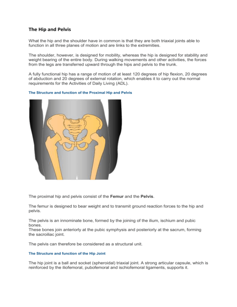
The Hip and Pelvis What the hip and the shoulder have in common is that they are both triaxial joints able to function in all three planes of motion and are links to the extremities. The shoulder, however, is designed for mobility, whereas the hip is designed for stability and weight bearing of the entire body. During walking movements and other activities, the forces from the legs are transferred upward through the hips and pelvis to the trunk. A fully functional hip has a range of motion of at least 120 degrees of hip flexion, 20 degrees of abduction and 20 degrees of external rotation, which enables it to carry out the normal requirements for the Activities of Daily Living (ADL). The Structure and function of the Proximal Hip and Pelvis The proximal hip and pelvis consist of the Femur and the Pelvis. The femur is designed to bear weight and to transmit ground reaction forces to the hip and pelvis. The pelvis is an innominate bone, formed by the joining of the ilium, ischium and pubic bones. These bones join anteriorly at the pubic symphysis and posteriorly at the sacrum, forming the sacroiliac joint. The pelvis can therefore be considered as a structural unit. The Structure and function of the Hip Joint The hip joint is a ball and socket (spheroidal) triaxial joint. A strong articular capsule, which is reinforced by the iliofemoral, pubofemoral and ischiofemoral ligaments, supports it. The two hip joints are linked together through the pelvis, via the sacroiliac and the lumbosacral joints, to the vertebral column. A fibrocartilage ring, called the acetabular labrum, deepens the acetabulum. Hip and Lumbar Spine angle in relation to Pelvis movement When the pelvis undergoes a movement, the hip and lumbar spine angle changes accordingly. Anterior Pelvis Tilt The Anterior Superior Iliac Spine (ASIS) of the pelvis moves anteriorly and inferiorly: forward and down reducing the anterior hip angle. This causes hip flexion and increases lumbar spine extension (hyperextension). The muscles that cause this motion are the Hip Flexors and the Back Extensors. When standing, the line of gravity of the trunk falls anterior to the axis of the hip joints, causing an anterior pelvis tilt. The Abdominals and the Hip Extensor muscles provide stability. Posterior Pelvis Tilt The Posterior Superior Iliac Spine (PSIS) moves posteriorly and inferiorly: back and down increasing the anterior hip angle. This causes hip extension and lumbar spine flexion. The muscles that cause this motion are the Hip Extensors and the Trunk Flexors. When standing, the line of gravity of the trunk falls posteriorly to the axis of the hip joints, causing posterior pelvis tilt. The Hip Flexors and Back Extensors provide dynamic stability, and the Iliofemoral Ligament provides passive stability. Lateral Pelvis Tilt When one side of the hip is higher than the other (hip hiking), adduction occurs on that side. On the side that is lower (hip drop), abduction occurs. In a standing position, the lumbar spine flexes laterally towards the side that is elevated. The muscles that are causing lateral pelvis tilt include the Quadratus Lumborum on the elevated side, and the Gluteus Medius on the lower side which acts in a reverse muscle pull down. In an asymmetric slouched posture, the trunk weight is shifted onto one leg, which allows the pelvis to drop on the other side. The Iliotibial Band provides support on the weight-bearing leg. When standing on one leg, hip adduction occurs causing the pelvis to drop on the unsupported side. Pelvis shifting When standing, a forward shift of the pelvis causes extension of the hip and lumbar spine. The upper lumbar vertebrae shift posteriorly, causing flexion, as can be seen with slouched or relaxed postures. The Iliofemoral Ligaments of the hip, the Anterior and Posterior Ligaments of the lower lumbar spine and Posterior Ligaments of the upper lumbar and thoracic spine hold this postural position. Little muscle action is therefore required. Pelvis rotation Pelvis rotation occurs around the leg that is fixed to the ground. The unsupported leg swings forward or backward along the pelvis. Forward rotation of the pelvis occurs when the unsupported side of the pelvis moves forward. At the same time, the trunk rotates to the opposite side and the femur of the supported leg rotates internally. Posterior (backward) rotation occurs when the unsupported pelvis moves backward. The trunk rotates to the opposite side and the femur on the stabilised side rotates externally. The Lumbopelvic rhythm when bending forward When you bend forward, a combined movement of the lumbar spine and the pelvis occurs. As you begin to bend forward, this movement starts from the head and upper trunk. The pelvis shifts backwards to keep the centre of gravity over the base of support, thus balancing the body. For approximately the first 45 degrees of forward flexion, the Extensor muscles of the spine maintain the balance of the body. The Posterior Ligaments become taut and the facet of the Zygapophyseal Joints come together, which provides stability for the Intervertebral Joints, and the muscles relax. When the movement has reached the point where all the vertebral segments are at full range, supported by the Posterior Ligaments and Facets, the pelvis begins to rotate forward: an anterior pelvis tilt. The glutes and hamstrings control this part of the movement. The pelvis will continue to rotate forward, until the muscles are at full length. The final Range Of Motion depends on the flexibility of the Back Extensors, the Fasciae and the Hip Extensors. To return to an upright position, the Hip Extensor muscles rotate the pelvis posteriorly, after which the Back Extensors extend the spine, beginning at the lumbar region and working its way upward. The Hip movement and muscle control during Gait Range of hip motion During normal gait cycle, a hip range of motion of 40 degrees flexion and extension can be observed, from 10 degrees of extension at terminal stance to 30 degrees flexion at midswing and initial contact. A lateral pelvic tilt and hip abduction/adduction occurs at 15 degrees: 10 degrees adduction at initial contact and 5 degrees abduction at initial swing. An internal/external rotation along with pelvic rotation totalling 15 degrees transverse plane motion can be observed, with the internal rotation peaking at the end of pre-swing. Muscle control during gait A number of muscles are in play during the gait cycle and its phases. The Hip Flexors control hip extension at the end of the stance phase; the swing phase is initiated when they contract. In case of a loss of flexor function, swing will be initiated by a compensatory posterior lurch of the trunk. If tightness of the flexors occurs, full hip extension is prevented during the 2nd half of the stance phase, therefore producing a shortened stride. This in turn may cause an increase in lumbar lordosis or forward trunk flexion when walking. The Hip Extensors control the flexor movement at initial foot contact, with the Glutes initiating hip extension. A loss of extensor function will cause the trunk to lurch posteriorly at foot contact. Tightness of the Gluteus Maximus results in a reduced range in the final stages of the swing phase, as the femur comes forward. To compensate for this, the pelvis is rotated to bring the leg through. The lower leg may externally rotate, caused by the external rotation of the muscle. Alternatively, the Gluteus Maximus may cause greater tension on the Iliotibial band, which may result in irritation to the lateral knee. The Hip Abductors control the Lateral Pelvic Tilt during the swing phase of the opposite leg. A loss of Gluteus Medius function causes a lateral shifting of the trunk over the same side during the stance phase, when the opposite leg swings through. Lateral shifting will also occur in the case of a painful hip, as it minimises the torque at the hip when it has to bear weight. The Tensor Fascia Latae also acts as an abductor and may affect optimal gait action if it becomes tight Muscles that can affect the Pelvis and Spine Reverse muscle action The muscles of the hip that cause pelvic movement through reverse action are: the Hip Flexors, for Anterior Pelvis Tilt the Hip Extensors, for Posterior Pelvis Tilt the Hip Adductors and Abductors, for Lateral Pelvis Tilt the Rotators, for Pelvic Rotation To minimise excessive pelvic movement, the abdominals, the erector spine, multifidus and quadratus lumborum are brought into play. Hip Muscle Imbalances and their effects Pain and discomfort are often caused by muscle imbalances. Overuse syndromes, soft tissue stresses, and joint pain develop as a result of continued abnormal responses. Decreased flexibility in the hip muscles or joints When muscle imbalances occur, weight bearing forces and movement are transmitted to the spine, rather than absorbed in the pelvis. Tight Hip Extensors will cause an increased lumbar flexion when the thigh flexes, and increased lumbar extension when the thigh extends. A decrease in hip flexion and incomplete hip extension during weight bearing will also place increased stress on the knee, as it cannot lock while the hip is flexed, unless the trunk is bent forward. Tight Abductors cause the opposite action. Tight Adductors cause Lateral Pelvis Tilt to the opposite side and a side bending of the trunk towards that side during weight bearing. Shortened Iliotibial Band, with shortened Tensor Fasciae Latae or Gluteus Maximus. An Anterior Pelvis Tilt Posture and Slouched Posture have a number of associated postural dysfunctions. The hip musculature imbalances with an Anterior Pelvis Tilt Posture are: a short Tensor Fasciae Latae and Iliotibial Band limited external rotation a weak and stretched Gluteus Medius (posterior fibres) and Piriformis an increased stress on the knee that is caused by excessive medial rotation of Femur during the 1st half of stance an associated compensation that includes medial rotation of the Femur, Genu Valgum, Lateral Tibial Torsion, Pes Plantas and Hallux Valgus The hip musculature imbalances with a Slouched Posture are: a short Rectus Femoris and Hamstrings a limited range of the hip rotators a weak, stretched Iliopsoas a weak and tight Gluteus Medius (posterior fibres) a weak and poorly developed Gluteus Maximus an associated compensation that includes Hip Extension, and occasionally medial rotation of the Femur, Genu Recurvatum, Genu Varum and Pes Valgus
