Chicken Wing Dissection
advertisement
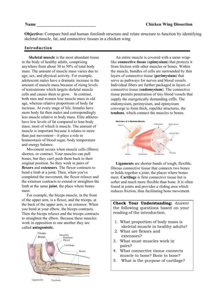
Name ___________________________ Chicken Wing Dissection Objective: Compare bird and human forelimb structure and relate structure to function by identifying skeletal muscle, fat, and connective tissues in a chicken wing Introduction Skeletal muscle is the most abundant tissue in the body of healthy adults, comprising anywhere from about 30 to 50% of total body mass. The amount of muscle mass varies due to age, sex, and physical activity. For example, adolescent males have a dramatic increase in the amount of muscle mass because of rising levels of testosterone which targets skeletal muscle cells and causes them to grow. In contrast, both men and women lose muscle mass in old age, whereas relative proportions of body fat increase. At every stage of life, females have more body fat then males and correspondingly less muscle relative to body mass. Elite athletes have low levels of fat compared to lean body mass, most of which is muscle. The amount of muscle is important because it relates to more than just movement—it plays a role in homeostasis of blood sugar, body temperature and energy balance. Movement occurs when muscle cells (fibers) shorten, or contract. Your muscles can pull bones, but they can't push them back to their original position. So they work in pairs of flexors and extensors. The flexor contracts to bend a limb at a joint. Then, when you've completed the movement, the flexor relaxes and the extensor contracts to extend or straighten the limb at the same joint, the place where bones meet. For example, the biceps muscle, in the front of the upper arm, is a flexor, and the triceps, at the back of the upper arm, is an extensor. When you bend at your elbow, the biceps contracts. Then the biceps relaxes and the triceps contracts to straighten the elbow. Because these muscles work in opposition to one another they are called antagonistic. Triceps Biceps An entire muscle is covered with a saran wraplike connective tissue (epimysium) that protects it from friction with other muscles or bones. Within the muscle, bundles of cells are surrounded by thin layers of connective tissue (perimysium) that serve as pathways for nerves and blood vessels. Individual fibers are further packaged in layers of connective tissue (endomysium). The connective tissue permits penetration of tiny blood vessels that supply the energetically demanding cells. The endomysium, perimysium, and epimysium converge to form thick, ropelike structures, the tendons, which connect the muscles to bones. Ligaments are shorter bands of tough, flexible, fibrous connective tissue that connects two bones or holds together a joint, the places where bones meet. Cartilage is firm connective tissue but is softer and much more flexible than bone. It is often found at joints and provides a sliding area which reduces friction, thus facilitating bone movement. Check Your Understanding: Answer the following questions based on your reading of the introduction. 1. What proportion of body mass is skeletal muscle in healthy adults? 2. What are flexors and extensors? 3. What must muscles work in pairs? 4. What connective tissue connects muscle to bone? Bone to bone? 5. What is the purpose of cartilage? Activity: Chicken Wings and Muscle Structure Chicken wings are homologous to the upper limb of humans; that is, they have many of the same structures due to their shared evolutionary history as vertebrates. Figure 1: Homologous Forelimb Bones in Vertebrates Figure 2: Chicken Wing CAUTION: Raw chicken may be contaminated by Salmonella. Keep your hands away from your face and mouth throughout this investigation. Wash your hands and wipe the desks after the dissection. 1. Examine the chicken skeleton and human skeleton and identify the humerus, ulna, radius, and wrist (carpal), hand (metacarpal) and finger bones (phalanges) on each (see figs. 1 and 2). 2. Use scissors to cut the skin lengthwise to the joint between the upper wing and lower wing. Carefully peel the skin from the wing. Examine the yellowish fat under the skin. What is its function? Which joint in your body corresponds to this joint in the chicken wing? 3. Remove the skin of the lower wing in the same way that you removed the skin from the upper wing. Leave the skin on the wing tip. Use scissors to carefully remove the skin from the joint between the upper and lower wing. Be careful not to cut any tendons or ligaments. How will you recognize tendons and ligaments? What is a tendon? What is a ligament? 4. Observe the thin, transparent, shiny layer covering the muscles. This is the deep fascia, or epimysium, which surrounds an entire muscle and separates it from its neighbors. The epimysium consists mostly of strong collagenous fibers, which support the skeletal muscle tissue. This thin but strong packaging enables the muscles to carry out their specific intended action both in isolation and in concert with other muscles. 5. Carefully insert the tip of the scissors under the thin connective tissue and remove some of the connective tissue to expose the skeletal muscle tissue underneath. 6. Use a dissecting needle to separate the pinkish muscles. Observe how the muscles are arranged in pairs on opposite sides of the bones. Figure 3 Bird Muscles Locate the flexors and extensors of the elbow joint. These would be the anterior (front) and posterior (back) muscle groups attached to the humerus. Name the primary elbow flexor. Name the primary elbow extensor. When muscles such as these act in opposition to one another, they are called . Notice the attachment points of the muscles. The origin, or fixed attachment, is nearer the chest, whereas the insertion, the point that moves during contraction, is on the forearm bones, distal to (away from) the elbow joint. 7. Straighten the chicken wing and hold it horizontally above the tray. Pull on each of the muscles and note the movement that each muscle causes. Turn the wing upside down and bend the joints. Again pull on each muscle and note how the bones move. 8. Cut through the middle of a muscle that you have identified as a flexor for the upper wing. What happens to the wing? 9. Cut through the middle of a muscle that you have identified as an extensor for the lower wing. What happens to the wing? 10. Bend and straighten the joint and observe how the bones fit together. The shiny, white covering of the joint surfaces is made of cartilage. What is the purpose of this cartilage? 11. Identify several white tendons connecting muscle and bone in upper and lower wing muscle groups. Where these tendons run over joints-- like the "elbow"--they are often in well-developed sheaths. What tissue comprises tendons? What is the source of the shiny, white color? 12. Locate blood vessels and nerves between muscle bundles and muscle bundles and bones. It is necessary to do some teasing with the dissecting probe to see these structures. Clean Up! 13. Place the chicken wing and the protective gloves in a plastic bag for disposal and wash hands thoroughly with soap and water. Carefully clean work area with disinfectant spray. Check Your Understanding: Answer the following questions based on your dissection of the chicken wing. 1. What structures of the chicken wing and human upper limb are homologous? Explain why they are homologous. 2. List five specific tissues that you examined in the chicken wing. Where are they located? 3. What is the role of blood vessels and nerves in skeletal muscle function? 4. What muscles flex and extend the elbow joint? What is the function of the action in chickens? In humans? Explain how differences in muscle and bone anatomy relate to the way of life of chickens (birds) and humans. Analysis 1. What type of tissue makes up the “meat” of a chicken? __________________________________________________________ 2. What type of tissue connects bone to bone? __________________________________________________________ 3. What type of tissue connects muscle to bone? __________________________________________________________ 4. What type of tissue acts as a cushion between bones? ___________________________________________________________ 5. Could the muscles function if they were not attached to the bone? Explain the interaction of bone and muscle. How do they work together to bring about movement? 6. Compare the muscles and bones of a chicken wing and the human arm. How they are the same, yet different. CHALLENGE Chicken (sketch here) Whale flipper Frog front arm Horse front leg Lion front leg Human arm Bat wing Chicken wing Bones in the upper limb (humerus, radius, ulna) Humerus (yes/no) Radius (yes/no) Ulna (yes/no) Humerus (yes/no) Radius (yes/no) Ulna (yes/no) Humerus (yes/no) Radius (yes/no) Ulna (yes/no) Humerus (yes/no) Radius (yes/no) Ulna (yes/no) Humerus (yes/no) Radius (yes/no) Ulna (yes/no) Humerus (yes/no) Radius (yes/no) Ulna (yes/no) Humerus (yes/no) Radius (yes/no) Ulna (yes/no) # of fingers Function of the Limb (Flying, swimming, grasping, etc) Answer: What does that tell us about our relationship to chickens, lions, horses, etc? How is this evidence for evolution?
