Novel LRBA Mutation in a Girl Presenting with Refractory Celiac
advertisement
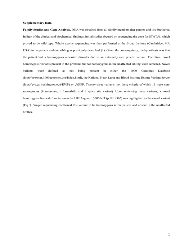
Supplementary Data Family Studies and Gene Analysis. DNA was obtained from all family members (her parents and two brothers). In light of the clinical and biochemical findings, initial studies focused on sequencing the gene for STAT5b, which proved to be wild type. Whole exome sequencing was then performed at the Broad Institute (Cambridge, MA USA) in the patient and one sibling as previously described (1). Given the consanguinity, the hypothesis was that the patient had a homozygous recessive disorder due to an extremely rare genetic variant. Therefore, novel homozygous variants present in the proband but not homozygous in the unaffected sibling were screened. Novel variants were defined as not being present in either the 1000 Genomes Database (http://browser.1000genomes.org/index.html), the National Heart Lung and Blood Institute Exome Variant Server (http://evs.gs.washington.edu/EVS/) or dbSNP. Twenty-three variants met these criteria of which 11 were nonsynonymous (9 missense, 1 frameshift, and 1 splice site variant). Upon reviewing these variants, a novel homozygous frameshift mutation in the LRBA-gene c.5505delT (p.Ile1836*) was highlighted as the causal variant (Fig1). Sanger sequencing confirmed this variant to be homozygous in the patient and absent in the unaffected brother. 1 Table1.Immunological features of the patient Patient Age references(2) ALC, /mm3 1600 >1500 AGC, /mm3 2100 >1500 IgG, mg/dl 1140 835-2094 IgA, mg/dl 327 67-433 IgM, mg/dl 34.46 47-484 IgE, kU/L 3.8 Isohemagglutinin titer Blood type O Rh + Anti A titer 1/16 Anti B titer 1/8 CD3+ CD16-CD56- 67 58-82 + CD3 CD4 + 43 26-48 + + 24 16-32 CD3-CD16+CD56+ 18 8-30 CD19+ 13 10-30 + 13 9-28 CD4+CD45RA+ 3 8-26 CD4+CD45RO+ 11 10-30 TCRγδ, 1.3 CD4+CD45RA+CD31+ 18.1 CD3 CD8 CD20 T cell activation response to PHA CD3+CD25+, % + + CD3 CD69 , % DHR test 83 46-89 41 50-76 Normal 2 Table2. B cell subsets of the patient + + CD 19 IgM-27 IgD - % Normal levels in healthy Turkish children(3) EUROCLASS(4) 12 12.9-45 6.5-29.2 9.8 5.1-11.8 7.2-30.8 86.3 62.2-76.2 2.6 3.2-5.9 1.1-6.9 6.1 2.8-10.2 0.6-3.5 (Switched memory B) CD19+IgM+27+IgD+ (Marginal Zone B) CD 19+IgM+27-IgD+ (Naive B) CD19+CD38 Low CD21 Low (Activated B) CD19+CD39 High IgM High (Transitional B) 3 Table 3. The identified mutations in LRBA deficiency. Chromosome Position Gene cDNA change Protein change 1 243336145 CEP170 c.T1276G p.F426V 4 8229354 SH3TC1 c.G1933T p.G645W 4 24801685 SOD3 c.A542T p.H181L 4 151727435 LRBA c.5505delT p.Ile1836* 5 132083419 CCNI2 c.C232T p.P78S 10 99342096 ANKRD2 c.G1018A p.A340T 11 64410191 NRXN2 c.C85T p.P29S 19 16265269 HSH2D c.G271C p.E91Q 19 36049477 ATP4A c.1365+2T>A NA 22 30775605 RNF215 c.G1118A p.R373H X 57618621 ZXDB c.140T>C p.L47P All genomic positions are based on human genome build 19. 4 REFERENCES 1. Dauber A, Stoler J, Hechter E, Safer J, Hirschhorn JN. Whole exome sequencing reveals a novel mutation in CUL7 in a patient with an undiagnosed growth disorder. J Pediatr. 2013;162(1):202-4 e1. 2. Ikinciogullari A, Kendirli T, Dogu F, Egin Y, Reisli I, Cin S, et al. Peripheral blood lymphocyte subsets in healthy Turkish children. Turk J Pediatr. 2004;46(2):125-30. 3. Cipe FE, Dogu F, Guloglu D, Aytekin C, Polat M, Biyikli Z, et al. B-cell subsets in patients with transient hypogammaglobulinemia of infancy, partial IgA deficiency, and selective IgM deficiency. J Investig Allergol Clin Immunol. 2013;23(2):94-100. 4. Wehr C, Kivioja T, Schmitt C, Ferry B, Witte T, Eren E, et al. The EUROclass trial: defining subgroups in common variable immunodeficiency. Blood. 2008;111(1):77-85. 5
