7. Epithelium and Junctions
advertisement
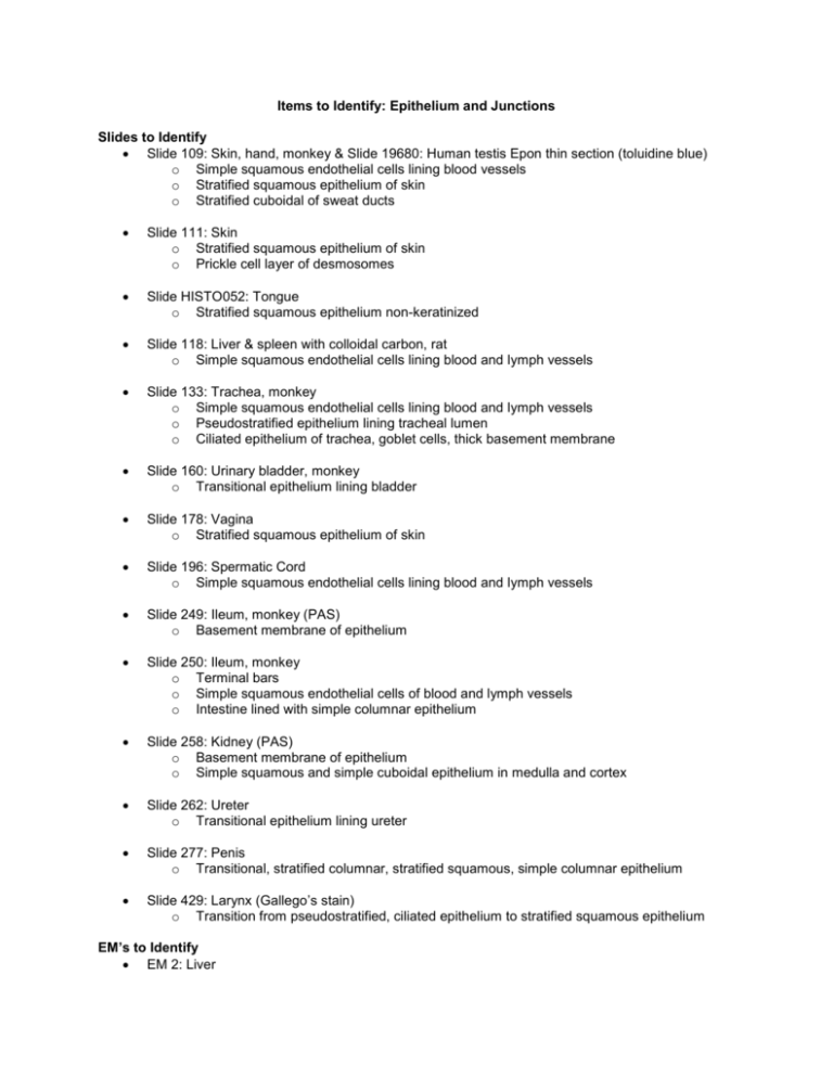
Items to Identify: Epithelium and Junctions Slides to Identify Slide 109: Skin, hand, monkey & Slide 19680: Human testis Epon thin section (toluidine blue) o Simple squamous endothelial cells lining blood vessels o Stratified squamous epithelium of skin o Stratified cuboidal of sweat ducts Slide 111: Skin o Stratified squamous epithelium of skin o Prickle cell layer of desmosomes Slide HISTO052: Tongue o Stratified squamous epithelium non-keratinized Slide 118: Liver & spleen with colloidal carbon, rat o Simple squamous endothelial cells lining blood and lymph vessels Slide 133: Trachea, monkey o Simple squamous endothelial cells lining blood and lymph vessels o Pseudostratified epithelium lining tracheal lumen o Ciliated epithelium of trachea, goblet cells, thick basement membrane Slide 160: Urinary bladder, monkey o Transitional epithelium lining bladder Slide 178: Vagina o Stratified squamous epithelium of skin Slide 196: Spermatic Cord o Simple squamous endothelial cells lining blood and lymph vessels Slide 249: Ileum, monkey (PAS) o Basement membrane of epithelium Slide 250: Ileum, monkey o Terminal bars o Simple squamous endothelial cells of blood and lymph vessels o Intestine lined with simple columnar epithelium Slide 258: Kidney (PAS) o Basement membrane of epithelium o Simple squamous and simple cuboidal epithelium in medulla and cortex Slide 262: Ureter o Transitional epithelium lining ureter Slide 277: Penis o Transitional, stratified columnar, stratified squamous, simple columnar epithelium Slide 429: Larynx (Gallego’s stain) o Transition from pseudostratified, ciliated epithelium to stratified squamous epithelium EM’s to Identify EM 2: Liver o Gap junction, desmosome, tight junction EM 2a: Liver- Gap junctions o Gap junction EM 3: Intestine (Basal) o Basal lamina EM 4: Intestine (Apical) o Tight junction, zonula adherens, terminal web EM 4a: Intestine – Occludens Junction o Tight junction structure EM 4b: Intestinal absorption cell (apex) o Junctions between intestinal absorptive cells EM 4c: Intestinal absorptive cells o Amplified lateral surface of intestinal absorptive cells EM 6b: Basal body – Cilia o Tight junction (zonula occludens), zonula adherens, desmosome (macula adherens) EM 8g: Epidermis o Desmosomes of epidermis EM 8h: Macrophage o Thickened basal lamina EM 10a: Capillary o Capillary endothelial cells EM 10f: Arteriolar wall o Thin basal lamina, tight junctions (zonula occludens) EM 16: Surface mucosa (stomach) o Basal lamina EM 17: Duodenum o Basal lamina
