Bmper_GCE_Strain_Report_042511
advertisement
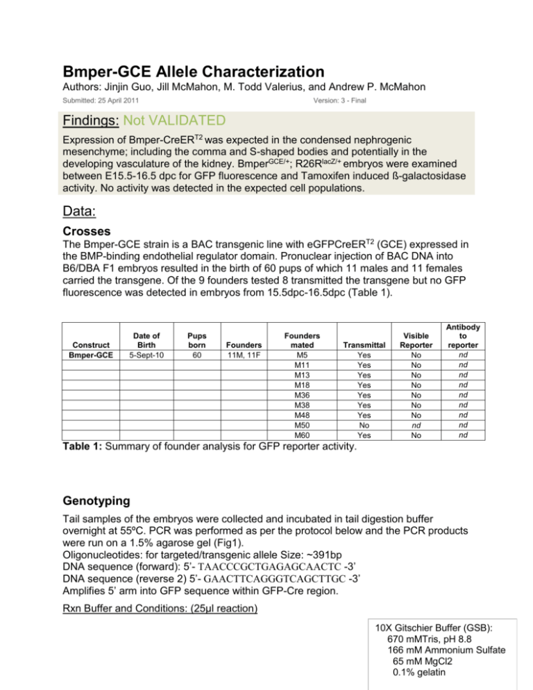
Bmper-GCE Allele Characterization Authors: Jinjin Guo, Jill McMahon, M. Todd Valerius, and Andrew P. McMahon Submitted: 25 April 2011 Version: 3 - Final Findings: Not VALIDATED Expression of Bmper-CreERT2 was expected in the condensed nephrogenic mesenchyme; including the comma and S-shaped bodies and potentially in the developing vasculature of the kidney. BmperGCE/+; R26RlacZ/+ embryos were examined between E15.5-16.5 dpc for GFP fluorescence and Tamoxifen induced ß-galactosidase activity. No activity was detected in the expected cell populations. Data: Crosses The Bmper-GCE strain is a BAC transgenic line with eGFPCreERT2 (GCE) expressed in the BMP-binding endothelial regulator domain. Pronuclear injection of BAC DNA into B6/DBA F1 embryos resulted in the birth of 60 pups of which 11 males and 11 females carried the transgene. Of the 9 founders tested 8 transmitted the transgene but no GFP fluorescence was detected in embryos from 15.5dpc-16.5dpc (Table 1). Construct Bmper-GCE Date of Birth 5-Sept-10 Pups born 60 Founders 11M, 11F Founders mated M5 M11 M13 M18 M36 M38 M48 M50 M60 Transmittal Yes Yes Yes Yes Yes Yes Yes No Yes Visible Reporter No No No No No No No nd No Antibody to reporter nd nd nd nd nd nd nd nd nd Table 1: Summary of founder analysis for GFP reporter activity. Genotyping Tail samples of the embryos were collected and incubated in tail digestion buffer overnight at 55ºC. PCR was performed as per the protocol below and the PCR products were run on a 1.5% agarose gel (Fig1). Oligonucleotides: for targeted/transgenic allele Size: ~391bp DNA sequence (forward): 5’- TAACCCGCTGAGAGCAACTC -3’ DNA sequence (reverse 2) 5’- GAACTTCAGGGTCAGCTTGC -3’ Amplifies 5’ arm into GFP sequence within GFP-Cre region. Rxn Buffer and Conditions: (25µl reaction) 10X Gitschier Buffer (GSB): 670 mMTris, pH 8.8 166 Ammonium Page 1 ofmM 5, printed 2/9/16. Sulfate 65 mM MgCl2 0.1% gelatin 10X GSB 25mM dNTP 10uM primer F 10uM primer R DMSO 2.5ul 1ul 1ul 1ul 2.5ul 2-mercaptoethanol Amplify Taq 5x cresol red dye Genomic DNA Total volume 0.125ul 0.2ul (5u/ul) 5ul 1ul 25 ul 94ºC 94ºC 55.5ºC 72ºC 72ºC 3min 1 cycle 30sec 30sec 35cycles 45sec 10min 1 cycle Fig1: Numbers 48, 52, 54, 55, 58, 59, 61 BmperGCE/+, P: BmperGCE/+ positive control, W: Wildtype control, N: Negative control. Cre-recombinase Activity The male founders were crossed with Rosa26RlacZ/+(R26R) mice to obtain BmperGCE/+; R26RlacZ/+ embryos. In order to activate β-gal reporter expression from the R26RlacZ/+allele, an intraperitoneal injection of Tamoxifen in corn oil was given to pregnant mice at 2mg/40g body weight or 2 doses at 2mg/40g body weight. Tamoxifen treated BmperGCE/+; R26RlacZ/+ embryos were dissected between 2-4 days post injection. UGS samples were processed for X-gal staining but only low levels of ß-galactosidase activity was detected in areas not consistant with the expected expression in either nephrogenic mesenchyme or vascular progenitor cells (Table 2 and Fig 2, 3)). Founders mated M5 Transmittal Yes ß-gal activity Tam (1X dose) ß-gal activity Tam (2X dose) 12.5-13.5 11.5-14.5 No No Correct Cre Activity No Page 2 of 5, printed 2/9/16. M11 Yes nd No No M13 M18 M38 M48 M60 Yes Yes Yes Yes Yes No nd No No No nd No nd nd No No No No No No Table 2. Summary of founder analysis for Cre-dependant ß-gal activity in Tamoxifen induced BmperGCE/+; R26RlacZ/+ embryos. Fig 2. Cre-dependant ß-gal activity in female BmperGCE/+; R26RlacZ/+ UGS.Two doses of 2mg/40g Tamoxifen, injected at 11.5 and 13.5dpc resulted in weak ß-gal activity on the surface of the kidney and reproductive tubes but not as expected in condensed nephrogenic mesenchymal derivatives. Page 3 of 5, printed 2/9/16. Fig 3. Cre-dependant ß-gal activity is not detected in the cortical region of BmperGCE/+; R26RlacZ/+ UGS.Two doses of 2mg/40g Tamoxifen, injected at 11.5 and 13.5dpc did not resulted in detectable ß-gal activity in condensed nephrogenic mesenchyme. Immunohistochemistry Immunohistochemistry was performed to examine if the eGFPCreERT2 allele was expressed in the expected Bmper domain. To test for Cre function, BmperGCE/+; R26RlacZ/+ UGSs from Tamoxifen injected mice were assayed. β-gal and GFP expression were examined by probing with rabbit anti-β-gal, chicken-anti-GFP and antirat-PECAM CD31 antibody. Whole UGSs were fixed in 4% paraformaldehyde at 4ºC 2 hours, washed 3 times in PBS, equilibrated in 30% sucrose overnight and then embedded in OCT and flash frozen on dry-ice. The UGSs were sectioned at 16um and probed with rabbit-anti-ßgal/Chicken-anti-GFP/rat-anti-PECAM CD31. ß-gal (Rabbit, MP Biomedicals, LLC, 55976, 1: 20000), GFP (Chicken, Aves Labs, Inc, GFP-1020, 1:500) anti-PECAM CD31 (Rat, BD Pharmingen, 553370, 1:1000) were incubated overnight at 4ºC and detected with secondary antibodies Alexafluor 488, 555, and 633 (Molecular probes). Neither GFP nor ß-gal positive cells were detected in the cortical region of BmperGCE/+; R26RlacZ/+ embryos at 15.5dpc (Fig 4). Page 4 of 5, printed 2/9/16. Fig 4. Neither GFP nor ß-gal positive cells are detected in condensed nephrogenic mesenchyme in BmperGCE/+; R26RlacZ/+ UGS at 15.5dpc. Page 5 of 5, printed 2/9/16.


