clinical study on non-traumatic perforations of small intestine
advertisement
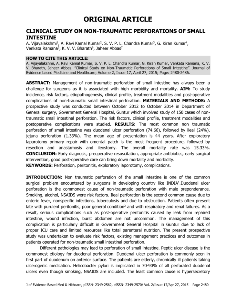
ORIGINAL ARTICLE CLINICAL STUDY ON NON-TRAUMATIC PERFORATIONS OF SMALL INTESTINE A. Vijayalakshmi1, A. Ravi Kamal Kumar2, S. V. P. L. Chandra Kumar3, G. Kiran Kumar4, Venkata Ramana5, K. V. V. Bharath6, Jaheer Abbas7 HOW TO CITE THIS ARTICLE: A. Vijayalakshmi, A. Ravi Kamal Kumar, S. V. P. L. Chandra Kumar, G. Kiran Kumar, Venkata Ramana, K. V. V. Bharath, Jaheer Abbas. ”Clinical Study on Non-Traumatic Perforations of Small Intestine”. Journal of Evidence based Medicine and Healthcare; Volume 2, Issue 17, April 27, 2015; Page: 2480-2486. ABSTRACT: Management of non-traumatic perforation of small intestine has always been a challenge for surgeons as it is associated with high morbidity and mortality. AIM: To study incidence, risk factors, etiopathogenesis, clinical profile, treatment modalities and post-operative complications of non-traumatic small intestinal perforation. MATERIALS AND METHODS: A prospective study was conducted between October 2012 to October 2014 in Department of General surgery, Government General Hospital, Guntur which involved study of 150 cases of nontraumatic small intestinal perforation. The risk factors, clinical profile, treatment modalities and postoperative complications were studied. RESULTS: The most common non traumatic perforation of small intestine was duodenal ulcer perforation (74.66), followed by ileal (24%), jejuna perforation (1.33%). The mean age of presentation is 44 years. After exploratory laparotomy primary repair with omental patch is the most frequent procedure, followed by resection and anastamosis and ileostomy. The overall mortality rate was 15.33%. CONCLUSION: Early diagnosis, preoperative resuscitation, appropriate antibiotics, early surgical intervention, good post-operative care can bring down mortality and morbidity. KEYWORDS: Perforation, peritonitis, exploratory laporotomy, complications. INTRODUCTION: Non traumatic perforation of the small intestine is one of the common surgical problem encountered by surgeons in developing country like INDIA1.Duodenal ulcer perforation is the commonest cause of non-traumatic perforation with male preponderance. Smoking, alcohol, NSAIDS were risk factors. Ileal perforation is the second common cause due to enteric fever, nonspecific infections, tuberculosis and due to obstruction. Patients often present late with purulent peritonitis, poor general condition2 and with respiratory and renal failures. As a result, serious complications such as post-operative peritonitis caused by leak from repaired intestine, wound infection, burst abdomen are not uncommon. The management of this complication is particularly difficult in Government General Hospital in Guntur due to lack of proper ICU care and limited resources like total parenteral nutrition. The present prospective study was undertaken to evaluate risk factors, existing management practices and outcomes in patients operated for non-traumatic small intestinal perforation. Different pathologies may lead to perforation of small intestine. Peptic ulcer disease is the commonest etiology for duodenal perforation. Duodenal ulcer perforation is commonly seen in first part of duodenum on anterior surface. The patients are elderly, chronically ill patients taking ulcerogenic medication. Helicobacter pylori is implicated in 70-90% of all perforated duodenal ulcers even though smoking, NSAIDS are included. The least common cause is hypersecretory J of Evidence Based Med & Hlthcare, pISSN- 2349-2562, eISSN- 2349-2570/ Vol. 2/Issue 17/Apr 27, 2015 Page 2480 ORIGINAL ARTICLE state i.e., Zollinger-ellisons syndrome. In modern era appropriate treatment of Helicobacter pylori eradication results in healing of uncomplicated duodenal ulcer. Infection is the commonest cause of ileal perforation is due to typhoid fever, tuberculosis3,4 nonspecific infections and lastly due to intestinal obstruction. The diagnosis is mainly by clinical examination, supported by x-ray abdomen showing free gas under diaphragm and ultrasound abdomen, leucocytosis5,6,4 was present in some cases with altered renal parameters. Radiological evidence of pneumoperitoneum could not be established in all cases. A decision of laporotomy done on basis clinical grounds and supplemented by investigations. In spite of recent advances in laparoscopy, simple closure with omental patch with peritoneal toilet is the effective procedure. Definitive ulcer surgery is not warranted in emergency situations and treatment with proton pump inhibitors, and Helicobacter pylori eradication therapy achieved good control over disease in follow up period. At laporotomy operative findings were noted. Intestinal perforation was managed by primary closure with omental patch, ileal primary closure, resection and anastamosis or by ileostomy. Peritoneal cavity was lavaged with normal saline. Tube drains were placed to drain pelvis and paracolic gutters. Post-operative antibiotics were used. Histological examination small bowel was 50% are due to enteric fever, 22.77% are due to tuberculosis, 22.22% are due to nonspecific infections7. Presence of granulomas in histopathological examination was suggestive of granulomatous inflammation with differential diagnosis of tuberculosis or crohn’s disease. Tuberculosis is more common and treated accordingly with anti tuberculous drugs. AIMS AND OBJECTIVES: To study incidence, risk factors, etiopathogenesis, clinical profile, treatment modalities and postoperative complications and also to assess morbidity and mortality. MATERIALS AND METHODS: The study is a prospective study in Department of General surgery Govt General Hospital, Guntur from October 2012 to October 2014. The history of present illness, duration of onset of peritonitis was noted. The drug history with special reference to NSAIDS and anti-tuberculous drugs were noted. History of evidence of typhoid fever and treatment for typhoid fever was also noted. Resuscitation time was found to be proportional to chronicity of disease. The diagnosis of perforation was made on clinical and radiological findings and was confirmed at operation. Specimen for histopathology was taken in selected cases, Triple antibiotic regimen such as cephalosporins, aminoglycosides, and metronidazole were tried in all cases and found to be effective. Specific chemotherapy for underlying disease was offered later proton pump inhibitors were used in all cases. All patients were explored with mid line incisions, complications and recovery were noted. Informed consent was taken from patients and study had been approved by ethical committee. J of Evidence Based Med & Hlthcare, pISSN- 2349-2562, eISSN- 2349-2570/ Vol. 2/Issue 17/Apr 27, 2015 Page 2481 ORIGINAL ARTICLE RESULTS: Sl. NO Etiology No. of Cases Percentage 1 Duodenal Perforation 112 74.66% 2 Jejunal perforation 02 1.33% 3 Ileal perforation 36 24.01% Total 150 100% Table 1: Distribution according to etiological factor Age No. of Cases Percentage <19 02 1.78 20-29 16 14.28 30-39 30 26.78 40-49 22 19.64 50-59 16 14.28 >60 26 2321 Table 2: Age wise distribution Etiology Duodenal perforation Jejunum Ileum Surgical intervention No. of Cases Percentage Simple closure with omental patch 108 72 Bilateral flank drains 04 2.66 Resection and anastamosis 02 1.33 Closure in 2 layers 24 16 Resection and anastamosis 04 2.66 Ilesotomy of perforated bowel loop 08 5.33 Table 3: Intervention wise distribution Age No. of Cases <40 years >/= 40 years 57 93 Morbidity Mortality Cases Percentage Cases Percentage 08 14.03 04 7.01 20 21.50 10 10.75 Table 4: Age Wise Morbidity and Mortality Size </= 0.5 cms 0.6 – 1 cms >1 cm Number of Cases 36 62 52 Morbidity Mortality Cases Percentage Cases Percentage 03 09 01 04 10 17 04 08 15 28 09 17 Table 5: Morbidity and mortality in relation to size of perforation J of Evidence Based Med & Hlthcare, pISSN- 2349-2562, eISSN- 2349-2570/ Vol. 2/Issue 17/Apr 27, 2015 Page 2482 ORIGINAL ARTICLE Duration Number <24 Hours >24 Hours 102 48 Morbidity Mortality Cases Percentage Cases Percentage 9 9 4 4 19 40 10 20 Table 6: Morbidity and mortality in relation to duration of symptoms DISCUSSION: In our study of all emergency admissions during the period are 1136 cases. Of these non-traumatic small intestinal perforation constitutes 13.20% of total emergencies. Duodenal ulcer is the commonest cause of non-traumatic perforation in our study as compared to Nair et al 1981, NN Mahendra et al 1988, DMC Rao 1999. Of all 150 cases, 112 cases are presented with duodenal perforation i.e., 74.66%. 36 cases are presented with ileal perforation i.e., 24% of 150 cases. 2 cases presented with jejunal perforation i.e., 1.33% of 150 cases. (Table 1) In our study duodenal perforation is more common in age group of 30-49 years, the youngest age was 17 years, eldest case was 68 years. The average age is 44 years.8 (Table 2) Non traumatic small intestinal perforation was found to be more common in males when compared to females which are attributed to smoking and alcoholism. Of all 112 cases of duodenal perforations included in study pneumoperitoneum was found in only 90 cases which account for 80.35% which is compared to study.9 Two cases of non-traumatic small intestinal perforation are included in study due to perforation of jejunal diverticulum. Of 36 cases of ileal perforation 18 cases are due to enteric fever, it constitutes 50%. The remaining 10 cases were due to tuberculosis, it constitutes 27.77%. And 8 cases were due nonspecific causes,it constitutes 22.22% of cases. Out of 112 cases 102 cases are operated with simple closure with omental patch. Ten cases whose general condition is poor, bilateral flank drains were kept as conservative managment. Out of 102 duodenal perforation operated cases 16 patients developed postoperative complications like wound infection, pneumonitis, burst abdomen, and enterocutaneous (fistula. 15 cases expired i e 13.3% of cases. (Table 3) Out of 36 cases of ileal perforation, 24 cases underwent closure in two layers using 2.0 10 silk. And 4 cases underwent resection and anastomosis and 8 cases underwent ileostomy.11,12 Out of 36 cases 8 cases expired and 16 patients developed post-operative complications. (Table3) Percentage of morbidity and mortality in cases below the age of 40 years is 14.03% and 7.01% respectively. (Table 4) Percentage of morbidity and mortality in cases above the age of 40 years 21.50% and 10.75% respectively. (Table 4) Mortality and morbidity are comparatively more in females in this study. This is attributed to elderly age of presentation, overweight and increased use of NSAIDS in females. J of Evidence Based Med & Hlthcare, pISSN- 2349-2562, eISSN- 2349-2570/ Vol. 2/Issue 17/Apr 27, 2015 Page 2483 ORIGINAL ARTICLE Percentage of mortality in duodenal perforation is 13.33% when compared to percentage of mortality in ileal perforation is 22.22% because of faecal contamination of peritoneal cavity leading to bacterial peritonitis, ileus and septicemia. Percentage of morbidity and mortality in cases of perforation size less than 0.5cms is 9% and 4%. (Table 5) Percentage of morbidity and mortality in cases of perforation of size0.5 to1cms is 17% and 8% respectively. (Table 5) Percentage of morbidity and mortality in cases of perforation of size more than 1cm is 28% and 17% respectively. Morbidity and mortality increases with size of perforation as the contamination of peritoneal cavity increases due to rapid outpouring of intestinal contents into peritoneal cavity leading to early progression of peritonitis to generalized peritonitis which result in greater morbidity and mortality. Percentage of morbidity and mortality in cases in time period of <24 hours of onset of symptoms are 9% and 4% respectively. (Table 6) Percentage of morbidity and mortality in cases presented in time period of >24 hours onset of symptoms are 40% and 20% respectively. Morbidity and mortality increases with duration of symptoms is due to outpouring of gastrointestinal contents into peritoneal cavity, distended paralyzed intestine, dehydration and electrolyte imbalance and establishment of generalized peritonitis. CONCLUSION: A clinical study on non-traumatic small intestinal perforation was conducted in Dept. of General surgery, government general hospital, Guntur for a period of 24 months from October 2012 to October 2014. Duodenal ulcer perforation was the commonest cause of non-traumatic small intestinal perforation with a male preponderance. More common in 30-49 years of age group. Smoking, alcohol, NSAIDS was aggravating factors. Perforation was the first manifestation of peptic ulcer disease in a small percentage. Radiological evidence of pneumoperitoneum could not be established in nearly one third of patients. Simple closure with omental patch with peritoneal toileting was very effective. Inspite of recent advances in closing duodenal perforation by laparoscopy and by other means, still simple closure with omental patch was widely practiced in the study group. Definitive ulcer surgery was not warranted in the emergency and treatment with proton pump inhibitors and Helicobacter pylori eradication achieved good control over the disease in the follow up period. Non traumatic perforation of jejunum is rare. Ileal perforations were mostly due to typhoid ulcer perforation. Closure in two layers was very much effective in small bowl perforation. The most common postoperative complication was wound infection. Deaths were due to septicemia. Morbidity and mortality in non-traumatic small intestinal perforation were found to depend on the age of the patient, sex of the patient, size of the perforation and duration of symptoms at the time of presentation, site of perforation, number of perforations and associated co-morbid conditions like hypertension, diabetes, and renal failure etc. J of Evidence Based Med & Hlthcare, pISSN- 2349-2562, eISSN- 2349-2570/ Vol. 2/Issue 17/Apr 27, 2015 Page 2484 ORIGINAL ARTICLE REFERENCES: 1. Sharma L, Gupta S, Soin AS, Sikora S, kapoor V, (1991) Generalised peritonitis in INDIA. The tropical spectrum. Jpn SURG 21; 272-277. 2. Ersumo T, W/meskel Y, Kottisso B (2005) Perforated peptic ulcer in Tikur Anbessa hospital; A Review of 74 cases. Ethiop Med J 43; 9-13. 3. KimchiNA, Brodie E, Shapiro M, Scapa E, Non traumatic perforation of small intestine. Report of 13 cases and review of literature. Hepato-gastroenterology. 2002; 49; 1017-1022 4. Wani RA, Parry FQ, Bhat NA, Wani MA, Bhat TH, Farzana F. Non traumatic terminal ileal Perforation. World J Emerg Surg 2006; 24; 1; 7. 5. Kapoor VK, Mishra MC, Ardhanari R, Chattopadhyay TK, Sharma LK, Typhoid enteric perforations. Jpn J Surg. 1985; 15; 205-208. 6. Noorani MA, Sial I, Mal V. Typhoid perforation small bowl astudy of 72 cases, JR Coll Surg Edinib. 1997; 42; 274-276. 7. Atamanalp SS, Aydini B, Ozturk G,Oren D, Basolu M, Yildirgam MI, Typhoid intestinal perforations; twenty six year experience. World j Surg. 2007; 3. 8. Plummer JM, McFarlane ME, Newnham Surgical management of perforated duodenal ulcer: The changing scene, west Indian Med J 2004; 53: 378-81. 9. Grassir, Romano S, Printo A, Romano LIEUR. J. Radiology 2004. 10. Ayite A, Dosseh DE, Kotakoa G, Tekou HA, James K(2006) Surgical treatment of the single non traumatic perforation of small bowl: excision – suture or resection – anastamosis. Ann Chir 131: 83-84. 11. Shah AA, Wani KA, Wazir BS (1999).The ideal treatment for typhoid enteric perforation: resection anastomosis. Int Surg 84: 35-38. 12. Ara C, Sogutlu G, Yildiz R, Kocak O, Isik B, Yilmaz S, Kirimlioglu V(2005) spontaneous small bowl perforations due to intestinal tuberculosis. J Gastrointestinal surg 9: 514-517. J of Evidence Based Med & Hlthcare, pISSN- 2349-2562, eISSN- 2349-2570/ Vol. 2/Issue 17/Apr 27, 2015 Page 2485 ORIGINAL ARTICLE AUTHORS: 1. A. Vijayalakshmi 2. A. Ravi Kamal Kumar 3. S. V. P. L. Chandra Kumar 4. G. Kiran Kumar 5. Venkata Ramana 6. K. V. V. Bharath 7. Jaheer Abbas PARTICULARS OF CONTRIBUTORS: 1. Assistant Professor, Department of Surgery, Guntur Medical College, Government General Hospital, Guntur. 2. Associate Professor, Department of Surgery, Guntur Medical College, Government General Hospital, Guntur. 3. Assistant Professor, Department of Surgery, Guntur Medical College, Government General Hospital, Guntur. 4. Post Graduate, Department of Surgery, Guntur Medical College, Government General Hospital, Guntur. 5. Post Graduate, Department of Surgery, Guntur Medical College, Government General Hospital, Guntur. 6. Post Graduate, Department of Surgery, Guntur Medical College, Government General Hospital, Guntur. 7. Senior Resident, Department of Surgery, Guntur Medical College, Government General Hospital, Guntur. NAME ADDRESS EMAIL ID OF THE CORRESPONDING AUTHOR: Dr. A. Vijayalakshmi, Assistant Professor, Department of Surgery, Guntur Medical College, Guntur. E-mail: vijayalakshmi126@gmail.com Date Date Date Date of of of of Submission: 13/04/2015. Peer Review: 14/04/2015. Acceptance: 15/04/2015. Publishing: 22/04/2015. J of Evidence Based Med & Hlthcare, pISSN- 2349-2562, eISSN- 2349-2570/ Vol. 2/Issue 17/Apr 27, 2015 Page 2486
