bit25563-sup-0001-SuppData-S1
advertisement
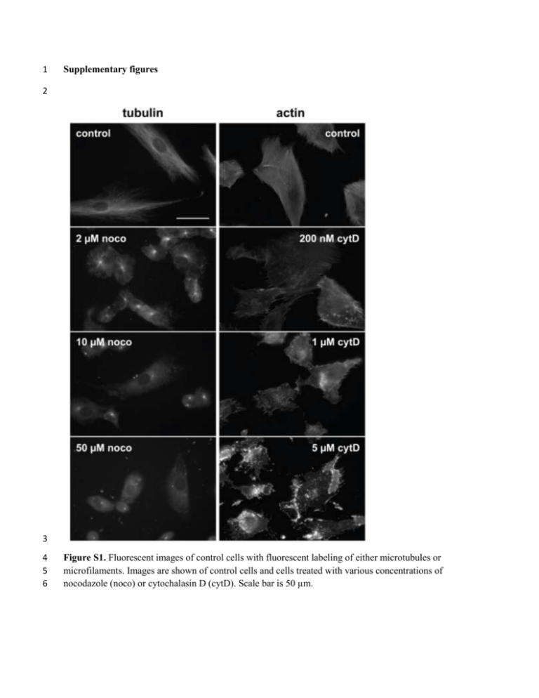
1 Supplementary figures 2 3 4 5 6 Figure S1. Fluorescent images of control cells with fluorescent labeling of either microtubules or microfilaments. Images are shown of control cells and cells treated with various concentrations of nocodazole (noco) or cytochalasin D (cytD). Scale bar is 50 µm. 7 8 9 10 11 12 13 14 15 16 17 18 19 20 Figure S2. Descriptive data of mitochondrial objects. A Histogram of size distribution of mitochondrial objects in a given cell for a given frame. Each bin corresponds to 10 pixels2. N = 242 mitochondrial objects. When area is restricted to be between 20 and 500 pixels for motility analysis, 176 mitochondrial objects remain. B Histogram of lifetimes of all mitochondrial objects for a given control cell. Each bin represents one interval (the time between two frames, which is 3 seconds), with the lowest bin representing zero intervals (an object which only appears in one frame). N = 3207 mitochondrial objects throughout the 5 minute time-lapse video. C Net distance in µm vs. lifetimes of all mitochondrial objects for a given control cell. There is a bin for each interval, with the first “bin” representing a lifetime of one interval (3 seconds). N = 1576 mitochondrial objects with a net distance of greater than zero (and lifetime > 0). 21 22 23 24 25 26 27 28 29 30 Figure S3. Combining cells in a given group preserves the lognormal distribution of net distances traveled by their mitochondria. A-F Histograms with fit normal distributions for each cell in the 10 µM nocodazole treated group. The center of the fitted distribution, corresponding to its geometric mean, is given in the upper right corner of the diagram. Normal probability plots are also given for each cell, with the R2 value and number of data points listed on each one. G Histogram with a fitted normal distribution for the pooled data set, along with the corresponding normal probability plot. 31 32 33 Figure S4. Cells after 30 minutes of treatment with 5 µM CCCP. Microtubules are fluorescently stained, and appear intact. Scale bar is 50 µm. 34 35 36 37 38 39 Figure S5. Image of the 1 µM cytochalasin D-treated cell shown in Video S6, with mitochondrial objects located in their final position and color coded according to the log value of their net distance traveled. The objects towards the top of the frame tend towards greater values, which is probably a result of the cell movement occurring in that region. The color scale is the same as in Fig. 4A. 40 41 42 43 44 45 46 47 48 49 50 51 52 53 54 55 56 57 58 Video captions Video S1. Video of the raw data taken of a sample control cell with fluorescent mitochondria. Scale bar for this and all other videos is 50 µm. This and all other videos are sped up to 20 frames (equivalent to 1 minute in real time) per second. Video S2. Pre-processed video of the same cell shown in Video S1. Video S3. Pre-processed video of a cell treated with 5 µM CCCP for 30 minutes. Video S4. Pre-processed video of a cell treated for 30 minutes with 50 µM nocodazole. Video S5. Pre-processed video of a cell treated with 5 µM cytochalasin D for 30 minutes. Video S6. A cell treated with 1 µM cytochalasin D for 30 minutes which shows excessive cell movement during the recording. The log values of the corresponding mitochondrial net distances are shown in Fig. S5.

