tpj12402-sup-0003-MethodS1-S3
advertisement
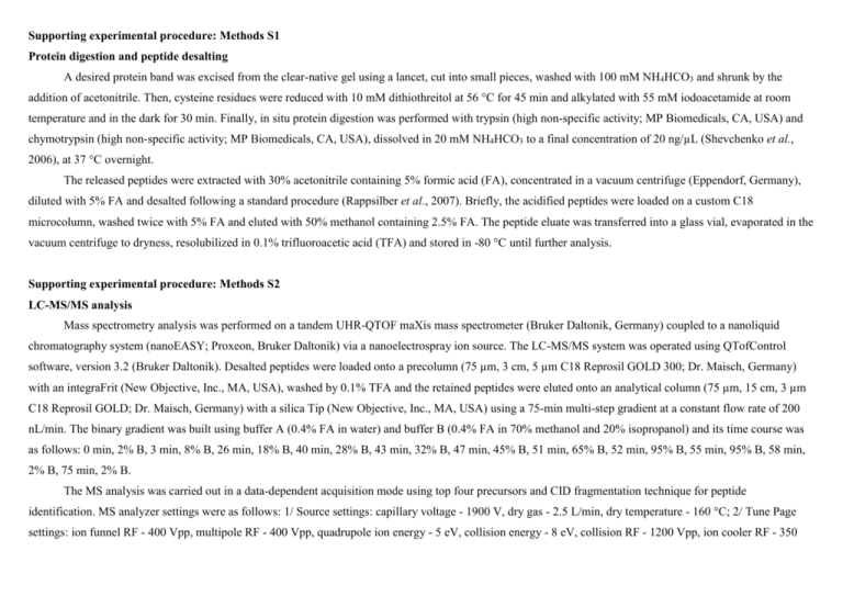
Supporting experimental procedure: Methods S1 Protein digestion and peptide desalting A desired protein band was excised from the clear-native gel using a lancet, cut into small pieces, washed with 100 mM NH4HCO3 and shrunk by the addition of acetonitrile. Then, cysteine residues were reduced with 10 mM dithiothreitol at 56 °C for 45 min and alkylated with 55 mM iodoacetamide at room temperature and in the dark for 30 min. Finally, in situ protein digestion was performed with trypsin (high non-specific activity; MP Biomedicals, CA, USA) and chymotrypsin (high non-specific activity; MP Biomedicals, CA, USA), dissolved in 20 mM NH4HCO3 to a final concentration of 20 ng/µL (Shevchenko et al., 2006), at 37 °C overnight. The released peptides were extracted with 30% acetonitrile containing 5% formic acid (FA), concentrated in a vacuum centrifuge (Eppendorf, Germany), diluted with 5% FA and desalted following a standard procedure (Rappsilber et al., 2007). Briefly, the acidified peptides were loaded on a custom C18 microcolumn, washed twice with 5% FA and eluted with 50% methanol containing 2.5% FA. The peptide eluate was transferred into a glass vial, evaporated in the vacuum centrifuge to dryness, resolubilized in 0.1% trifluoroacetic acid (TFA) and stored in -80 °C until further analysis. Supporting experimental procedure: Methods S2 LC-MS/MS analysis Mass spectrometry analysis was performed on a tandem UHR-QTOF maXis mass spectrometer (Bruker Daltonik, Germany) coupled to a nanoliquid chromatography system (nanoEASY; Proxeon, Bruker Daltonik) via a nanoelectrospray ion source. The LC-MS/MS system was operated using QTofControl software, version 3.2 (Bruker Daltonik). Desalted peptides were loaded onto a precolumn (75 µm, 3 cm, 5 µm C18 Reprosil GOLD 300; Dr. Maisch, Germany) with an integraFrit (New Objective, Inc., MA, USA), washed by 0.1% TFA and the retained peptides were eluted onto an analytical column (75 µm, 15 cm, 3 µm C18 Reprosil GOLD; Dr. Maisch, Germany) with a silica Tip (New Objective, Inc., MA, USA) using a 75-min multi-step gradient at a constant flow rate of 200 nL/min. The binary gradient was built using buffer A (0.4% FA in water) and buffer B (0.4% FA in 70% methanol and 20% isopropanol) and its time course was as follows: 0 min, 2% B, 3 min, 8% B, 26 min, 18% B, 40 min, 28% B, 43 min, 32% B, 47 min, 45% B, 51 min, 65% B, 52 min, 95% B, 55 min, 95% B, 58 min, 2% B, 75 min, 2% B. The MS analysis was carried out in a data-dependent acquisition mode using top four precursors and CID fragmentation technique for peptide identification. MS analyzer settings were as follows: 1/ Source settings: capillary voltage - 1900 V, dry gas - 2.5 L/min, dry temperature - 160 °C; 2/ Tune Page settings: ion funnel RF - 400 Vpp, multipole RF - 400 Vpp, quadrupole ion energy - 5 eV, collision energy - 8 eV, collision RF - 1200 Vpp, ion cooler RF - 350 Vpp, transfer time - 85 µs, pre-pulse storage - 7 µs; 3/ MS/MS settings: auto MSMS - on, 4 precursor ions, threshold for switching from MS to MSMS mode 5000 cts, active exclusion after 5 spectra for next 30 s, excluded mass range of precursors - 50-350 amu and 1500-2200 amu. All data were collected in a mass range of 50-2200 amu with the acquisition time of 500 ms for MS and 250-750 ms for MS/MS according to the precursor intensity. Supporting experimental procedure: Methods S3 Protein data analysis Raw data were processed and transformed into mgf files with DataAnalysis v.4.2 SP4 (Bruker Daltonik, Germany). The mgf files were subsequently uploaded to ProteinScape v.2.2 and searched against a custom protein database with Mascot search engine (v.2.2.07; in-house server; Matrix Science, UK) (Perkins et al., 1999). The database consisted of TAIR10 Arabidopsis database (The Arabidopsis Information Resources; v.20101214; 35386 sequences), common protein contaminants (247 sequences), PSI-NDH supercomplex homologs (93 sequences) related to the taxonomy of Hordeum vulgare found by a BLAST search (UniProtKB; www.UniProt.org) and shuttled versions of all protein sequences. The Mascot search was performed with no enzyme specificity in two steps. The first search was done with wide mass tolerances for both the precursor and fragment ions (±50 ppm and ±0.1 Da, respectively). Then, a list of high-confident peptide identification was used for re-calibration of precursor and fragment masses in the corresponding mgf files and the Mascot search was repeated with narrower mass tolerances (±5 ppm and ±0.03 Da for precursors and fragments, respectively). For the search, carbamidomethylation of cysteine was set as a fixed modification and N-terminal protein acetylation, deamination of asparagine and glutamine and oxidation of methionine were set as variable modifications. A minimum ion peptide score of 15 with a minimum peptide length of 7 amino acids at a significance threshold of p<0.05 and a false discovery rate of < 1% were used to validate the proteins. Finally, all peptide and protein assignments were re-validated manually. Complete lists of identified proteins for trypsin and chymotrypsin digestion are shown in Table S1 and S2, respectively. More detailed reports of each identified protein for both used digestions (Supporting Data S1 and S2, respectively) are also included. References Perkins, D.N., Pappin, D.J., Creasy, D.M. and Cottrell, J.S. (1999) Probability-based protein identification by searching sequence databases using mass spectrometry data. Electrophoresis 20, 3551-3567. Rappsilber, J., Mann, M. and Ishihama, Y. (2007) Protocol for micro-purification, enrichment, pre-fractionation and storage of peptides for proteomics using StageTips. Nat. Protoc. 2, 1896-906. Shevchenko, A., Tomas, H., Havlis, J., Olsen, J. and Mann, M. (2006) In-gel digestion for mass spectrometric characterization of proteins and proteomes. Nat. Protoc. 1, 2856-2860.


