Introduction to Bioinformatics
advertisement
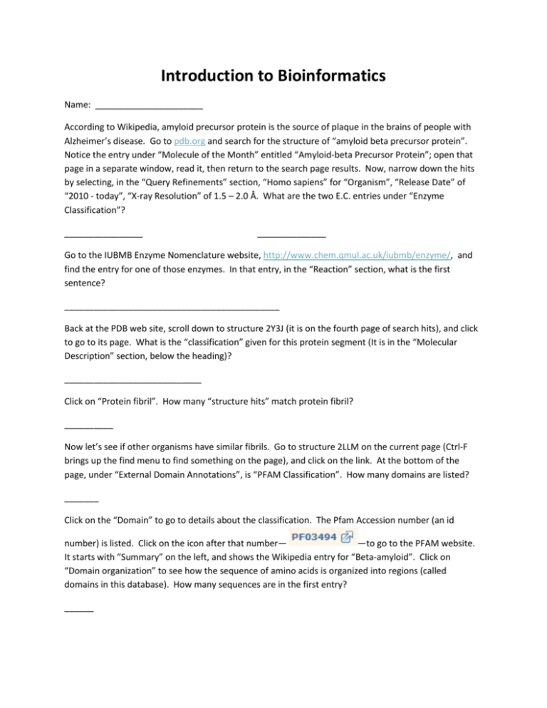
Introduction to Bioinformatics Name: ______________________ According to Wikipedia, amyloid precursor protein is the source of plaque in the brains of people with Alzheimer’s disease. Go to pdb.org and search for the structure of “amyloid beta precursor protein”. Notice the entry under “Molecule of the Month” entitled “Amyloid-beta Precursor Protein”; open that page in a separate window, read it, then return to the search page results. Now, narrow down the hits by selecting, in the “Query Refinements” section, “Homo sapiens” for “Organism”, “Release Date” of “2010 - today”, “X-ray Resolution” of 1.5 – 2.0 Å. What are the two E.C. entries under “Enzyme Classification”? ________________ ______________ Go to the IUBMB Enzyme Nomenclature website, http://www.chem.qmul.ac.uk/iubmb/enzyme/, and find the entry for one of those enzymes. In that entry, in the “Reaction” section, what is the first sentence? ____________________________________________ Back at the PDB web site, scroll down to structure 2Y3J (it is on the fourth page of search hits), and click to go to its page. What is the “classification” given for this protein segment (It is in the “Molecular Description” section, below the heading)? ____________________________ Click on “Protein fibril”. How many “structure hits” match protein fibril? __________ Now let’s see if other organisms have similar fibrils. Go to structure 2LLM on the current page (Ctrl-F brings up the find menu to find something on the page), and click on the link. At the bottom of the page, under “External Domain Annotations”, is “PFAM Classification”. How many domains are listed? _______ Click on the “Domain” to go to details about the classification. The Pfam Accession number (an id number) is listed. Click on the icon after that number— —to go to the PFAM website. It starts with “Summary” on the left, and shows the Wikipedia entry for “Beta-amyloid”. Click on “Domain organization” to see how the sequence of amino acids is organized into regions (called domains in this database). How many sequences are in the first entry? ______ If the mouse cursor is placed on the sequence, the parts are described. Click on “Show all sequences with this architecture”. (A scroll bar is now present on the right.) Let’s compare the sequences of amino acids in this protein from three different species: homo sapiens, and any two others of your choice. To do this, the sequence of amino acids is needed. Click on “A4 HUMAN”, then, on the left side of the page, click on “Sequence”, and, under the formatted sequence, click on “Show the unformatted sequence.” The unformatted sequence will be pasted in to http://www.ebi.ac.uk/Tools/msa/clustalo/, so open that site (control-click works in Microsoft Word). The data must be entered in FASTA format. FASTA – Fast Alignment Search Tool-All (since it works on both nucleotide and amino acid sequences). Associated with this software is a style of formatting a nucleic acid or protein sequence. It is important because many bioinformatics programs require that the sequence be in FASTA format. The FASTA format has a title line for each sequence that begins with a “>” followed by any needed text to name the sequence. The end of the title line is signified by a paragraph mark (hit the return key). Bioinformatics programs will know that the title line isn’t part of the sequence if you have it formatted correctly. The sequence itself does NOT have any returns, spaces, or formatting of any kind. The sequence is given in one-letter code. An example of two proteins in correct FASTA format is shown below: >Homo sapiens like spinach MTEYKLVVVGAGGVGKSALTIQLIQNHFVDEYDPTIEDSYRKQVVIDGETCLLDI LDTAGQEEYSAMRDQYMRTGEGFLCVFAINNTKSFEDIHHYREQIKRVKDSEDVP MVLVGNKCDLPSRTVDTKQAQDLARSYGIPFIETSAKTRQGVDDAFYTLVREIRK HKEKMSKDGKKKKKKSKTKCVIM >Mouse poop MLPGLALLLLAAWTARALEVPTDGNAGLLAEPQIAMFCGRLNMHMNVQNG KWDSDPSGTKTCIDTKEGILQYCQEVYPELQITNVVEANQPVTIQNWCKR GRKQCKTHPHFVIPYRCLVGEFVSDALLVPDKCKFLHQERMDVCETHLHW HTVAKETCSEKSTNLHDYGMLLPCGIDKFRGVEFVCCPLAEESDNVDSAD AEEDDSDVWWGGADTDYADGSEDKVVEVAEEEEVAEVEEEEADDDEDDED So, make up an appropriate title and paste your data into the Clustal Omega – Multiple Sequence Alignment window. Do this for each of at least three species. (Don’t do the same three species that anyone else does.) Click the “Submit” button. When your results are displayed, click the “Show colors” button (that just colors amino acid residues that have similar properties). You can make some sense out of the line with asterisks, etc. in it, but we will cover that even more in the class. If a warning appears, it may be because java needs to be updated on that computer; either give permission for the program to run, or update java (Control panel; programs; java; update tab). Let’s look at a “phylogenetic tree” of the data. Click on “Phylogenetic Tree”, which is near the top of the screen. A “phylogram” is shown. The numbers are an estimate of how similar the sequences are. Paste the phylogram below. (To do that, click in the browser to activate it, open “Snipping Tool” in Programs/Accessories). Paste it into Word. The picture should now be in Word. Email me this document (cking@troy.edu).
