Mouse Mammary Cell Flow Cytometry
advertisement
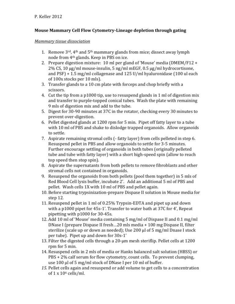
P. Keller 2012 Mouse Mammary Cell Flow Cytometry-Lineage depletion through gating Mammary tissue dissociation 1. Remove 3rd, 4th and 5th mammary glands from mice; dissect away lymph node from 4th glands. Keep in PBS on ice. 2. Prepare digestion mixture: 10 ml per gland of ‘Mouse’ media (DMEM/F12 + 2% CS, 10 µg/ml mouse-insulin, 5 ng/ml mEGF, 0.5 µg/ml hydrocortisone, and PSF) + 1.5 mg/ml collagenase and 125 U/ml hyaluronidase (100 ul each of 100x stocks per 10 mls). 3. Transfer glands to a 10 cm plate with forceps and chop briefly with a scissors. 4. Cut the tip from a p1000 tip, use to resuspend glands in 1 ml of digestion mix and transfer to purple-topped conical tubes. Wash the plate with remaining 9 mls of digestion mix and add to the tube. 5. Digest for 30-90 minutes at 37C in the rotator, checking every 30 minutes to prevent over-digestion. 6. Pellet digested glands at 1200 rpm for 5 min. Pipet off fatty layer to a tube with 10 ml of PBS and shake to dislodge trapped organoids. Allow organoids to settle. 7. Aspirate remaining stromal cells (- fatty layer) from cells pelleted in step 6. Resuspend pellet in PBS and allow organoids to settle for 3-5 minutes. Further encourage settling of organoids in both tubes (originally pelleted tube and tube with fatty layer) with a short high-speed spin (allow to reach top speed then stop spin). 8. Aspirate the supernatants from both pellets to remove fibroblasts and other stromal cells not contained in organoids. 9. Resuspend the organoids from both pellets (pool them together) in 5 mls of Red Blood Cell lysis buffer, incubate 2’. Add an additional 5 ml of PBS and pellet. Wash cells 1X with 10 ml of PBS and pellet again. 10. Before starting trypsinization-prepare Dispase II solution in Mouse media for step 12. 11. Resuspend pellet in 1 ml of 0.25% Trypsin-EDTA and pipet up and down with a p1000 pipet for 45s-1’. Transfer to water bath at 37C for 4’, Repeat pipetting with p1000 for 30-45s. 12. Add 10 ml of ‘Mouse’ media containing 5 mg/ml of Dispase II and 0.1 mg/ml DNase I (prepare Dispase II fresh…20 mls media + 100 mg Dispase II, filter sterilize (scale up or down as needed); Use 200 µl of 5 mg/ml Dnase I stock per tube). Pipet up and down for 30s-1’ 13. Filter the digested cells through a 20-µm mesh steriflip. Pellet cells at 1200 rpm for 5 min. 14. Resuspend cells in 2 mls of media or Hanks balanced salt solution (HBSS) or PBS + 2% calf serum for flow cytometry, count cells. To prevent clumping, use 100 µl of 5 mg/ml stock of DNase I per 10 ml of buffer. 15. Pellet cells again and resuspend or add volume to get cells to a concentration of 1 x 10⁶ cells/ml. P. Keller 2012 Staining for flow cytometry 1. Apply all antibodies to 200K cells (200 µl of 1 x 10⁶ cells/ml in HBSS/PBS + 2% CS +/- DNase I) for 20 min at 4C or 10-15 min at RT in the dark. 2. Wash 1-2 times with 3 mls of HBSS/PBS + 2% CS per tube. 3. Resuspend all with 200-250 µl of HBSS/PBS + 2% CS and keep on ice in the dark until flow. Samples: 1. Unstained 2. Isotype (APC-IgG2b and FITC-IgG2a or APC-IgG2b, PerCP-Cy5.5-IgG2a and FITC-IgG1) 3. FITC-CD49f or FITC-CD61 alone 4. PE-CD45 or PE-CD31 alone 5. PerCP-Cy5.5-CD49f alone (if using) 6. APC-CD24 alone 7. Experimental stain (note, it is best to make an antibody master mix to avoid lengthy pipetting or uneven staining) a. FITC-CD49f, PE-Lineage (PE-CD45, PE-CD31, PE-Ter119), APC-CD24 b. FITC-CD61, PE-Lineage (PE-CD45, PE-CD31, PE-Ter119), PerCP-Cy5.5 CD49f, APC-CD24 Antibody APC-CD24 FITC-CD49f PerCP-Cy5.5-CD49f FITC-CD61 PE-CD31 PE-CD45 PE-Ter119 APC-IgG2b FITC-IgG2a PerCP-Cy5.5-IgG2a FITC-IgG1 Amount for 200K cells 1 µl of 1:20 dilution 2 µl 1 µl 2 µl 2 µl 1 µl of 1:20 dilution 1 µl 1 µl of 1:20 dilution 2 µl 1µl 2 µl While at Calibur-order of operations Run unstained and set up machine for FSC, SSC, voltages for FL1-FL4 Run FITC-CD49f or FITC-CD61 and compensate out of FL2 Run PE-CD45 and compensate out of FL1 and FL3 Run PerCP-Cy5.5-CD49f and compensate out of FL2 and FL4 Run APC-CD24 and compensate out of FL3 Run and collect Isotype Run and collect Experimental stain(s) P. Keller 2012 Gating strategy: 1. On the isotype: Gate FSC/SSC for ‘cells’ between ~150 and 950 on the graph (approximately—you want to leave out debris which usually clusters in the corner below 100-200 and the stuff that is off the plot on the top edge). Apply the “Cells” gate to all other samples…check to make sure this gate makes sense (i.e. the FSC/SSC clouds aren’t shifted) 2. On the isotype: under the cells gate, make a histogram in the PE channel (FL2) and gate everything less than ~101.5 as ‘Lineage neg’ (or where this makes sense; should be 99% or so of cells). 3. On the isotype: under the cells, lin- and live gates, gate your isotypes as needed (usually put them at 101.1 or less for both) 4. Apply all of the gates to your stained experimental samples. Collect data from the cells gated for “cells, lin-, live” and either quad or circle gates for 24, 49f and 61. Antibody APC-CD24 FITC-CD49f PerCP-Cy5.5CD49f FITC-CD61 PE-CD31 PE-CD45 PE-Ter119 APC-IgG2b FITC-IgG2a PerCP-Cy5.5IgG2a FITC-IgG1 Vendor (Cat#) eBiosciences (17-0242-80) StemCell Tech (10111) BioLegend (31367) BD Biosciences (553346) eBiosciences (12-0311-83) BD Biosciences (553081) eBiosciences (12-5921-81) eBiosciences (17-4031-81) BD Biosciences (553929) BioLegend (400531) BD Biosciences (553443) Clone Isotype Concentration M1/69 IgG2b 0.2 mg/ml GoH3 IgG2a unknown GoH3 IgG2a 0.2 mg/ml 2C9.G2 IgG1 0.5 mg/ml 390 IgG2a 0.2 mg/ml 30-F11 IgG2b 0.5 mg/ml TER119 IgG2b 0.2 mg/ml eB149/10H5 - 0.2 mg/ml R35-95 - 0.5 mg/ml RTK2758 - 0.2 mg/ml A85-1 - 0.5 mg/ml

