Microwave processing and immunolocalization
advertisement
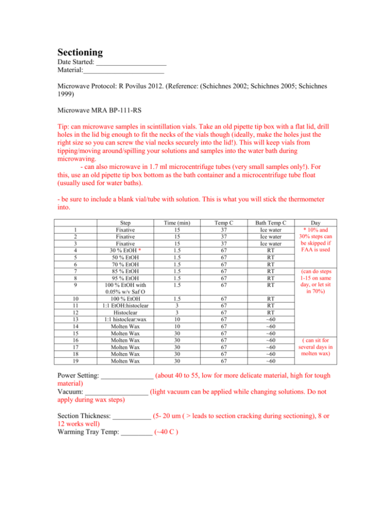
Sectioning Date Started: ____________________ Material:_______________________ Microwave Protocol: R Povilus 2012. (Reference: (Schichnes 2002; Schichnes 2005; Schichnes 1999) Microwave MRA BP-111-RS Tip: can microwave samples in scintillation vials. Take an old pipette tip box with a flat lid, drill holes in the lid big enough to fit the necks of the vials though (ideally, make the holes just the right size so you can screw the vial necks securely into the lid!). This will keep vials from tipping/moving around/spilling your solutions and samples into the water bath during microwaving. - can also microwave in 1.7 ml microcentrifuge tubes (very small samples only!). For this, use an old pipette tip box bottom as the bath container and a microcentrifuge tube float (usually used for water baths). - be sure to include a blank vial/tube with solution. This is what you will stick the thermometer into. 1 2 3 4 5 6 7 8 9 10 11 12 13 14 15 16 17 18 19 Step Fixative Fixative Fixative 30 % EtOH * 50 % EtOH 70 % EtOH 85 % EtOH 95 % EtOH 100 % EtOH with 0.05% w/v Saf O 100 % EtOH 1:1 EtOH:histoclear Histoclear 1:1 histoclear:wax Molten Wax Molten Wax Molten Wax Molten Wax Molten Wax Molten Wax Time (min) 15 15 15 1.5 1.5 1.5 1.5 1.5 1.5 Temp C 37 37 37 67 67 67 67 67 67 Bath Temp C Ice water Ice water Ice water RT RT RT RT RT RT 1.5 3 3 10 10 30 30 30 30 30 67 67 67 67 67 67 67 67 67 67 RT RT RT ~60 ~60 ~60 ~60 ~60 ~60 ~60 Day * 10% and 30% steps can be skipped if FAA is used (can do steps 1-15 on same day, or let sit in 70%) ( can sit for several days in molten wax) Power Setting: _______________ (about 40 to 55, low for more delicate material, high for tough material) Vacuum: __________________ (light vacuum can be applied while changing solutions. Do not apply during wax steps) Section Thickness: ___________ (5- 20 um ( > leads to section cracking during sectioning), 8 or 12 works well) Warming Tray Temp: _________ (~40 C ) Section Mounting: ______________________________________________________________________________ ______________________________________________________________________________ _____________________________________________________________________________ (use prepared gelatin/egg white subbed, poly-L-lysine coated, or Fisher Probe on Plus slides, float pieces of ribbon on water on slide, transfer to warming tray. After ~15 min and ribbon has expanded, removed water with syringe/transfer pipette/kimwipe. Let sit overnight at RT or on warming tray, or at least let sit on warming tray until all water has evaporated, ~3-4 hours) Deparaffin/Rehydration: ______________________________________________________________________________ ______________________________________________________________________________ ________________________________________________________________________ (3 of steps: histoclear, 15 min, then 2 (10 min) steps each of: 100% EtOH, 95% EtOH, 70% EtOH, rinse with a few changes of dH20). Do not let slides dry out to proceed with immunolocalization) Immunolocalization, section: (Gong 2006; Moctezuma 1999; Sauer 2010)***see Gong 2006 for recipes for BS, HWB, and LWB solutions*** - have all solution prepared, keep an eye out for step(s) that need prewarming of solutions. - wash step times and number are suggestions. Can be adjusted. - how to actually apply incubation solutions and wash steps to slides: - for wash steps, lay slides section-face-up in a flat bottomed container (square petri dishes work great! Can fit 3 slides, uses only 10-15 mL wash solution per step). Add enough wash solution to barely cover slides. Cover and place on shaking table. - for incubation solutions (like prot K, 1’ aB, 2’ aB, DAPI), lay slides section-face-up in humid chamber (water-tight plastic containers with moistened paper towel at the bottom and some sort of configuration to keep slides from actually touching the moist paper towel), pipette some amount (600 ul or less) of incubation solution in each slide, cover each slide with a slide-sized piece of parafilm (sterile slide down). - for diluting the antibodies, can use 1x PBS, 1% BSA BS solution, 3% BSA BS solution - Controls: -without 1’ aB -without 2’ aB -1’ aB pre-incubated with compound that you think it should be binding to specifically -positive control with 1’ and 2’ aB with material for which you already know the compound is present… ideally tissue from the same organism, but can also be nitrocellulose membrane or gelatin block spotted with known compound(s) the 1’ aB should react with - if you are getting really high background, consider pre-absorbing your 1’ aB (see Gong 2006) - Permeabilization (check which ones that apply): prewarmed 1 ug/ml proteinase K (promega) in 1x PBS at 37 C for 30 min rinse with 1x PBS, 10 min at RT rinse with 1x PBS, 10 min at RT rinse with 1x PBS, 10 min at RT 10% DMSO (v/v) at RT for 60 min rinse with 1x PBS, 10 min at RT rinse with 1x PBS, 10 min at RT rinse with 1x PBS, 10 min at RT 1:20, Triton x100 : 1x PBS at RT for 60 min rinse with 1x PBS, 10 min at RT rinse with 1x PBS, 10 min at RT rinse with 1x PBS, 10 min at RT 0.1% Triton x100 and 2.0% Drislease (Sigma) in 1x PBS at 37 C for 60 min (Barton 2010) rinse with 1x PBS, 10 min at RT rinse with 1x PBS, 10 min at RT rinse with 1x PBS, 10 min at RT Postfixation: 10% FAA in 1x PBS, 15 min at RT rinse with 1x PBS, 10 min at RT rinse with 1x PBS, 10 min at RT rinse with 1x PBS, 10 min at RT Blocking: incubate in BS for 45 min at RT Rinse briefly with LWB Primary antibody (___________________________________) - Dilution: ___________________________ - Incubate overnight (16 hours) at 4 C Rinse vigorously with HWB, 20 min at RT Rinse vigorously with HWB, 20 min at RT Rinse vigorously with HWB, 20 min at RT Secondary antibody (___________________________________) - Dilution: ___________________________ - incubate 2 hours in dark at 37 C Rinse vigorously with LWB, 20 min at RT Rinse vigorously with LWB, 20 min at RT rinse with 1x PBS, 10 min at RT rinse with 1x PBS, 10 min at RT If AP conjugated 2ndard antibody was used: incubate with 200 uL Western blue (promega) in the dark at RT, check every 10 min until blue color is observed (~20 min?) DAPI – Sectioned material (Friedman Lab protocol) Rinse material with 1x PBS, 10 min at RT Incubate with DAPI solution (1:4, 1x PBS : DAPI(1ug/ml)) for 1 hour at RT in the dark Rinse with 1x PBS, 15 min at RT Rinse with 1x PBS, 15 min at RT Mount in dH20, coverslip (can seal with clear nailpolish), observe References: Barton D, Overall R (2010) Cryofixation rapidly preserves cytoskeletal arrays of leaf epidermal cell revealing microtuble co-alignments between neighbouring cells and adjacent actin and microtubule buncles in the cortex. Journal of Microscopy 237, 79-88. Gong H, Peng Y, Zou C, et al. (2006) A simple treatment to significantly increase signal specificity in immunohistochemistry. Plant Molecular Biology Reporter 24, 93101. Moctezuma E (1999) Changes in Auxin Patterns in Developing Gynophores of the Peanut Plant (Arachis hypogaea L.). Annals of Botany 83, 235-242. Sauer M, Friml J (2010) Immunolocalization of Proteins in Plants. In: Plant Developmental Biology (eds. Hennig L, Köhler C), pp. 253-263. Springer Science+Business Media, LLC. Schichnes D, Nemson J, Ruzin S (2002) Microwave Paraffin Techniques for Botanical Tissues. In: Microwave Techinques and Protocols (eds. Giberson R, Demaree R). Humana Press, Inc, Totowa, NJ. Schichnes D, Nemson J, Ruzin S (2005) Microwave protools for plant and animal paraffin microtechnique. Microscopy Today, 50-53. Schichnes D, Nemson J, Sohlberg L, Ruzin S (1999) Microwave protocols for paraffin microtechnique and in situ localization in plants. Microscopy and Microanalysis 4, 491-496.
