lecture_2015-03-131426202135211165676055021e177ac20
advertisement
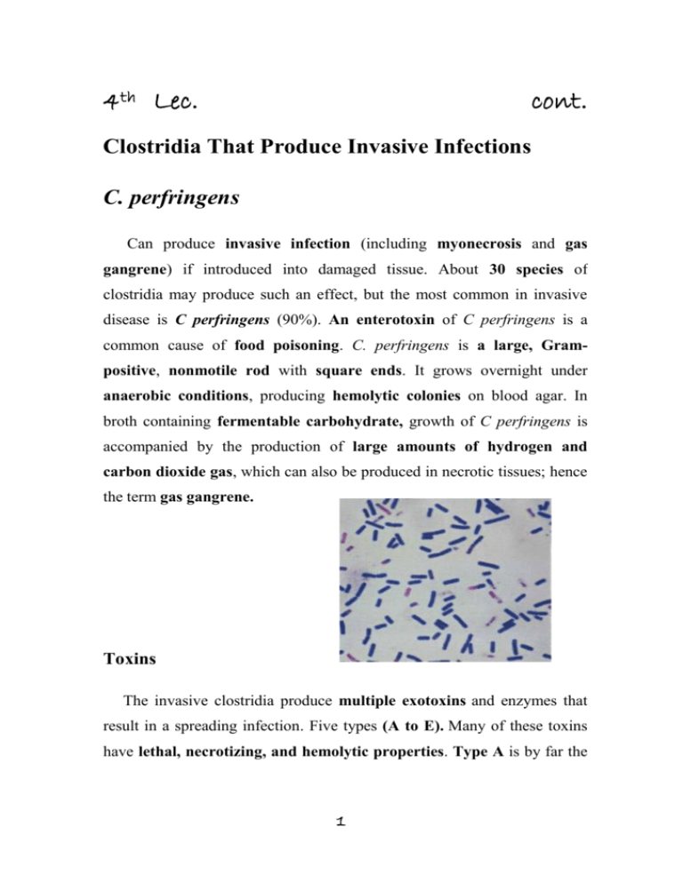
4th Lec. cont. Clostridia That Produce Invasive Infections C. perfringens Can produce invasive infection (including myonecrosis and gas gangrene) if introduced into damaged tissue. About 30 species of clostridia may produce such an effect, but the most common in invasive disease is C perfringens (90%). An enterotoxin of C perfringens is a common cause of food poisoning. C. perfringens is a large, Grampositive, nonmotile rod with square ends. It grows overnight under anaerobic conditions, producing hemolytic colonies on blood agar. In broth containing fermentable carbohydrate, growth of C perfringens is accompanied by the production of large amounts of hydrogen and carbon dioxide gas, which can also be produced in necrotic tissues; hence the term gas gangrene. Toxins The invasive clostridia produce multiple exotoxins and enzymes that result in a spreading infection. Five types (A to E). Many of these toxins have lethal, necrotizing, and hemolytic properties. Type A is by far the 1 most important in humans and is found consistently in the colon and often in soil. The most important exotoxin is: 1. The α-toxin, a phospholipase that hydrolyzes lecithin and sphingomyelin (an important constituent of cell membranes), thus disrupting the cell membranes of various host cells, including erythrocytes, leukocytes, and muscle cells. 2. The ϴ-toxin has similar hemolytic and necrotizing effects. It alters capillary permeability and is toxic to heart muscle. This poreforming toxin is closely related to streptolysin O 3. DNase and hyaluronidase 4. Collagenase that digests collagen of subcutaneous tissue and muscle. 5. Enterotoxin: a powerful enterotoxin produces by some strains especially when grown in meat dishes. When more than 108 vegetative cells are ingested and sporulate in the gut, enterotoxin is formed. The enterotoxin is a protein (MW 35,000) that may be a nonessential component of the spore coat; it is distinct from other clostridial toxins. Which, when attached to enterocyte membranes, causes an increase in intracellular calcium and altered membrane permeability. This leads to loss of cellular fluid, electrolytes and macromolecules and marked hypersecretion in the jejunum and ileum. It induces intense diarrhea in 6–18 hours. Much less frequent symptoms include nausea, vomiting, and fever. This illness is similar to that produced by B cereus and tends to be self-limited. 2 Pathogenesis In invasive clostridial infections, spores reach tissue either by contamination of traumatized areas (soil, feces) or from the intestinal tract. The spores germinate at low oxidation-reduction potential; vegetative cells multiply elaborating α-toxin, ferment carbohydrates present in tissue, and produce gas. The distention of tissue and interference with blood supply, together with the secretion of necrotizing toxin and hyaluronidase, favor the spread of infection. Tissue necrosis extends, providing an opportunity for increased bacterial growth, hemolytic anemia, and, ultimately, severe toxemia and death. In gas gangrene (clostridial myonecrosis), a mixed infection is the rule. In addition to the toxigenic clostridia, proteolytic clostridia and various cocci and gram-negative organisms are also usually present. C perfringens occurs in the genital tract of 5% of women. clostridial uterine infections followed instrumental abortions. Clostridium sordellii has many of the properties of C perfringens. C sordellii has been reported to cause a toxic shock syndrome after medical abortion with mifepristone and intravaginal misoprostol. Endometrial infection with C sordellii is implicated. Clostridial bacteremia is a frequent occurrence in patients with neoplasms. In New Guinea, C perfringens type C produces a necrotizing enteritis (pigbel) that can be highly fatal in children. Immunization with type C toxoid appears to have preventive value. Clinical Findings From a contaminated wound (eg, a compound fracture, postpartum uterus), the infection spreads in 1–3 days to produce crepitation in the subcutaneous tissue and muscle, foul-smelling discharge, rapidly 3 progressing necrosis, fever, hemolysis, toxemia, shock, and death. At times, the infection results only in anaerobic fasciitis or cellulitis. C perfringens food poisoning usually follows the ingestion of large numbers of clostridia that have grown in warmed meat dishes. The toxin forms when the organisms sporulate in the gut, with the onset of diarrhea—usually without vomiting or fever—in 6–18 hours. The illness lasts only 1–2 days. Diagnostic Laboratory Tests Specimens consist of material from wounds, pus, and tissue. 1. Gram-stained smears The presence of large gram-positive rods suggests gas gangrene clostridia; spores are not regularly present. 2. Material is inoculated into chopped meat-glucose medium and thioglycolate medium and onto blood agar plates incubated anaerobically produce hemolysis, and colony form. 3. The growth from one of the media is transferred into milk. A clot torn by gas in 24 hours is suggestive of C perfringens. 4. Once pure cultures have been obtained by selecting colonies from anaerobically incubated blood plates, they are identified by biochemical reactions (various sugars in thioglycolate, action on milk). 5. Lecithinase activity is evaluated by the precipitate formed around colonies on egg yolk media. 6. Final identification rests on toxin production and neutralization by specific antitoxin in lab animals. C perfringens rarely produces spores when cultured on agar in the laboratory. 4 Treatment Until the advent of specific therapy, the most important treatment is prompt and extensive surgical debridement (amputation)of the involved area and excision of all devitalized tissue, in which the organisms are prone to grow. Administration of antimicrobial drugs, particularly penicillin, is begun at the same time. Hyperbaric oxygen may be of help in the medical management of clostridial tissue infections. It is said to "detoxify" patients rapidly.Antitoxins are available against the toxins of C perfringens, Clostridium novyi, Clostridium histolyticum, and Clostridium septicum, usually in the form of concentrated immune globulins. Polyvalent antitoxin (containing antibodies to several toxins) has been used. Although such antitoxin is sometimes administered to individuals with contaminated wounds containing much devitalized tissue, there is no evidence for its efficacy. Food poisoning due to C perfringens enterotoxin usually requires only symptomatic care. Prevention & Control Early and adequate cleansing of contaminated wounds and surgical debridement, together with the administration of antimicrobial drugs directed against clostridia (eg, penicillin), are the best available preventive measures. Antitoxins should not be relied on. Although toxoids for active immunization have been prepared, they have not come into practical use. Prevention of food poisoning involves good cooking hygiene and adequate refrigeration 5 Clostridium Difficile & Diarrheal Disease Pseudomembranous Colitis C difficile is a Gram-positive rod that readily forms spores. It is present in the stool of 2% to 5% of the general population, sometimes at higher rates among hospitalized persons and infants. Frequent cause of antibiotic-associated diarrhea AAD. Toxin Administration of antibiotics results in proliferation of drugresistant C difficile that produces two large polypeptide toxins, A and B, with similar structure (45% homology). Toxin A, a potent enterotoxin that also has some cytotoxic activity, binds to the brush border membranes of the gut at receptor sites. Toxin B is a potent cytotoxin. Both toxins are found in the stools of patients with pseudomembranous colitis. Not all strains of C difficile produce the toxins, and the tox genes apparently are not carried on plasmids or phage. Pathogenesis Alteration of the colonic flora with antimicrobics (particularly ampicillin, cephalosporins, clindamycin and more recently the fluoroquinolones) favors C difficile which can grow and increased in numbers that make toxin more effective. The enterotoxic properties of the A toxin seem to dominate in watery or bloody diarrhea cases. The fits better picture with the action of the cytotoxic B toxin is the inflammatory plaques of colonic mucosa, which may coalesce into an 6 overlying "pseudomembrane" composed of fibrin, leukocytes, and necrotic colonic cells. Plaques and microabscesses may be localized to one area of the bowel and the patient frequently has associated abdominal cramps, leukocytosis, and fever. Diagnosis Pseudomembranous colitis is diagnosed by detection of one or both toxins in stool and by endoscopic observation of pseudomembranes or microabscesses in patients who have diarrhea and have been given antibiotics. Treatment The disease is treated by discontinuing administration of the offending antibiotic and orally giving either metronidazole or vancomycin. Antibiotic-Associated Diarrhea The administration of antibiotics frequently leads to a mild to moderate form of diarrhea, termed antibiotic-associated diarrhea. This disease is generally less severe than the classic form of pseudomembranous colitis. As many as 25% of cases of antibioticassociated diarrhea may be associated with C difficile. 7
