blood supply of brain - PBL-J-2015
advertisement
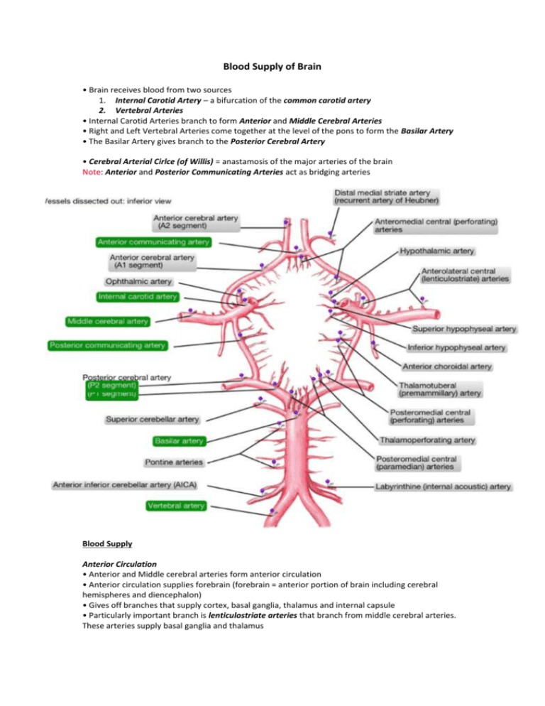
Blood Supply of Brain • Brain receives blood from two sources 1. Internal Carotid Artery – a bifurcation of the common carotid artery 2. Vertebral Arteries • Internal Carotid Arteries branch to form Anterior and Middle Cerebral Arteries • Right and Left Vertebral Arteries come together at the level of the pons to form the Basilar Artery • The Basilar Artery gives branch to the Posterior Cerebral Artery • Cerebral Arterial Cirlce (of Willis) = anastamosis of the major arteries of the brain Note: Anterior and Posterior Communicating Arteries act as bridging arteries Blood Supply Anterior Circulation • Anterior and Middle cerebral arteries form anterior circulation • Anterior circulation supplies forebrain (forebrain = anterior portion of brain including cerebral hemispheres and diencephalon) • Gives off branches that supply cortex, basal ganglia, thalamus and internal capsule • Particularly important branch is lenticulostriate arteries that branch from middle cerebral arteries. These arteries supply basal ganglia and thalamus Posterior Circulation • Consists of branches arising from posterior cerebral arteries, basilar and vertebral arteries • Posterior circulation supplies posterior cortex, midbrain and brainstem • Important branches are the Anterior Inferior Cerebellar Artery (comes of basilar artery) and Posterior Inferior Cerebellar Artery (comes of vertebral arteries) which supply regions of the pons and medulla The Internal Capsule • The internal capsule is a unique location where a large number of motor and sensory fibers travel to and from the cortex. Damage of any kind in this location will cause some relatively unique findings that can allow you to localize the lesions to the internal capsule by exam alone. • Location – From diagram above, can see it separates the caudate and lentiform nuclei • Infarcts to lenticulostriate arteries may cause motor and sensory deficits due to the fact the internal capsule has both motor and sensory fibres running through it

