Chest trauma fact sheet
advertisement
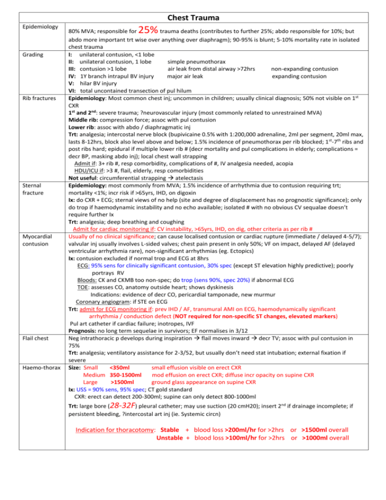
Chest Trauma Epidemiology Grading Rib fractures Sternal fracture Myocardial contusion Flail chest Haemo-thorax 25% 80% MVA; responsible for trauma deaths (contributes to further 25%; abdo responsible for 10%; but abdo more important trt wise over anything over diaphragm); 90-95% is blunt; 5-10% mortality rate in isolated chest trauma I: unilateral contusion, <1 lobe II: unilateral contusion, 1 lobe simple pneumothorax III: contusion >1 lobe air leak from distal airway >72hrs non-expanding contusion IV: 1Y branch intrapul BV injury major air leak expanding contusion V: hilar BV injury VI: total uncontained transection of pul hilum Epidemiology: Most common chest inj; uncommon in children; usually clinical diagnosis; 50% not visible on 1st CXR 1st and 2nd: severe trauma; ?neurovascular injury (most commonly related to unrestrained MVA) Middle rib: compression force; assoc with pul contusion Lower rib: assoc with abdo / diaphragmatic inj Trt: analgesia; intercostal nerve block (bupivicaine 0.5% with 1:200,000 adrenaline, 2ml per segment, 20ml max, lasts 8-12hrs, block also level above and below; 1.5% incidence of pneumothorax per rib blocked; 1 st-7th ribs and post ribs hard; epidural if multiple lower rib # (decr mortality and pul complications in elderly; complications = decr BP, masking abdo inj); local chest wall strapping Admit if: 3+ rib #, resp comorbidity, complications of #, IV analgesia needed, acopia HDU/ICU if: >3 #, flail, elderly, resp comorbidities Not useful: circumferential strapping atelectasis Epidemiology: most commonly from MVA; 1.5% incidence of arrhythmia due to contusion requiring trt; mortality <1%; incr risk if >65yrs, IHD, on digoxin Ix: do CXR + ECG; sternal views of no help (site and degree of displacement has no prognostic significance); only do trop if haemodynamic instability and no echo available; isolated # with no obvious CV sequalae doesn’t require further Ix Trt: analgesia; deep breathing and coughing Admit for cardiac monitoring if: CV instability, >65yrs, IHD, on dig, other criteria as per rib # Usually of no clinical significance; can cause localised contusion or cardiac rupture (immediate / delayed 4-5/7); valvular inj usually involves L-sided valves; chest pain present in only 50%; VF on impact, delayed AF (delayed ventricular arrhythmia rare), non-significant arrhythmias (eg. Ectopics) Ix: contusion excluded if normal trop and ECG at 8hrs ECG: 95% sens for clinically significant contusion, 30% spec (except ST elevation highly predictive); poorly portrays RV Bloods: CK and CKMB too non-spec; do trop (sens 90%, spec 20%) if abnormal ECG TOE: assesses CO, anatomy outside heart; shows dyskinesis Indications: evidence of decr CO, pericardial tamponade, new murmur Coronary angiogram: if STE on ECG Trt: admit for ECG monitoring if: prev IHD / AF, transmural AMI on ECG, haemodynamically significant arrhythmia / conduction defect (NOT required for non-specific ST changes, elevated markers) Pul art catheter if cardiac failure; inotropes, IVF Prognosis: no long term sequelae in survivors; EF normalises in 3/12 Neg intrathoracic p develops during inspiration flail moves inward decr TV; assoc with pul contusion in 75% Trt: analgesia; ventilatory assistance for 2-3/52, but usually don’t need stat intubation; external fixation if severe Size: Small <350ml small effusion visible on erect CXR Medium 350-1500ml mod effusion on erect CXR; diffuse incr opacity on supine CXR Large >1500ml ground glass appearance on supine CXR Ix: USS = 90% sens, 95% spec; CT gold standard CXR: erect can detect 200-300ml; supine can only detect 800-1000ml Trt: large bore (28-32F) pleural catheter; may use suction (20 cmH20); insert 2nd if drainage incomplete; if persistent bleeding, ?intercostal art inj (ie. Systemic circn) Indication for thoracotomy: Stable + blood loss >200ml/hr for >2hrs or >1500ml overall Unstable + blood loss >100ml/hr for >2hrs or >1000ml overall Pneumothorax Pneumomediastinum Pul contusion Tracheobronchial inj Diaphragm inj Gas embolism Aortic inj Indication for thoracoscopy: haemothorax failed to resolve after 3/7 Sucking chest wound = allows air to pass in AND out; will occur preferentially through this if opening >2/3 diameter of trachea resp failure; in penetrating chest trauma, appearance of pneumothorax on CXR may be delayed, so rpt at 6hrs and 12hrs Ix: CXR: up to 25% missed on initial CXR Supine CXR: air will collect ant-inf-medial etched diaphragm / mediastinum, deep sulcus sign USS: use linear transducer, loss of sliding lung sign; >90% sens, >95% spec Small = At mid clavicular point or 4th IC space ant axillary line Medium = at mid axially line Large = at post axillary line Trt: if tension, finger thoracostomy better than needle (prone to occlusion) then immediate chest drain If IPPV and cardiac arrest bilat pleural decompression If IPPV + unequal AE + Sa <90% on 100% O2 / SBP <100 despite IVF immediate unilateral pleural decompression If sucking, immediate occlusive dressing taped on 3 sides then chest drain If small, no haemothorax, no other significant inj, and IPPV unlikely trt conservatively If mod / large do IDC, don’t do needle decompression Tension pneumomediastinum = incr JVP but normal BS, rare +++ Ix: CXR: air stripe, prominent heart border, dark line along superior surface of central diaphragm making it look continuous from L – R hemithorax; supine CXR: 20% sens CT: >95% sens, 85% spec; indicated if suggestion of GI inj or decr LOC Always investigate GI tract if penetrating inj Trt: conservative; if tension – decompression at suprasternal notch Due to blunt trauma / GSW alveolar rupture, fluid transudation into alveolar spaces decr lung compliance (lowest at 3/7); CXR and examination changes usually occur within 6hrs (can take 24hrs to develop; aspiration and infarction take longer to develop than contusion); if opacity becomes more circumscribed over days – 2/52, consider lung haematoma; severe if >20% lung vol ( 80% develop ARDS, 50% develop pneumonia); if IPPV, use low vol, low p techniques to avoid barotrauma; usually recover over 3/52 Rare; 65% penetrating; 80% blunt occur within 2.5cm of carina; consider if pneumomediastinum, subC emphysema, persiste nt air leak from ICC, segmental lung collapse, haemoptysis Ix: CXR (subC / mediastinal emphysema, pneumothorax, deviated ETT tip, distention / migration of ETT balloon; bronchoscopy Trt: 1Y closure Penetrating: L sided more common; may be v small defect; CT (95% sens and spec), laparoscopy (100% sens) Blunt: more common with lateral torso trauma; 90% from MVA, 10% delayed diagnosis by 48hrs, 80% have abdo inj, 50% have pelvic #, 33% mortality, 70% L sided, usually posterior, rarely of tendinous portion; 50% present with delayed rupture ( obstruction, infarction, fistula); defect enlarges with time; herniation of omentum, transverse colon, stomach, SI CXR: elevated hemidiaphragm, pleural effusion, lower rib #, diaphragm shadow doesn’t reach chest wall, hemithorax opacity and displacement of mediastinum despite ICC, gas / NG in bowel filled chest; collapse of lower lung fields; inhomogenous mass in hemithorax; displaced mediastinum away from inj CT (50% sens for R, 78% sens for L); laparoscopy (90% sens); DPL (50% sens); MRI Arterial: due to communication between pul veins and airways artery, gas enters pul vein when airway p > venous p; usually due to penetrating trauma; FND, air bubbles on fundoscopy, CV collapse after IPPV, gas in ABG’s Pulmonary: iatrogenic from CVL insertion; gas in heart decr CO; trt with 100% O2 and IVF Usually high energy inj; side impact incr risk; 50% will have had episode of low BP; 30% have normal chest examination; decr pulse p in legs in 30% Classification: intimal tear = intima only; tear = intima and muscularis (60% strength); rupture = complete I = intramural haematoma, limited intimal flap II = subadventitial rupture, altered shape of aorta III = aortic transection with active bleeding / aortic obsturction with ischaemia Location: 65-90% in isthmus (prox descending, between origin L subclavian and site of attachment of ligamentum arteriosum; 5-10% ascending aorta / arch (rapidly fatal), 10% distal, 15% multiple sites Ix: CXR: supine CXR 90% sens, 30% spec; erect CXR 95% sens, incr spec Wide upper mediastinum (90% sens, 10% spec; >8.5cm in supine film, >6cm in erect), blurring of upper descending aorta and L side of arch, loss of aortic knuckle, incr R paratracheal stripe >4mm; incr L paratracheal stripe >5mm; L apical cap; massive haemothroax, tracheal / oesophageal deviation (to R of T4 spinous process); opacification of aortopul window; depression of L main bronchus to below 40deg from horizontal; deviation of oesophagus to R at T4 CT chest angiography: 95% sens, 80% spec, NPV 99-100% Aortic angiography: if mediastinal haematoma seen on CT, but no aortic inj Myocardial lac and periC tamponade Oesophageal perf Assessment Investigation Management Transoesophageal echo: 100% sens, 98% spec; CI if oesophageal inj; good if too unstable for CT Trt: if unstable (SBP <90) OT (mortality >85%) Stenting is up and coming If stable + CAD / >55yrs / intimal tear only conservative mng with control HTN as for dissection Prognosis: 85% pre-hospital death; if reach hospital, 85% survive 1hr, 70% 6hrs, 50% 24hrs, 25% 1/52, 10% 4/12; 99% die without repair Assoc with penetrating inj; hypotension, decr HS, incr JVP (Beck’s triad); suggested by PEA in absence of hypoV or tension Ix: echo (indication for ED thoracotomy if FAST +ive and SBP <70 unresponsive to IVF) Mng: urgent surgical intervention; see indications for ED thoracotomy Rare; usually lower 1/3; assoc with tracheal, T3-4 inj; delayed diagnosis in 2/3; from blunt, 5% mortality, 25% infectious complications (mediastinitis); usually see pleural effusion on L; Ix with gastrograffin swallow (70% sens) or gastroscopy; trt with NG, Abx, acid suppression, OT (mortality related to time to OT) No pain / tenderness / auscultatory abnormality = 100% NPV for significant chest injury Decr BS = 50-85% sens, 90% spec for haemothorax; percussion less sens 85% sens, 95% spec for pneumothorax CT chest: pros: more sens than XR at detecting lung contusion, pneumothorax, mediastinal haematoma, rib #’s; can do CT angiogram; angiography not needed if no mediastinal haematoma; non-invasive; cheap CXR: pros: erect film can view haemathorax 200-300ml Cons: supine film (mediastinum widened, may miss small haem/pneumothorax (800-1000ml needed); miss 50% ant / lat rib #’s TOE: pros: can be done in resus, quick, minimally invasive, low complication rate Cons: requires sedation, limited info on distal ascending aorta / aortic arch Thoracotomy required in 10% blunt trauma Indications for ED thoracotomy: in extremis (in cardiac arrest) likely to arrest before reaching OT + vital signs present in ED Indicated in penetrating chest trauma (30% survival rate) Seldom indicated in blunt truma (only 1% survival) Do L thoracotomy regardless of findings (then extend to R if needed) long ant 5th ICS incision retractor release of pericardial tamponade (incise vertically infront of phrenic nerve) suture cardiac lacs control bleeding lung tissue (use aortic clamp, or clamp pul hilum) clamp descending aorta (if terminally hypotensive) internal cardiac massage (1 hand infront, 1 hand behind heart; futile if >10mins) Indications for OT thoracotomy: cardiac tamponade; vascular inj at thoracic outlet; massive air leak from IDC; massive / continuing haemothorax (>200ml/hr or >1500ml if stable, >100ml/hr or >1000ml if unstable); mediastinal transversing penetrating inj; oesophageal / tracheal / bronchial / gt vessel inj When to stop resus: irreparable inj (eg. Blunt cardiac trauma); vol replacement not achieved within 15mins of thoracotomy; heart in non-viable rhythm after 30mins Notes from: Dunn, computer notes, Cameron
