Gohara PARTNERSHIPS-Elisa copy
advertisement
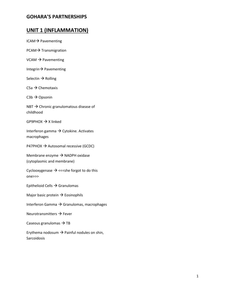
GOHARA’S PARTNERSHIPS UNIT 1 (INFLAMMATION) ICAM Pavementing PCAM Transmigration VCAM Pavementing Integrin Pavementing Selectin Rolling C5a Chemotaxis C3b Opsonin NBT Chronic granulomatous disease of childhood GP9PHOX X linked Interferon gamma Cytokine. Activates macrophages P47PHOX Autosomal recessive (GCDC) Membrane enzyme NADPH oxidase (cytoplasmic and membrane) Cyclooxygenase <<<she forgot to do this one>>> Epithelioid Cells Granulomas Major basic protein Eosinophils Interferon Gamma Granulomas, macrophages Neurotransmitters Fever Caseous granulomas TB Erythema nodosum Painful nodules on shin, Sarcoidosis 1 GOHARA’S PARTNERSHIPS UNIT 2 (VASCULAR & HEART) Dz of intima—atherosclerosis Dz of media—montenberg ? spelled wrong. Occurs usually in the leg and would see calcified BV’s on xray high sensitivity C reactive protein - risk of dev coronary artery dz Tear chest pain radiating to the back—aortic dissection Anti-endothelium Ab- Kawasaki Smoking and inflamed V. A. N.—Beurger Neuropathy—Churg Straus Risk for PE- thrombophlebitis Polymyalgia rheumatica—temporal arteritis hyalinization-- benign hypertension Inflamm of aortic arch and branches—Takyasu hyperplastic arteriolar sclerosis—malignant HTN (early stages) Hep B—PAN onion skinning—malignant hypertensions (will see very plump and swollen endothelial cells on electromicroscopy) In the advanced stages will see onion skinning Tree bark--- syphilis intial trigger for atherosclerosis—endothelial cell injury Strawberry tongue—Kawasaki Asthma—Churg Straus Persistent fever in child w/ rash—Kawasaki LDL receptor mutation—hyperlipidemia 2a intima thickening—SMC migration Weak brachial pulses—takyasu TGFbeta: inhib growth (so prevent cap, thickening etc) Trousseau sign—migratory thrombophlebitis assoc with malignancy (tumor) PDGF - bad guy main player in formation of atherosclerosis Coronary artery aneurysm—Kawasaki lipoprotein a - bad guy Mid systolic click—mitral prolapse defect in fibrillin-1: marfan sx Hep B- PAN mutation in TGF receptor- Loez-Deitz Anti proteinase 3—cANCA Prolapse mitral valve- marfans Sinusitis—Wegener 25% risk of rupture—aneurysm greater than 6 cm Leg pain plus smoking—Beurger tree bark appearance—syphilis Hyperplastic arteriolar sclerosis—malignant HTN DeBakey type I—asc and desc. Aortic dissection Medial cystic Necrosis—Marfan Mid-systolic click- mitral valve prolapse!!! Polymyalgia rheumatic—giant cell artritis 2 GOHARA’S PARTNERSHIPS Inflamm of A. V. N. w/ microabsesses—Beurgers high lipoprotein a (u checked homocysteine levels and they were normal and hes not HTN Vignettes before ischemic heart dz lecture --35 y/o man w/ perasthesias in his L foot (mononeuropathy) for 3 weeks --puritic rash on chest for 3 months --asthma 6 months ago Dx: Churg Strauss Tests: pANCA and will see eosinophilia on CBC 65 y/o woman with dysphagia, cough, and hoarseness of voice for a year. Numbness and tingling in her hands and feet. Widow of a sailor during Vietnam war. Dx: Syphilis with thoracic aortic aneurysm Tx: check for syphilis and do an echo for thoracic aneurysm 42 y/o drug user w/ h/o Hep B dx’d 6 months ago. Presents w/ productive cough, sputum is tinged with blood and urine is pink Dx: PAN DOESN'T INVOLVE LUNGS! So either Wegener or microscopic polyangitis ---do pANCA or cANCA (Wegener) 45 y/o man rushes to ER after he collapses. Pulseless but EKG revealed abnormal electric signalspulses parodoxicus—no pulse but see signals on EKG ALWAYS MEANS CARDIAC TEMPONADE probably from MI 55 y/o has higher sensitivity CRP, blood glucose, lipid profile is normal, not a smoker, he is active. He is at high risk for atherosclerosis b/c he has 3 GOHARA’S PARTNERSHIPS Unit 3 (HEART & KIDNEY) Heart Failure Cells—macrophages with hemosiderin seen in the alveoli of the lungs Alpha actin mutation—dilated cardiomyopathy Cardiac Fibers disarray—hypertrophic cardiomyopathy (aka idiopathic hypertrophic subaortic stenosis) SLE—Libman sacs endocarditis Trousseau sign—hypercoagulability and marantic endocarditis Osler Nodes—painful on the pulp of the fingers Roth Spots—hemorrhagic spots on the retina Splinter hemorrhages—Infective endo Janeway lesion-painless on the soles and palms Anti-angiogenic cleavage product of prolactin— peripartum dilated cardiomyopathy Immunologic complication in infective endocarditis—glomerulonephritis Dystrophin mutation—dilated cardiomyopathy Mid systolic click—mitral valve prolapse Cor pulmonale—R sided heart failure due to lung problems (L side might be fine) Elevated LDH5 in CHF—happens in R sided heart failure and happens when dilatation of the sinusoids and the hepatocytes get compressed and they necrosis around the central vein area and release LDH5 Elevated BNP—heart failure Alcohol—dilated cardiomyopathy Mutation of Beta-myosin heavy chain— hypertrophic cardiomyopathy Staph Aureus—normal valves Strep Viridians—abnormal valves Marantic endocarditis—trousseau sign and pancreatic tumor and other malignancy Caterpillar nucleus—Anitschkov Cells Fibrillous pericarditis—post MI (Dressler’s Syndrome), RF, and uremic pericarditis associated with chronic renal failure Syncope—aortic stenosis 35 y/o woman comes in w/ a facial rash and has arthralgias, she dies in a car accident what will you see in her heart?—will see lesions on both sides of the valves b/c she has Libman Sac’s endocarditis (associated w/ SLE) 18 y/o boy present w/ fever and tachycardia with fever 5 days after a tooth extraction—temp is 102 and has a new murmur and a hemorrhagic spot on his retina the boy has infective endocarditis so do a blood culture and echo. Since it was a tooth extraction and he has no underlying heart dz HACEK bacteria are probably most likely the cause. Both sides of the valve—Libman-Sacks Dz Stenosis –syncope Fish mouth mitral valve—rheumatic fever (RF) Macolumn patch—it is in the endocardium and is associated with RF 84 y/o man w/ 2 episodes of syncope while playing golf—dx is aortic stenosis, dx by echo, and pathogenesis is wear and tear and severe atherosclerosis polyvinyl chloride—liver hemangiosarcoma (industrial occupation hazard) 4 GOHARA’S PARTNERSHIPS Kaposi Sarcoma—HHV8 Myxoma—dizziness and loss of consciousness, positional, problems with blood flow and part of carney sx Rhadbomyoma—associated with tuberous sclerosis Radiation therapy—adhesive mediastino pericarditis (chronic) and also cavernous hemangioma Chemotherapy—dilated cardiomyopathy (adriomyosin toxicity??) Low C3 and C4 in serum thing of activation of the classical complement pathway—Post Strep glomerularneph Impetigo—post strep Humps—post strep Occurs in renal transplant—Berger sx—IgA Nephropathy Palpable rash- Henoch-schonlein (HS) Purpura serum IgA-- Henoch-schonlein (HS) Purpura Normal Bleeding Time-- Henoch-schonlein (HS) Purpura RBC Casts—nephritic syndrome Hep B—PAN C ANCA-Wegner Antibodies to 3 chain of collagen type IV—Good pastures syndrome Linear IgA deposits—good pastures (see antiGlomerular BM) good pastures--see anti-Glomerular BM 1. 40 y/o man presenting w/ hemoptysis hematuria, and acute renal failure for 3 days. What does he have? What tests? Should we do a renal biopsy? --He has good pastures sx—why not wegner? b/c it is very acute and wegner doesn't behave like that. Run serologic tests for anti-IgM. Will do a renal biopsy and on LM you will see crescents and IF you will see LINEAR IgG 2. IVDU, skin rash, hematuria, HTN, eosinophilia. Ddx and renal morphology? PAN b/c eosinophilia and IVDU may suggest HepB. And you will see necrotizing vasculitis in the kidney 3. 25 y/o man is asx and notices blood in his urine. What is ddx? IgA nephropathy. 4. 10 y/o boy presents w/ palpable red rash on butt, abd pain, and arthralgia. Ddx? Henochschonlein (HS) Purpura. Skin findings? IgA deposits. Serologic tests? High serum IgA 5. Hematuria and HTN, his BUN and Creatinine are elevated and RBC casts in his urine (nephritic). What would you like to ask? Did he have a sore throat or impetigo (skin infection). What blood tests? ASO and C3 and C4 (will be low). On LM? Mesangial cell prolif and necrosis. IF? Lumpy bumpy deposits. EM? Humps on subepithelium Full house IF SLE Low C3 In nephritic is either post-strep or SLE Berger-IgA IgG deposits in tubular basement membrane SLE IgA deposits in the skin—HS purpura PAN does not involve the lung!! Eosinophilia- PAN 5 GOHARA’S PARTNERSHIPS Schistocytes and hematuria HUS, TTP, malignant HTN, scleroderma Fibrin crescent 45 y/o woman presents w/ tight skin on her face and dysphagia, and her hands swell when she reaches into her freezer and shes also hypertensive. DDx? Scleroderma. Test? ANA (high titer speckled) and SCL 70. Renal bx? Sclerosis. Spikes on EM or jones stain Membraneous nephritis Apple green bifuringence amyloid Hep Cmembranoprolif dz type 1, and cryoglobulinemia Hep B nephritic (PAN), nephrotic (membranous nephritis) If you have a pt w/ pulmonary fibrosis in the context of the Glom Dz 2 lecture Scleroderma Anti-smith SLE Selective proteinuria lipod nephrosis Nephoritic/nephritic combo SLE, membranoprolif dz, focal segmental glomerulosclerosis KW lesions Diabetes Mesangialization Membranoprolif type 1 Ribbon like deposits in lamina densa on EM membranoprolif type 2 Malignant lymphoma membranous dz Subendothelial deposits SLE Full house IF SLE Oval fat bodies they are tubular cells filled up with lipid droplets (lipidurea—spilling lipids in the urine) nephrotic sx Cortical medulla glomeruli focal segmental glomerular sclerosis Associated with HIV focal segmental glomerular sclerosis collapsing type **cytokines damage to podocytes (visceral epithelial cells) lipoid nephrosis If a patient comes in and says they notice that their urine if FOAMY means they are spilling large amounts protein into their urine so check the urine for proteins and quantify how much they are spilling 5 y/o presents with itchy vesicular rash and foamy urine for 6 days. Urinalysis (UA) showed 4+ bodies and oval fat bodies and when you quantitate he has 7 gm/24 hours Patient has membraneous nephritis related to posion ivy! 58 y/o woman with cervical lymphadenopathy and the LN become painful when she drinks wine. She presents with bilateral pitting edema and her serum cholesterol is 400. UA shows 4+ protein LN painful when drinking wine points to Hodgkins lymphoma (so malignant lymphoma) nephrotic syndrome Membraneous nephritis 45 y/o surgeon w/ h/o Hep C has nephrotic syndrome. Dx? What will you see on renal bx? Dx is Membranoproliferative dz type 1. On LM will see 1. Lobular proliferation of mesangial cells 2. Thick BM and if you did Jones/silver stain you will see split BM. On immunofluourescence will show IgG and C3, C3 will be around the lobules not the around the papillary loops?. EM will see mesangialization. Deposits in tubular BM SLE 6 GOHARA’S PARTNERSHIPS Papillary necrosis analgesic use for prolong period of time and large amounts Sterile pyuria chronic renal papillary necrosis 58 y/o lady with rheumatoid arthritis for 25 years she presents w/ renal colic (Renal colic is a type of abdominal pain commonly caused by kidney stones. It is intense flank pain that radiates down the ureter b/c it is contracting trying to expel something and the pain radiates down to the groin) and turbid urine. Her UA shows many neutrophils but no bacteria sterile pyuria. Dx? Chronic interstitial nephritic tubular papillary necrosis. She has RA which means she’s probably been taking analgesics for a long time so ask her about analgesic use. 70 y/o male with constipation for 4 weeks presents to the ER w/ back pain and renal failure. DDx? Constipation is important hypercalcemia is associated w/ constipation so patient probably has multiple myeloma. Confirm this with immunoglobulin electrophoresis in the serum and urine both and in the Bone marrow. Renal changes? In the glomeruli Amyloid and light chain. Tubules will see obstruction, destruction and calcification. In the interstitium will see infiltrates. Sickle cell dz and renal probs papillary necrosis Papillary necrosis sickle cell dz, diabetes, chronic analgesic abuse/use, severe pyelonephritis w/ obstruction. Probs w/ -3 chain of type 4 collagen good Pasteur Pulmonary fibrosis and renal dz scleroderma GI, skin and kidney probs in children HS purpura GI, skin and kidney probs in adults PAN Humps post strep Subendothelial deposits SLE Onion skinning malignant HTN Hyalinization benign HTN Tubulorexis?--> ischemic tubular necrosis Calcium oxylate crystals in the urine antifreeze which is ethylene glycol Staghorn stone proteus mirabilis, splits urea and calcium phosphate crystal deposition. Complication of staghorn stone is hydronephrosis and eventually pyonephrosis (full of pus) Damage to visceral epithelial cells of the glomerulus lipoid nephrosis Hep C-membranoprolif type 1 HIV- collapsing focal segmental glomerulonephrosis Full house fluorescence—SLE Muddy brown casts ATN Sterile pyuria—papilary necrosis Eosinophils in the urine acute interstitial nephritis Poison ivy—membraneous nephritis Fibrin crescents rapidly progressive glomerulonephritis Berry aneurysm adult polycystic kidney Esophageal atresia dysplastic kidney Radial arranged cysts infantile cystic dz Autosomal dominant adult polycystic kidney dz Increased PTH renal osteodystrophy 7 GOHARA’S PARTNERSHIPS Metabolic acidosis assoc w/ high anion gap chronic renal failure 8 GOHARA’S PARTNERSHIPS UNIT 4 (RESPIRATORY AND NEURO) Charcot-Leyden crystals eosinophil major basic protein (this protein damages the epithelium) OBSTRUCTIVE LUNG DISEASES Hypertrophy of the SM of the bronchioles and bronchi asthma Cyanosis chronic bronchitis ADAM-33 & YKL40 associated w/ asthma Smoking emphysema (centriacinar) and chronic bronchitis Azospermia cystic fibrosis Steatorrhea cystic fibrosis 1 trypsin deficiency can develop liver cirrhosis b/c this enzyme is produced in the liver sweat chloride cystic fibrosis one sided pneumonectomy other side hyperinflation Foul smelling sputum bronchiectasis (cystic fibrosis can lead to bronchiectasis!!) emphysema in pt less than 40 y/o emphysema 1 trypsin deficiency DIFFUSE INTERSTITIAL LUNG DISEASES cor pulmonale chronic bronchitis flat diaphragm emphysema barrel chest emphysema panacinar emphysema 1 antitrypsin deficiency ‘ 65 y/o male w/ dyspnea and productive cough w/ clear sputum for 6 weeks. He reports having walking pneumonia w/ productive cough that lasted for 3 months last year. Ddx: chronic bronchitis and want to ask him if he’s a smoker. What do you expect to see when you examine his head and neck blueish hue in his face and you will see distended jugular vein b/c cor pulomale (R-sided HF) 3/5 y/o black women present w/ painful nodules on her legs, eyes itchy and watery and recurrent viral infection. She has sarcoidosis: erythema nodosum, conjunctivitis, recurrent viral infection. What do you want to ask this lady? Does she have a DRY COUGH. LABS: Ca and ACE. 65 y/o shipyard worker presents w/ dyspnea and fatigue for 6 months. ASBESTOS. If the dyspnea and fatigue are caused by MESOTHELIOMA Amphibole fibers 65 y/o lady has silicosis after she retires from a glass factory. She also has h/o of RA she has CAPLAN syndrome 55 y/o man presents w/ interstitial pulmonary fibrosis after being on anti-arrythmic med AMIODORONE 55 y/o lady w/ h/o back pain for 5 years she presents with recurrent attacks of severe shortness of breathe and wheezing for the past 3 months. Salient features of this vignette: Wheezing= obstruction; Back pain: taking NSAIDS. So NSAIDS can ppt intrinsic asthma. So ask the woman what meds shes on. Alveolitis—restrictive lung dz Recurrent respiratory viral infection intrinsic asthma Raynauds phenom and pulmonary fibrosis scleroderma Hyperplasia of the submucosal glands chronic bronchitis and asthma Curshman spiral seen in the sputum in asthmatic patients epithelial cells trapped in mucous Caplan Syndrome—RA and pneumoconiosis Amiodorone pulmonary fibrosis, microvesciular steatosis of the liver (fat infiltration of the liver w/ small fat droplets), and hypothyroidism Honeycomb lung restrictive lung dz Tb and silicosis inhibits ability of the macrophages to kill/phagocytose mycobacteria 9 GOHARA’S PARTNERSHIPS g-IFN granulomas (b/c it activates the macrophages) Steatorrhea CF amphibole fibers mesothelioma Salty sweat CF hypercalcemia sarcoidosis Eosinophil MBP Epithelia damage Charcot-Leyden crystals pigeon dropping hypersensitivity pneumonia egg shell calcifications silicosis Hyperinflation vs Emphysema Emphysema has alveolar wall destruction ADAM-33 asthma PIS alpha1 antitrypsin Antibodies to GM-CSF alveolar proteinosis (acquired) Increased Reed index chronic bronchitis Recurrent pulm infection, male infertility, fatty diarrhea cystic fibrosis TB Silicosis Caplan RA & w/ any Pneumoconioses Mental changes hypercalcemia sarcoidosis A 55 y/o lady w/ a h/o RA for 20 years presents w/ severe shortness of breath for 6 months. Chest x ray reveals honeycomb lung. No occupational exposure. Ask about meds. Prob not just rheumatoid lung because don’t get honeycomb lung (you see nodular lesions w/ rheumatoid lung). Probably on methotrexate. Methotrexate can cause diffuse fibrosis of the lung and liver toxicity. Female w/ unexplained liver enzymes and she has dyspnea on exertion. Panacinar empysyma. Usually in lower lobes. (Centriacinar emphysema is usually in upper lobes) Young Male, sudden onset sharp right chest pain. Decreased breath sounds. Sharp chest pain means plural irritation of some kind. Decreased breath sounds means there’s a barrier between you and his lungs. Possibly spontaneous pneumothorax because of paraseptal emphysema (usually peripheral). Could also be pleural effusion. Serositis SLE Aspirin Intrinsic asthma Thyroid dysfunction amiodarone can cause HYPO or hyper Mesothelioma Amphibole. Asbestos fibers ACE Sarcoidosis Activated macrophages Gamma interferon. Granulomas. Anergy Sarcoidosis. Depletion of peripheral T cells that are consumed within the granuloma. Situs inversus Kartageners Quarts Silicosis Serpentine Abestos (good fiber) Anthracosis Carbon particles in lymph node Chitinase YKL40. Related to asthma Wheezing obstruction Metalloproteinase ADAM33. Related to Asthma Crackles something in alveoli Leukotriene Asthma RAST check for specific IgE. Extrinsic asthma/allergy test. Recurrent respiratory infections & male infertility Sick cilia. If GI probs too, Cystic Fibrosis (CF) Loose fibrous plug in airway good prognosis. BOOP 10 GOHARA’S PARTNERSHIPS DIP Desquamated interstitial pneumonia. Misnomer. Macrophages. Associated w smoking Beta Blockers bronchospasm Eosinophilia Churgg strauss & Extrinsic asthma Calciuria Sarcoidosis Erythema nodosum sarcoidosis Cyanosis chronic bronchitis. The Blue bloaters ATELECTASIS Peanut aspiration causing complete obstruction of the airway absorption atelectasis Ab to GM-CSF acquired alveolar proteinosis Plexogenic arteriopathy primary pulmonary htn Hyaline membrane/ARDS (these are used interchangeably) Lines of Zahn. Usually seen in premortem thrombi Increased LDH3 pulmonary infarction Uremia ARDS Pleural plaques Asbestos exposure Bone marrow emboli CPR incidental finding Fat emboli w/ long bone fracture Head injury mostly assoc. w/ pulmonary edema and ARDS. This is mostly a neurogenic edema (not increased permeability) Hypoxia refractory to O2 ARDS Gelatinous sputum alveolar proteinosis 11 GOHARA’S PARTNERSHIPS UNIT 5 (GI) Chronic irritation of vocal cords Singer’s nodules Stress aphthous ulcers (canker sores) Plummer Vinson syndrome iron deficiency anemia, chylosis, glossitis, upper esophageal web Swollen gums AML5 & Dilantin Glossitis pernicious anemia, Kawasaki, and plummer-vinson syndrome Macroglossia Adults = amyloid, Children = cretinism Smoking Carcinoma of larynx and oral cavity EBV Nasopharyngeal carcinoma Anti Ro Sjogren syndrome (intrauterine fetal heart block) Stevens Johnson syndrome vesicular rash of skin and oral mucosa and is usually associated w/ antibiotic therapy Koplik spots measles Ludwig angina pancytopenia, gingivitis, and inflammation extending to the neck causing cellulitis of the neck Orchitis Mumps lymphoid stroma (as associated w/ salivary gland tumors) Warten Myxoid stroma (as associated w/ salivary gland tumors) pleomorphic adenoma Malorie Weiss Syndrome longitudinal tear of mucosa of esophagus following forcible vomiting (assn. w/ heavy drinking) If perforates Boerhaave Epiphrenic diverticulum above LES. Fluid accumulates in outpouching and when person lies down, a large amount of fluid can regurgitate upward Portal hypertension esophageal varices (due to dilation of collaterals – namely venous plexuses in lower esophagus) Closely associated with alcoholic cirrhosis of liver Diminished myenteric ganglia achalasia Chagas disease achalasia, dilatation of 3 different organs: heart, esophagus, colon Barrett’s Esophagus Metaplastic changes at the lower end of the esophagus. Intestinal metaplasia (looks like intestinal mucosa) Medical student presents to health center on June 12 due to whitish painful lesion on buccal surface of lip. Worried because she is taking her step 1 exam on June 16. Diagnosis: Aphthous ulcer (canker sore) associated with stress Erythema multiforme early malignancies, drugs, 65 yo lady has received multiple antibiotics for collagen vascular disease. pneumonia. Comes to ER with red vesicular lesions of lip, face, oral cavity. Diagnosis: Stevens Johnson Pseudomembrane Candida (can scrap it off) Syndrome (precursor of Steven Johnson is erythema multiforme if doesn’t involve buccal Mucormycosis diabetes mucosa) Fibrous thickening of the gingivae phenytoin (Dilantin) Risk of lymphoma Sjogren 12 GOHARA’S PARTNERSHIPS 35 y/o Male presents with fatigue and pallor for 6 months. Physical exam reveals cracked lips and red beefy tongue. Hemoglobin of 8 (anemic). MCV is 70 (microcytic anemia). Differential: Plummer Vinson. Upper endoscopy should show upper esophageal web Urea breath test H pylori 35 yo female presents with hematemesis following birthday party where had 6 mixed drinks and 4 beers. Vomited 3 times on the way home. Diagnosis: Mallorie Weiss 35 yo female who has had watery diarrhea for 1 year. 6-7 bowel movements per day. 2 episodes of hematemesis. Upper endoscopy shows multiple gastric and duodenal ulcers. Differential: Zollinger Ellison. Tests to run: Gastrin level. Will be elevated. 45 yo female presents with problems with her balance for 6 months. And numbness of her fingers. On physical exam she is pale and has loss of vibratory sensations. Differential: Numbness means neurological issue. Also problems with 16 year old boy receiving chemo for ALL. Develops balance—if you ask her to close her eyes and walk pain and dysphagia. Endoscopy reveals small she will tilt to one side. Possibly pernicious anemia shallow ulcers in esophagus. Diagnosis: Herpetic because they usually have subacute combined ulcers (he’s immunosuppressed from the chemo so degeneration. If you suspect it tests to run: CBC to prone to infections and #1 infections in the see if anemic or not. Will find macrocytic anemia. Check serum B12 – will be low. Upper endoscopy esophagus are from Herpes) will show atrophic gastritis. Run antibodies for anti 45 yo female presents with cc of food sticking intrinsic and antiparietal. On peripheral smear, when she eats. Barium swallow shows marked might see hypersegmented neutrophils (>5 dilatation of esophagus. Diagnosis: Achalasia segments) 5 yo boy from Kenya presents with difficulty swallowing and breathing. Exam reveals a mass in his nasopharynx showing necrosis. Diagnosis: Nasopharyngeal carcinoma caused by EBV Olive like mass congenital pyloric stenosis Anti H,K ATPase atrophic gastritis Gastrinoma Zollinger Ellison syndrome Cushing ulcer acute gastric ulcer usually associated with head injury Protein losing gastropathy Menetrier’s disease Krukenberg tumor metastatic gastric carcinomas to the ovaries from the signet ring (gastric type) type of gastric caner Nitrite preservatives gastric carcinoma 70 yo Japanese immigrant male presents with hematemesis and epigastric pain for 12 months and an enlarged left supraclavicular node. Diagnosis: Gastric carcinoma. The node is the Virchow node. Endoscopy may show: a flat lesion, an excavated lesion, a fungating lesion, linitis plastica w/ a very thick stomach wall. If patient was a female who came in with an ovarian mass in addition to the other symptoms, ovarian mass would be a Krukenberg tumor 55 yo male presents with loss of appetite and pedal edema for 8 months. Feels full after eating small amounts of food. Differential: protein losing gastropathy. Feels full because stomach lumen is smaller because rugae are hypertrophic. Endoscopy will show thickened rugae. This is Menetriere disease. E cadherin mutation gastric cancer 13 GOHARA’S PARTNERSHIPS 65 yo female with history of epigastric pain and barrett’s esophagus. Scope shows small nodule in stomach. Biopsy reveals homogeneous lymphocytic population. Differential: MALT lymphoma. Need to look for H pylori (urea breath test) and 11:18 translocation with MALT. Radiation treatment is usually very successful. in last four months. Ask about diet (I like sandwiches but they make me feel worse). Patient has celiac sprue. Rash is dermatitis herpetiformis. Tests to run: antigliadin, antiglutaminase, antiendomysial. Biopsy of skin: want to do IF to see IgA present in dermal papillae of the skin lesion PAS positive rods Whipple disease 7 yo male presents with watery diarrhea for 3 days. 2 of his best friends at school have had the same problem. Diagnosis: Norwalk virus 60 yo male presents with bloating, diarrhea and 3 week old baby presents with projectile vomiting arthralgias for 6 months. Hyperpigmented patches for 2 days. Diagnosis: hypertrophic pyloric stenosis. on hands and neck. Mini-mental status exam reveals some confusion. Diagnosis: whipple. Tests: Will see peristalsis and will feel olive mass biopsy of small bowel will show PAS positive rods 51 yo bipolar patient presents with loss of within macrophages in the lamina propria appetite, sense of gastric fullness, and abdominal x-ray reveals a large mass in his stomach. 25 yo medical students presents with steatorrhea Differential: Bezoar—probably trichobezoar. Scope and abdominal pain 3 weeks after returning from will show ball of hair. Ultrasound is even better – spring break where he vacationed with his friends can see the lumen and actually see the ball inside in the Grand Cayman. Diagnosis: Giardia (or could the stomach be tropical sprue). Tests: look at stool Acanthocytes abetalipoproteinemia Antigliadin Celiac sprue Osmotic diarrhea lactase deficiency Bloody, painful, low volume diarrhea dysentery Strangulated obstruction of artery and veins Incarcerated obstructed vein only 11:18 translocation MALT (can also occur in small intestine) Rotavirus diarrhea in infants Obstructive jaundice tumor of ampulla 40 yo AA female presents with chronic complaint of bloating and excessive gas especially after eating pizza. Diagnosis: lactase deficiency 31 yo Caucasian male with vesicular erythematous rash on left elbow. Rash is pruritic. Lost 15 pounds After an office party 5 people start vomiting. Ate eggs and chicken salad. Diagnosis: staph aureus food poisoning 2 day old infant vomits after every feeding and develops a fever, pulmonary infiltrates with difficulty breathing. Dx: esophageal fistula w/ aspiration. Why not pyloric obstruction? B/c that does not present until 2-3 weeks; can also have aspiration w/ pyloric obstruction. 35 y/o lady presents w/ dysphagia for 6 months. You do endoscopy and you see narrowing of the esophagus and bx reveals severe fibrosis. You also expect to see scleroderma and stricture. In systemic scerloderma the patient would also be complaining of dyspnea, shortness of breath, raynauds, tight or leathery skin. On PE you can find out the patient has HTN (prob malignant) that the patient may not be aware of. Scl70 is associated w/ 14 GOHARA’S PARTNERSHIPS systemic scleroderma. If she had CREST (not the systemic disease) you would she telangiectasia in the eyes and maybe on her nose and she would still have raynauds. If she had CREST you would speckled ANA pattern and anti-centromere antibodies. 35 y/o HIV+ male presents w/ bad taste in his mouth and yellowish white lesion on his tongue which can be scraped off. He asks if he’s going to develop cancer of his tongue. Dx: oral thrush (candida). It is not leukoplakia b/c leukoplakia cannot be scrapped off. Boerhaave syndrome lacerations (Mallory Weiss syndrome) w/ perforated esophagus 29 y/o missionary from Guatemala presents w/ dysphagia, HF, and constipation. PE shows he has pedal edema and bilateral pulmonary crackles. Tests: BNP is high, EKG shows LV hypertrophy. Echo shows dilated cardiomyopathy. He remembers that 6 years ago he had a febrile illness for 10 days and he had a swollen face at the time. Dx: Chagas Dz—dilated esophagus (achalasia), the dilated cardiomyopathy, and constipation is due to toxic megacolon. Older gentleman w/ DM, HTN, hyperlipidemia. He comes into the ER w/ severe abdominal pain and bloody stools. Dx: Ischemic bowel. 45 y/o banker (type A personality) presents w/ severe epigastric pain boring to his back and black tarry stool (melena). Dx: Peptic ulcer. Remember Type A personalities are more prone to develop peptic ulcers. 25 y/o male presents w/ bilateral testicular pain following a viral illness. Dx: Mumps. Transmural inflammation Crohns disease Crypt abscess ulcerative colitis Fistula Crohn Stricture Crohn Skip Lesions Crohn risk of carcinoma UC Antibiotics pseudomembraneous colitis Failure to pass meconium Hirschsprung Toxic megacolon UC Lady who has facial palsy and swelling of her L Ischemic bowel Chagas cheek. Dx: salivary gland tumor of some kind. So bx Pseudomembraneous colitis Hirschsprung of the mass reveals glands embedded in myxoid stroma. Dx: pleomorphic adenoma. Melanosis of the lip Peutx-Jegher 40 y/o man presents w/ regurgitation of large amts Hypokalemia villous adenoma of fluids during his sleep. Dx: Epiphrenic Diverticulum, which is right above the LES. Microvesicular fat amiodarone, pregnancy, tetracycline, Reye’s syndrome Baby presents w/ abdominal distention, abdominal tenderness, & absent bowel sounds also blood in Fatal in pregnancy Hepatitis E stools. X ray reveals distended loops of small Hep B surface antibody body positive, surface bowel. Dx: intussusception. Also may have ischemia b/c of obstruction from the one segment antigen negative, core antibody negative vaccine going into the other segment. 15 GOHARA’S PARTNERSHIPS Anti smooth muscle antibody Type 1 autoimmune hepatitis SPINK1 familial pancreatitis, Cancer in the ovaries Anti kidney, liver, microsome antibody Type 2 autoimmune hepatitis Fat necrosis lipase Fulminant hepatitis Type A and B Pseudocystchronic pancreatitis, no epithelial lining Fatty infiltration with hepatitis Hep C Trousseau sign10% of pts w/ pancreatic CA Low hepsidin with hemochromatosis C2A2Y mutation. You have Iron in hepatocytes—Causes damage and cirrhosis cystic fibrosispancreatitis viscous secretions Iron in kuppfer cells hemosiderosis Empysema + liver disease alpha 1 antitrypsin deficiency Obstructive jaundice (extra hepatic)CA head of the pancreas Obstructive jaundice carcinoma in head of pancreas Trousseau sign marantic endocarditis Elevated serum alpha fetoprotein hepatocellular carcinoma Young males presents with pain and jaundice and onion skinning. History of IBD. Imaging beading of Onion skinning primary sclerosis cholangitis. IBD pancreatic ducts plus pANCA association Amyloid type 2 diabetes PPARgamma diabetes Primary biliary cirrhosis antimitochondrial type 2. Islet hyperplasia in new born diabetes antibody, pruritis, granulomas, low CD4 type. Oral contraceptives adenoma, cholestasis, rarely budd chiari syndrome Black liver Dubin Johnson syndrome Adipokines increase sensitivity to insulin HbA1C glycemic control Osmotic diuresis DKA Vinyl chloride angiosarcoma Absent UGT Crigler Najjar type 1 23 y/o female presents with severe RUQ pain. Has been using OCCP (oral contraceptive pills) for 3 years. Diagnosis: ruptured adenoma of the liver 10 y/o male. Develops fever. Given aspirin. 2 days later and starts vomiting and becomes lethargic. Unresponsive on physical exam. ALT elevated, ammonia high, sugar very low. Dies in ER. Autotopsy shows microvesicular fatty liver Diagnosis: Reye’s syndrome 16 GOHARA’S PARTNERSHIPS UNIT 6 Acanthosis increased thickness of the epidermis Spongiosis edema of the epidermis (lichen planus, lupus) Wheal (hives) urticaria (which is a type I hypersensitivity reaction) Target lesions erythema multiforme mycoplasma Granular IgG and C3 deposits immune complexes at dermal epidermal junction (lupus). If you see these lesions in the lesion and in normal skin (SLE). If you only seen in the lesion, it’s discoid lupus (DLE) Progressive systemic sclerosis anti-SCL70 (aka DNA topoisomerase) KNOW BOTH NAMES Panniculitis another term for erythema nodosum Erythema nodosum sarcoidosis, IBD Leukocytoclastic vasculitis fragmented PMNs. Compared to Microangiopathic hemolytic anemia where we have schistocytes Koebner phenomenon new lesions develop at sight of injury (old lesions). Seen in Lichen planus, psoriasis Abnormal elastic fibers (elastosis) senile aging, sun damage Hydropic degeneration of basal layer Civatte lesions lichen planus (a chronic dermatoses) Pigment incontinence chronic dermatosis (Lichen planus and lupus) Monro’s microabscess lymphocytes within the epidermis. Psoriasis Auspitz sign bleeding when scratch because of the thinning of the skin. Psoriasis Blistering diseases suprabasilar vesicle and IgG binding to desmoglein 3 giving us INTERcellular IgG deposits is Pemphigus vulgaris. Also has Nikolsky’s sign (blister expands when you push on it) Subepidermal blister and antibodies to hemidesmosomes and linear IgG and C3 at DE junction bullous pemphigoid Celiac disease IgA at tip of dermal papillae called Dermatitis herpetiformis Morphea limited scleroderma 17
