Karling TG (1961) Zur Morphologie, Entstehungsweise
advertisement
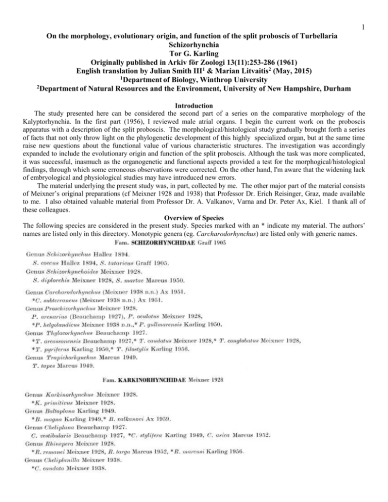
1 On the morphology, evolutionary origin, and function of the split proboscis of Turbellaria Schizorhynchia Tor G. Karling Originally published in Arkiv för Zoologi 13(11):253-286 (1961) English translation by Julian Smith III1 & Marian Litvaitis2 (May, 2015) 1Department of Biology, Winthrop University 2Department of Natural Resources and the Environment, University of New Hampshire, Durham Introduction The study presented here can be considered the second part of a series on the comparative morphology of the Kalyptorhynchia. In the first part (1956), I reviewed male atrial organs. I begin the current work on the proboscis apparatus with a description of the split proboscis. The morphological/histological study gradually brought forth a series of facts that not only throw light on the phylogenetic development of this highly specialized organ, but at the same time raise new questions about the functional value of various characteristic structures. The investigation was accordingly expanded to include the evolutionary origin and function of the split proboscis. Although the task was more complicated, it was successful, inasmuch as the organogenetic and functional aspects provided a test for the morphogical/histological findings, through which some erroneous observations were corrected. On the other hand, I'm aware that the widening lack of embryological and physiological studies may have introduced new errors. The material underlying the present study was, in part, collected by me. The other major part of the material consists of Meixner’s original preparations (cf Meixner 1928 and 1938) that Professor Dr. Erich Reisinger, Graz, made available to me. I also obtained valuable material from Professor Dr. A. Valkanov, Varna and Dr. Peter Ax, Kiel. I thank all of these colleagues. Overview of Species The following species are considered in the present study. Species marked with an * indicate my material. The authors’ names are listed only in this directory. Monotypic genera (eg. Carcharodorhynchus) are listed only with generic names. 2 Morphology and Histology The Turbellaria Kalyptorhynchia are characterized by a true sheathed proboscis at the anterior end of the body. This proboscis is a closed muscular bulb whose distal portion is surrounded by a proboscis sheath that generally opens at the exact anterior tip of the body. The Kalyptorhychia fall naturally into two groups—Eukalyptorhynchia and Schizorhynchia. In the first-named group, the proboscis-bulb is unipartite and ends distally in an invaginable, more or less conical structure—the apical cone (Fig 61). In the Schizorhynchia, the proboscis comprises two dorsoventrally opposing halves that are proximally either broadly connected or only just touching each other. The proboscis therefore resembles forceps in a vertical position. In the family Schizorhynchidae, the proboscis halves (tongues, lips) are unarmed or only bear tiny teeth (Figs 50, 51). In the family Karkinorhynchidae, the proboscis-halves (basal pieces, muscle bulbs, hook supports) each bear a stout cuticular hook (Figs 52, 53). The most extreme development of these hooks occurs in the family Diascorhynchidae. Here, the muscle bulbs are lacking and two tubular, muscular glandular sacs are attached (Fig 54). With Schizorhynchus coecus, Hallez (1894) described the first representative of the Schizorhynchia. A second species, S. tataricus, was shortly thereafter described by Graff (1905). After the species-rich microfauna of marine sands was discovered, our knowledge of the Schizorhynchia, which form a characteristic element in this habitat, increased rapidly (first through Beauchamp, 1927 and Meixner 1928, 1938). Additional results on this group were later provided by Marcus (1949, 1950, 1952), Karling (1949, 1950, 1956), and Ax (1951, 1956, 1959). A comparative compilation concerning the morphology of the Schizorhynchia is, however, presently lacking. Here, I hope to fill this gap with a complete (inasmuch as possible) comparison of the characteristic proboscis structures of these worms. The size of the proboscis correlates in a characteristic relationship to its morphology and function. The hook-bearing proboscides (Karkinorhynchidae and Diascorhynchidae) are relatively small (1/50 to 1/20 of the body length), the unarmed proboscides of the Genera Thylacorhynchus, Proschizorhynchus, and Trapichorhynchus relatively enormous (1/6 to 1/5 of the body length), whereas the weakly-differentiated proboscides in the primitive genera Schizorhynchus, Schizorhynchoides and Carcharodorhynchus occupy an intermediate position with corresponding lengths of 1/20 to 1/10. The absolute dimensions are mainly of taxonomic interest. In the family Schizorhynchidae two types of probscides can be distinguished, exemplified by both of Meixner’s families Schizorhynchidae and Thylacorhynchidae (1928, p 237-238): in the genera Schizorhynchus, Schizorhynchoides, Carcharodorhynchus, Proschizorhynchus and Trapichorhynchus, the tongues are somewhat hemicircular in cross-section (Figs 7, 8), in the genus Thylacorhynchus, they are lip-shaped, broad, flat, and highly folded (Figs 1, 2). I have shown that the latter type can be related to the former by transitional intermediates and that the rest of the organization does not support a division of the Family Schizorhynchidae (1950, pp. 24-29). On practical grounds, one can however, speak of a Schizorhynchus-type and a Thylacorhynchus-type when considering only the proboscis-tongues. In the Schizorhynchus-type, the tongues are generally pointed and the tips very mobile. Marcus (1949, p. 42) describes the tongues of Trapichorhynchus as rounded, but further direct observation is not available. In the Thylacorhynchus-type, the anterior margin should be rounded according to Meixner (op. cit.). This information is not conclusive, however as in all cases where I was able to study the margins of the tongues on live animals, the margins were clearly lobed. In a specimen of T. arcassonensis the number of triangular lobes was 3 on each lip – the same as in a specimen of T. conglobatus (Swedish West coast, identity uncertain), and in a specimen of T. pyriferus (Danish North-sea coast) with 4-5 lobes per lip (Figs 3, 4). According to Graff (1905, p. 146), the posterior portions of the tongues in Schizorhynchus tataricus “are only connected to one another by a small bridge”. Probably this is an error in observation, as in a later figure of the same species (1904-1908, Fig 11), a very stout “bridge” is drawn by the same author. This connection is so wide in Carcharodorhynchus and Thylacorhynchus that the proboscis is posteriorly cup-shaped. The transition to the Thylacorhynchus-type can be derived by strong flattening of the tongues in the former—closest to the T. pyriferus subtype with its relatively thick and solid tongues. Also belonging to this subtype are T. pyriferus and T. arcassonensis (Fig 1). Another subtype with almost leaf-shaped, thin, and strongly folded lips contains T. conglobatus (Fig. 2) and presumably, T. caudatus and T. filostylis (comp Karling 1950, p. 25, figs 3A-C and 6; Meixner’s fig 11, 1928, is a serial cross-section series through an immature specimen of indeterminate species—in any case, not T. conglobatus). 3 According to Graff (1905, p. 147), the tongues of Schizorhynchus tataricus are surrounded by a sheath of “fine circular and thicker longitudianal fibers,” whereas “loosely arranged longitudinal muscles half-fill the internal spaces of the tongues”. Later workers have concluded that the tongues are filled with vertical muscle fibers. The fibers are inserted on both ends to a fixed structureless membrane, which surrounds the tongues on all sides, and the fibers are closelyarranged in regular circular and longitudinal rows. In the Schizorhynchus-type, the fibers are relatively long medially, and shorter laterally (Figs 7, 8), while all of the fibers in the Thylacorhynchus-type are very short (Figs 1, 2). The number of fibers has increased dramatically in the latter type. Some details of the histological structure of the tongues can be considered here. At the nodus, the lips are penetrated by a fine tube, the nodal-pore (Fig 16). Therefore, this is not just a “grave-shaped depression of the inner membrane” (Karling 1950, p. 18). This pore allows necks of glands lying in the brain area to extend through the nodus, a fact that is particularly clear in an illustration of Trapichorhynchus (Marcus, 1949, fig 44a). Such glands occur in at least Carcharodorhynchus, Trapichorhynchus and Thylacorhynchus spp. (Figs 15, 17; c.f. Karling op. cit. p 6-7, Fig 2-3; Marcus 1949, pp. 34-35, Fig 44). Common to all schizorhychids is a plasmatic muscle-free reticulum in the tongues around the nodal pore (Fig 16). The proboscis sheath inserts equally around the lobed proboscis margins of Thylacorhychus spp., but both dorsally and ventrally there is a median pocket of variable depth. In squeeze-preparations, rounded structures occur at the tonguemargins, especially at the tips of the lobes, which in favorable sections appear as small (in T. pyriferus approximately 3µm long), weakly cuticularised bumps (Figs 4, 5). In the remaining genera, the tips of the tongues protrude far from the proboscis sheath. These free tips are especially long in the genus Proschizorhynchus (1/2 to 2/3 of the proboscis-length, see Meixner 1928 p. 240, and Karling 1950, p. 18), distally finger-shaped, in cross-section circular, and are surrounded by a layer of thin epithelium with a strong basement membrane (Figs 6, 7). Fine longitudinal muscles run along the outer edge of the tips under the epithelium and run posterior to the juncture (edge of the proboscis pocket) nearly to the nodus. In these free “fingers”, the vertical fibers in P. gullmarensis are concentrated into two lateral rows, and one (sometimes appearting to be two) fluid-filled space fills the median portion (Fig 6). This fluid may not only facilitate the mobility of the fingers but also the erection thereof. 4 On the inner side of the tongues, the epithelium usually appears as a structureless, nuclear-free membrane, often torn off. Among the Schizorhynchus-types, however, the epithelium, especially proximally in a median longitudinal strip, can be relatively thick (Figs 12, 13). Both Trapichorhynchus and Carcharodorhynchus show noteworthy differentiations of the inner epithelium. In the former species, the proboscis cavity is proximally covered with a nuclei-rich, tall epithelium that posses (dermal?) erythrophil glands (Marcus 1949, p 34, fig 44; in litt). In Carcharodorhynchus, the the lateral epithelium between the tongues (“Zwischenhaut”) and lateral epithelium of the tongues are clearly epithelial with nuclei, and there are cuticular hooks, clearly derivatives of this epithelium, from which the genus gets its name (Figs 9-14). The first description of this genus comes from Meixner (1938, p 137). This genus shows “a very remarkable improvement of the tweezers design: the split halves of the proboscis in this new species are provided on each side of their inner surface with four longitudinal rows of alternately-placed cuticular teeth”. A sketch of this proboscis was published by Ax (1951, fig 31c). My own work provides some additional information. The teeth are placed laterally on the lips, near the lateral epithelium between the tongues, but missing distally, resulting in two U-shaped bands. They are arranged alternately in 10 longitudinal rows on either side, and comprise a circular 1-1.5µm wide (original says 1-15µm; this is probably a typo) basal disc bearing a straight or slightly caudally-curved tooth, 1.5µ long (Fig 14). Because of their small size, they easily escape notice; they are best seen in sections tangential to the rows of teeth. In my opinion, these teeth should not be homologised with the teeth on the proboscis-margin in Thylacorhynchus (see above and page 21). The surface epithelium of the tongues thus shows variation within the schizorhynchids; it may be well-developed with nuclei, membranous, or provided with cuticular hooks. 5 Already in Schizorhynchus coecus Hallez (1894, p3 Fig 3) recognized two large lateral proboscis glands at the base of the proboscis. In S. tataricus, Graff (1905, p 147, Figs 21, 23, 25) described and illustrated the proboscis glands, as “multicellular sacs, surrounded by a fine simple epithelium and fine secretory ducts, that are a little longer than the proboscis”. These glands were later observed in various schizorhynchids. In Schizorhynchoides diplorchis and S. martae, they each possess six cells and are enclosed by a sheath of fine muscles (Meixner 1928, p 241; Marcus 1950, p 37). Such a sheath is lacking in Carcharodorhynchus, which is characterized by just two bundles of cells (Figs 10 & 16). In Thylacorhynchus caudatus (?) the glands are pear-shaped and contain four cells each (Meixner op. cit. p. 244). In T. pyriferus the sac is split on both sides into two lobes containing up to 8 cells each (Karling 1950 p. 6). In a new set of serial sections of a well-extended animal of this species, neither the splitting of the gland sacs nor the presence of free cells near the gland cells could be confirmed. An occasional splitting of the gland sacs could possibly be caused by the lateral proboscis retractors. The supposed free “mouth-glands (Mundwinkeldrüsen)” of Thylacorhynchus conglobatus (op. cit. p 13, Fig 6B) have been shown on inspection to be multicellar, indistinctly wrapped gland sacs. The findings in two species of Proschizorhynchus are instructive. In P. helgolandicus, the glands are spherical diverticula of the middle epithelial layer (Zwischenhaut), and comprise glandular epithelia with a tightly fitting outer muscle layer. The secretion is released into the proboscis cavity by large openings in the diverticula (Figs 8, 18). This construction is most like the condition in Schizorhynchus tataricus (see above). This is wholly different in P. gullmarensis: here, the Zwischenhaut is connected proximally to a muscular tube which continues into a glandular pad that surrounds the entire base of the proboscis (Fig 19-21). In this pad, the gland cells form four clusters that in squeeze preparation, appear as four separate bundles (Karling 1950, p. 18, Fig 10F-H). Lateral glandular sacs are allegedly missing in Trapichorhynchus. Perhaps we can find their homologues in the inner erythrophil glands of this species (see above; page 4). 6 Let us now consider the proboscis hooks in Karkinorhynchidae and Diascorhynchidae. A lateral view of the hooks of the family Karkinorhynchidae reveals that they are bent uniformly, as in Karkinorhynchus primitivus (Fig 28, 29) and Baltoplana spp (Figs 22, 46; see Karling 1949, fig 4A-D). They are very often basally expanded and distally thin and needle-like with a marked kinking between the two sections (Figs 23, 38), where sometimes, a difference between the dorsal and ventral hook can be detected (Beauchamp 1927, fig 2II; Karling 1949 fig 4E; Marcus 1952 figs 83, 93). The hooks can also bear subsidiary teeth, as in Rhinipera marcusi (one pair, fig 23) and Cheliplanilla caudata (two pairs, figs 24, 25; Meixner 1938 fig 32B; in an example of this species from Esberg, Danish North Sea coast, side pieces (“Nebenapparate”) were missing). The hooks are trough-shaped basally and distally closed. In Karkinorhynchus, the cuticle of the hook is relatively thin and surrounds the distal part of each tongue, where they appear to strengthen the tongues as an end-cone epithelium. Distally, the inner part of the channel closes to form a tube, while the free edges of the trough remain (Figs 31-33). The accessory teeth (see above) of Cheliplanilla are derivatives of these free hook edges. In other species, the closed hook-tips are round in cross-section as in Baltoplana magna (Karling 1949 fig 5A-C). By extending and strengthening the hook bases, the special form of the hooks in Diascorhynchus, with curved endtooth and almost equally broad basal portions, can be derived (Fig 26). Only the end teeth of the channel-shaped hooks project freely into the proboscis sheath (Fig 54). Already an illustration by Meixner (1928, fig 17b) shows that the hooks are dissimilar, with one larger hook having prominent muscle insertions at the border between the two sections and more curved end-tooth. The dorsal hook is quite similar in all species, bilaterally similar, and with the base broader than that of the ventral hook. In a closed proboscis, the tip of this hook lies inside the tip of the longer ventral hook. In the latter, the muscle insertions are located asymmetrically, especially in Diascorhynchus lappvikensis, where one is drawn out into a rectangular lobe and shifted rostrally (Fig 26). If we disregard the proboscis hooks, the proboscis apparatus in Karkinorhynchus is strikingly reminiscent of that of schizorhynchids. Meixner noted of the proboscis that it possessed “approximately 20µm long cuticular hooks and “two long gland-sacs with long ducts” (1928, p 245, fig 12). Later, he illustrated two cross-sections of the proboscis of this species and described the outer longitudinal muscles along the tongues “that attach to the outer bases of the hooks at the rear and surround the horseshoe-shaped split halves” (1929, p 781 fig 12a, b; see below page 14). Karkinorhynchus exhibits a relatively primitive type of hook-bearing proboscis, that informs our understanding of the more specialized probscides in the karkinorhynchids and diascorhynchids. The muscular tongues are still well-developed, clearly flattened, with numerous short radially (vertically) arranged fibers in regular longitudinal and transverse rows (Figs 28, 29, 34, 35, 7 40). At the nodus, they are broadly connected and exhibit a nodal pore and a muscle-free reticulum. The similarity with, for example Carcharodorhynchus among schizorhynchids is almost complete. Proboscis abductors (divaricators) insert posterior to the nodus. A nuclear-free membrane lines the inner surface of each tongue. Notable structures are the two lateral gland sacs (Figs 30, 35, 39, 40). Each sac comprises a distal pear-shaped secretory container and a proximal, nearly spherical, syncytial glandular ball with 3-5 nuclei. Fine muscle fibers surround the secretory container and form a sphincter around the constricted juncture between the two sections, while the nuclei-free plasma of both containers sometime protrude irregularly into the lumen of the proboscis (Fig 30). The two glandular balls are enclosed by a solid membrane, which extends into short proximal projections. Fixator fibers probably insert here; their existence could not be determined with certainty due to the lack of material. Other karkinorhynchids known to me lack lateral gland sacs but possess various lateral accessory apparati. The tongues (or bulbs) in the proboscides of remaining karkinorhynchids are more or less reduced in comparsion to those of Karkinorhynchus. In Baltoplana spp., they are attached posteriorly, while in the other species, they appear as two more or less cylindrical tongues, posteriorly in contact. The number of longitudinal rows of radial muscles inside the tongues is only known in a few species; in Baltoplana spp., 4 double rows (after Karling 1949, p. 7, “four longitudinal rows”), in Cheliplanilla caudata 2 (double?) rows. In Rhinepera targa (Marcus 1952 p. 110, fig 93) the muscular tongues are rudimentary. Hook abductors on the outside of the tongues should be a shared feature of karkinorhynchids (see above, Karkinorhynchus). In Baltoplana magna, these comprise numerous fine fibers, that are more or less clearly divided into two groups; on the other hand, in Cheliplanilla caudata there are only two thick fibers. In the latter species, the postrostral fiber framework is fused into a strong, vertical muscular plate, from which divaricators and proboscis retractors originate (Fig 27). An epithelial membrane—a derivative of the proboscis-sheath--is tightly applied to the muscular bulbs and inserts at the back of the hooks.. Accordingly, the whole proboscis is free in the proboscis sac, and the same goes for Rhinepera remanei (Figs 36, 37 and Meixner 1928 f. 14 & 15), Cheliplana stylifera (Karling 1949, fig 6B), and probably various other species. In contrast, the inner layer of the proboscis sheath is not tightly applied to the proboscis bulbs in Baltoplana spp (Figs 45, 46), which is also the situation in Karkinorhynchus (see above). In regard to the proboscis hooks in Diascorhynchus glandulosus, Beauchamp (1927, p. 8) writes “they are equally mounted on a recurved piece”. The attached sketch shows however, that here we are not dealing with a muscular tongue as in the karkinorhynchids, but rather a curved muscle of hook divaricators, as was later described for D. borealis and D. serpens (Fig 54, see Meixner 1928, p 249; Karling 1949, p 29). Topographically similar and certainly homologous divaricators are found in karkinorhynchids (see above); however, these are, in the Diascorhynchidae, considerably enhanced. Probably without counterparts in the karkinorhynchids are the occlusors (adductors) that arise from the lateral and median bulges on the hooks and insert in the postrostral muscular septum (Karling, op. c. p. 31). The most characteristic feature of the proboscis in Diascorhynchus is the presence of two cylindrical glandular tubes, referred to as a “Diascus”. Meixner (1928, p 239, 249-250) and Karling (1949, p. 31) establish the manner in which the tubes open laterally, the presence of a single cell nucleus in the tube wall and the attachment of the tubes to the body wall by a single muscle fiber. The latter author describes the histological structure of the tube: lined by a thin epithelium (each with a single nucleus) and sheathed in a single layer of longitudinal muscle fibers and an outer layer of myocyte nuclei. He further states that the tubes are only a container for the secretions; posteriorly, they each receive their secretion from one (or 2?) large gland cells. I now will consider the lateral apparatus, which is found in the majority of karkinorhynchids (see above). In Baltoplana magna, I have described four soft sensory and secretory fingers, which at their termini have stiff “pseudocilia” (1950, p7, figs 4, 5). Re-examination show that only the lateral fingers represent autonomous entities (Fig 22) and that the dorsoventrally opposing ones are short and only (not in all preparations) papilliform projections from the epidermis of the muscle bulbs. The lateral fingers are likely to contain two secretory outlets. In most species, two pointed lateral lobes are present (Beauchamp 1927, p7 fig 2II; Meixner 1928 p246 fig 14-15; Marcus 1952, p110 figs 83, 93). They are weakly cuticularized and often difficult to see. In Cheliplana stylifera, I have only seen these on the everted proboscis of a strongly squeezed specimen (Fig 38). In profile, the lobes of Rhinepera remanei are of a characteristic curved form (Fig 37; comp Meixner, l.c.), that nevertheless is not constant; everted, they are distinctly finger-like and weakly cuticularized. In Cheliplanilla caudata, Meixner described the lateral apparatus as rods with a forked tip and transversely positioned basal piece (1938, p 32 fig 32B). My findings complement this information somewhat (Fig 24, 25, 27). The rods are 2429µm long, uniformly rod-shaped in lateral view, tapering (in dorsoventral view) toward the tip with the distal end curved slightly inward. Basally, the rods are forked, with characteristic side-lobes, near the muscular bulbs, and arising just a short distance anterior to their articulation. Insasmuch as they appear hollow, they serve as a secretory tube for glands in the postrostral bulb (see below). In one preparation of Rinepera marcusi a pair of rods like those in Cheliplanilla (Fig 23) occurred. 8 Meixner has briefly described a characteristic median bulb that is attached to the rear of the proboscis in Rhinepera remanei (1928, pg 246, fig 14-15). To avoid confusion with the proboscis bulb of the Eukalyptorhynchia, I designate it here as a “postrostral bulb” or proboscis support. According to Meixner, this is composed of a multicellular syncytium and is (according to the drawings) somewhat cylindrical. It is described as “surrounded by thick longitudinal and circular muscles and penetrated by two proboscis retractor muscles.” Protractors also originate in the bulb. In Baltoplana magna and Cheliplana stylifera, I overlooked the bulb (1949, p.8), and assumed that it was only a cylindrical protrusion that surrounded the deeper proboscis sheath on all sides. Marcus opposed my view, and described and illustrated a similar 9 postrostral bulb in R. remanei, R. targa, and C. asica (1952, Figs 84, 85, 94, 95). This has compelled me to re-examine the morphology and evolution of this structure. Cheliplanilla possesses a well-differentiated, sharply delimited postrostral bulb (Figs 27, 41-44). The wall of the bulb is a direct continuation of the basemement membrane and attached musculature of the proboscis sheath, and consists of a membrane with externally attached muscle fibers. Posterior to the nodus, circular muscle fibers are especially apparent; further posteriorly, a network of skewed (spiral) fibers appears (Fig 27). So far it appears that no cell processes pierce the septum, and that large gland cells lie within (Figs 43-44). (I have not studied the innervation of the proboscis apparatus). However, retractors do pierce the septum—a dorsal and a ventral pair in a more rostral position than the lateral muscles (see below, page 268---p. 12 ). The lumen of the proboscis sheath extends caudally only to the nodus; the sheath accordingly does not enclose the postrostral bulb, and hence, does not have the character of a tissue projection 10 (“Gewebezapfen”). There are sometimes spaces in the bulb, particularly between the septum and the lateral retractors (Figs 43, 44). In some cases, it is likely that these spaces are continuations of the proboscis lumen, which, on the lateral sides only, sometimes extends far caudally. The anatomy of the postrostral bulb in Rhinepera remanei closely matches that of Cheliplanilla (Fig 36). In the best set of longitudinal sections (on which Meixner’s fig 14, 1928 was based), spaces around the lateral retractors occur as they do in Cheliplanilla. What Meixner designated as proboscis protractors (Rp) is thus probably the central portion of the septum separated from the bulb, whose fibers sometimes extend obliquely forward. Despite the illustration in Meixner, this septum does not extend to the body wall, but lies anterior to and closely against the spaces in the postrostral bulb. The illustration of Cheliplana asica by Marcus must be interpreted likewise (1952, fig 85). Leaving the relationship between Cheliplanilla caudata and Rhinepera remanei, I return again to Baltoplana magna and compare it to Baltoplana valkanovi. It appears that although a postrostral bulb is present in both species, it is only weakly developed and rarely -- as in Fig 47 (comp. Karling 1949, fig 4B)—cylindrical in appearance. In this case, the bulb can be interpreted as a projection delimited posteriorly by a thin septum (somewhat better developed in B. valkanovi than in B. magna), through which cell-bodies are sometimes sunken. The double-walled nature of the septum is characteristic. I was able to show that this wall is composed only of the muscular tube of the proboscis sheath and 11 accompanying basement membrane. These conditions appear most clearly in preparations where the bulb is strongly stretched transversely, and when the – often indistinctly apparent – somewhat hemispherical posterior wall of the nodus is closely appressed (Fig 45, 48). The lumen of the proboscis sheath extends only to about the nodus, where the sheath epithelium gives rise to the epithelia of the muscle bulbs and accessory apparatus, respectively. At this point, the body wall muscle splits off from the epithelium and gives rise to a folded muscular tube with net-like fibers. Here, in the deeper part of the sheath, the epithelium is membranous and probably elastic. The proboscis retractors are formed from a gathering of fibers from the musculature of the proboscis sheath and fibers that cross the tissue around the nodus and insert at various locations along the proboscis. The dorsal and ventral retractors often run in straight lines from the basal piece to the body wall (Fig 46, 47; comp. Karling 1949 fig 4A); here, the hooks are folded back. My material of Cheliplana stylifera is not sufficient to determine the histological construction of the postrostral bulb. It is likely somewhat similar to the construction in Cheliplanilla and Rhinepera remanei. The postrostral bulb must therefore, be treated as a largely individual organ, as in Meixner and Marcus (see above). The evolution of this structure will be dealt with later (page 14). The foregoing description of the morphology of the proboscis in the narrow sense must be supplemented by a summary of the construction of the proboscis sheath and of the origin of its associated glands and muscles to give an idea of how the apparatus as a whole might function. The proboscis sheath of the Schizorhynchia is small and weakly differentiated. It inserts dorsally and ventrally rather far anteriorly on the probscis tongues – in the family Schizorhynchidae, toward the distal ends of the lips, in the family Karkinorhynchidae on the hook bases, and in the family Diascorhynchidae, on the border between the distal teeth and the basal pieces of the hooks –but continues laterally as a connecting tissue between the proboscis tongues. The epithelium of the sheath is membranous and weakly cuticularized in most representatives of the Karkinorhynchidae and Diascorhynchidae. In the Schizorhynchidae, it is better developed and contains nuclei that are concentrated in a more or less thickened lateral epithelium between the tongues (Figs 7, 13). A pair of nuclei is always found laterally near the hook 12 bases in Karkinorhynchus (Figs 30, 34). A sphincter at the proboscis pore is always present. Moreover, the musculature of the sheath comprises a longitudinal fiber layer whose fibers in part, diverge into the body wall in very different ways (Fig 27, 30, 46-48). When these fibers run anteriorly from the wall of the sheath, they can simply be regarded as dilators derived from the body wall muscle. The same can be said of the protractors that periodically arise at the juncture (see, for example, Diascorhynchus serpens, Karling 1949, p. 29, fig 14A). Muscles arising from the sheath and running posteriorly on the other hand, include the proboscis retractors (see below) or short integumental retractors that create a ring-shaped depression around the proboscis pore in a variety of species (for example, Baltoplana magna, Karkinorhynchus primitivus, Cheliplanilla caudata, Proschizorhynchus gullmarensis and Diascorhynchus borealis; comp Figs 29, 30, 39 and Meixer 1928, fig 18; Karling op. cit. fig 10H). At least these latter fibers are clearly derivatives of the body wall muscles. If we disregard the sheath dilators, the muscles moving the proboscis comprise retractors and protractors, of which the former are strongly developed. The retractors show a great similarity across the various types of probscides, inasmuch as they usually occur in lateral, dorsal, and ventral pairs. In the family Schizorhynchidae, short, but fiber-rich lateral bundles are characteristic (Figs 19-21; see Meixner 1928 and Karling 1950). They usually originate far posteriorly, near the nodus (see Karling op. cit. fig 10G); however, in Thylacorhynchus, they are located somewhat more anteriorly, near the “Mundwinkeln,” (corners of the mouth) where they influence the shape of the lateral glands. The dorsal and lateral retractors in Thylacorhynchus spp. are divided into an anterior and posterior system, and by their contraction, contribute to a particular wrinkling of the proboscis tongues (Fig 63, see Karling op cit Figs 3A-B, 6A-B and 7F). Sheath dilators located far posteriorly can occasionally be confused with anterior proboscis retractors. Even in Karkinorhynchus there is a portion of the retractor muscle fibers that arise at the juncture near the hook bases. In Baltoplana the proboscis retractors form three paired bundles (see Karling 1949, p. 8., f. 5e). The lateral bundles divide into two groups of fibers. In Cheliplanilla (see above, p. 9), the dorsal and ventral muscles are each composed of one very thick fiber, whereas the lateral muscles comprise two fibers each (Figs 41, 42). Peripherally, each fiber divides into fine branches. Rhinepera remanei, as nearly as can be seen, is like Cheliplanilla with regard to the retractors. After passing through the septum, retractors can extend anteriorly when the proboscis is withdrawn far posteriorly, and can therefore, appear to be protractors (Fig 36; comp. Marcus 1952, figs 94, 95). Probably, the functions of these muscles are multiple (see below, p. 19). In all karkinorhynchids, the lateral retractors insert more posteriorly than the dorsal and ventral retractors. The retractors in Diascorhynchus spp. are highly specialized. I have described a dorsal and a ventral pair of retractors in D. serpens (Fig 54; see 1950, pp 29-31, figs 13, 14A-B). Each muscle consists of two fibers “an anterior one that arises posterior to the sheath wall at the back of the hooks and a posterior one that arises in the fiber-web at the back of the proboscis”. This is remarkably similar to the construction in schizorhynchids and karkinorhynchids, where it is demonstrated that some fibers arise on hook bases and others, more caudally. Lateral retractors are lacking in Karkinorhynchus and Diascorhynchus. Proboscis protractors arising from the proboscis halves or from the postrostral bulb (in karkinorhychids) are lacking or are composed of very fine individual fibers (Fig 27; see also Karling 1950, p. 20, fig 10H). Also, fibers arising from the muscular layer of the proboscis sheath can function as dilators or protractors (see above). A dorsoventral septum behind the brain also is likely to be of importance for the movement of the proboscis. This was first described by Meixner for Proschizorhynchus oculatus, Schizorhynchoides diplorchis, Thylacorhynchus caudatus, and Diascorhynchus borealis (1928, pp 24—250, fig 7). I have found this septum, probably composed of connective tissue fibers, in various Schizorhynchidae, especially prominent in Carcharodorhynchus (Fig 17). Diascorhynchus serpens possesses a similar septum. I have searched in vain for a similar septum in karkinorhynchids. I will return to the function of this septum below. In all Schizorhynchidae, I have found glands that open laterally into the proboscis lumen (exception: Trapichorhynchus, see page 260 --- p. 5 ). A comparison of these glands follows below (p. 271). The secretion of these glands appears often appears grainy and eosinophilic, however, in other cases, it stains only weakly. In Thylacorhynchus spp., I have described medially located “subrostral glands” that “probably open in a medioventral pocket of the ventral lip” (1950, p7 and 13). A precise follow-up shows that these glands open through fine ducts into a medioventral pocket of the proboscis sheath (see p. 13). This is confirmed by the absence of structures on the ventral lip that could receive the secretion of these glands. Glands opening at the nodus also occur sometimes, at least in schizorhychids (see p 250). Frontal glands with cyanophilic secretions open into the proboscis pore or immediately outside it (Figs 17, 27, 47). These could be a common property of all Schizorhynchia. 13 Evolution and further differentiation Graff already made an attempt to derive the schizorhynch from the conorhynch (1904-08, p. 2090). Because of his incorrect understanding of the former type of proboscis (p. 256—p. 3 ), his attempt is now of no interest. In the families Placorhynchidae and Gnathorhynchidae, the proboscis contains muscular bundles or plates composed of crossed lamellae similar to the proboscis halves of the schizorhynch. Meixner repeatedly touches on the question of the origins of the schizorhynch, by invoking its similarities with the conorhynch of known families. However, his descriptions of these similarities vary widely. In 1938, he wrote in part (p. 33) that the differentiations are to be construed as “most likely analagous; their structures are independent of each other” however “the formation of the gripping lips on the proboscis of the Placorhynchidae leads to the split proboscis of Schizorhynchia” (p 136). Even in 1929, he already was of the correct opinion that the muscle fibers in the schizorhynch compare with the internal longitudinal muscle of the eukalyptorhynch proboscis” (p 781). Comparing the eukalyptorhynch proboscis with a protruded end cone with a schizorhynch with specialized construction, for example, in a Proschizorhynchus-, Thylacorhynchus-, or Baltoplana spp., the difference between the two types appears completely unreconcilable. However, in the Eukalyptorhynchia, one can often see the proboscis in an invaginated state, where it appears cup-like (see, for example Graff 1882, t. 11, fig 2 or Meixner 1938, fig 25). If we posit that the cup-shape is the normal resting state of the proboscis, the step to the split proboscis of, for example, Carcarodorhynchus, is not a particularly long one. This latter proboscis is proximately, signficantly cup-like, and therefore, it follows that the muscular bulbs (tongues, lips) of the schizorhynch are derived from the vertical-radial muscle columns (the inner longitudinal muscles) of the conorhynch (Fig 49-50). The persistent invagination of the end cone is related to the functional changes in the proboscis (see p. 17). The suction cup is transformed to a gripping apparatus; the radial symmetry is transformed completely to bilateral symmetry; the dorsoventrally opposed parts grow at the cost of the lateral parts to produce a pincer. However, the cup-form is maintained and further differentiated in the proboscis of Thylacorhynchus; in the genus Proschzorhynchus, the cup form is transformed to the pincer type (Fig 51). It is therefore necessary to homologize the muscular tongues of the schizorhynch proboscis with the end bulb retractors of the conorhynch proboscis, probably deriving the divaricators (p. 7) that lie outside of the muscular tongues from the longitudinal muscles of the conorhynch that converge outside of the septum at the nodus (lm-di). In the conorhynch, the proboscis sheath generally inserts into a sharply-delimited juncture (ju) and gives rise to the epidermis of the terminal cone (“Endkegelhaut”). In the schizorhynch, we find the homologue of the Endkegelhaut in the membrane surrounding the muscular tongues, whereas the proboscis sheath continues imperceptibly into the lateral epithelium between the tongues (Zwischenhaut). The juncture becomes accordingly a sinuous, hard-tofollow line. In the transition of the conorhynch into the schizorhynch, many structures necessary for the function of the former were lost or strongly modified. The circular muscles lying against the septum are important for movement of the conorhynch (Fig 49A). In the schizorhynch, these are always missing. The nodal glands opening on the terminal bulb 14 again open through the nodus into the proboscis lumen (Fig 49-50; see page 13). In most Schizorhynchia these glands are absent. In the conorhynch proboscis—especially in the family Zonorhynchidae—so-called juncture glands are differentiated. In the schizorhynch proboscis secretory activity is concentrated into the lateral quadrants, and accordingly the lateral glandular sacs of the Schizorhynchia can be homologized with the juncture-glands. At this point, it cannot be determined if the glands opening into the ventral side of the proboscis sheath in Thylacorhynchus spp. should similarly be regarded as modified juncture glands or if they should be homologized with the so-called sheath glands of various Eukalyptorhynchia. Even in the conorhynch proboscis cuticular hooks can arise at the juncture (Family Gnathorhynchidae). It seems possible that these structures are the result of the secretory epithelium at the juncture (see Karling 1953, p. 500). In this regard the cuticular hooks in the family Gnathorhynchae may be homologized with those of the Karkinorhynchidae and Diascorhynchidae. However, among Schizorhynchia, the hook-less schizorhynch proboscis arose first, and from this the armed proboscis arose secondarily (see below). The muscular structure of the proboscis in Gnathorhynchidae is developed in a strongly divergent manner from that in the schizorhynch proboscis. I have found that cuticular hooks can occur in the form of small hooklets in the split proboscis of schizorhynchids (p. 4). One can imagine that from these, two large hooks originated—similarly to the cirri of the male genital organs (see Karling 1956 p. 203). The presence of an almost ideal transition form from unarmed to armed proboscis – Karkinorhynchus primitivus – makes a different mode of evolution likely. I have (see above, p. 6) found striking convergences between Karkinorhynchus and Schizorhychidae in the construction of the proboscis (Fig 50-52). This is true for the construction of the tongues and lateral gland sacs, the lack of a postrostral bulb and the so-called “accessory apparatus”, the presence of nuclei in the sheath epithelium, and for some features in the formation of the locomotory musculature. The hooks are thin-walled and simply appear as a hardening of the tongue tips. From Karkinorhynchus, one can easily proceed to the other karkinorhynchids and partly to the diascorhynchids. The former taxon has as its main features the development of various types of accessory apparati in the proboscis and the differentiation of a postrostral bulb, as well as a gradual regression of the muscular bulbs (Fig 53). For Diascorhynchus, we assume a further development of the lateral gland sacs of Karkinorhynchus into the diascus (p. 7) and a complete regression of the muscular tongues (Fig 54). In fact, the difference in the construction of the lateral gland sacs and the diascus is relatively insignificant. Note, for example, the distal, muscular secretion sacs and the proximally arising fixator muscles (see page 7). The amplification of the hook bases parallels the regression of the muscular bulbs. The hook divaricators are amplified and result in a strong u-shaped sheet of muscle. The hook occlusors of the proboscis in Diascorhynchus (Karling 1949, p. 31, f.14G), as we will see below (p. 19) emerged from the radial muscles of the basal piece. A point posterior to the hook bases appears as a nodus where the occlusors and divaricators join. Its location is fixed in part by the proboscis retractors (p. 12). Here, I will consider again the lateral apparatus and postrostral bulb of the proboscis in the proboscis of Karkinorhynchidae (Figs 55-57). Even in Karkinorhynchus, the secretion of the lateral glandular sacs accumulates in uneven lateral epithelial lobes before it is discharged into the proboscis lumen. Baltoplana takes a step further by the development of sensory fingers through whose tips the secretion is discharged. I have shown above (p. 7) further differentiation of these lateral apparati. In the formation of these structures and of the dorsoventral seizing apparatus, we find remarkable analogies to penial structures (see Karling 1956). We find that with glands or cuticular structures – often with both – evolutionary differentiation can occur from an undifferentiated epithelium. On the basis of the present material, the evolution of the postrostral bulb is not completely elucidated, although the main lines are in my opinion, already clear. I have found that the post-rostral bulb in Baltoplana spp. sometimes appears as a tissue projection supporting the proboscis, defined at the rear by a septum. One could imagine that the deep portion of the proboscis sheath was primarily filled by epithelial cell bodies (Fig 58A), which later degenerated with the exception of an elastic surface layer, leaving an empty muscular tube (58B). (One can compare this to a similar deep fold in the copulatory organ of two Polycystis species: Karling 1956, p. 233, figs 40, 45). Accordingly, the vertical septum was a new formation, comparable to the often present septum behind the brain (p 11). The evolution towards Cheliplanilla and so on comprises a reduction in the inner wall of the fold, whereas a strong adhesion of the outer wall to a strengthened vertical septum around the exit points of the retractors follows (C). Accordingly, the bulb was mostly a mesenchymatous structure. This hypothesis, I call the Zapfen Theory (Projection Theory). However, in Baltoplana, the cytoplasm of the lateral apparatus and that of the epithelium covering the proboscis bulb is continuous with the tissue of the postrostral bulb, exactly as in Karkinorhynchus, where the cytoplasm of the lateral epithelial projections between the tongues are continuous with that of the lateral glandular sacs. Hence, it is evident that the post-rostral bulb and the lateral glandular 15 sacs are homologous, and are derivatives of the proboscis sheath. The tissue of the bulb is thus of epithelial, not mesenchymal origin, and the Zapfen Theory must accordingly be rejected; the bulb is formed by the fusion of lateral glandular sacs (Figs 55-57). Various characteristics can be explained by this hypothesis. Karkinorhynchus and Diascorhynchus have lateral glandular sacs, yet have no postrostral bulb; all known karkinorhynchids lack lateral glandular sacs with the exception of Karkinorhynchus, whereas a postrostral bulb is present. In sagittal sections, the lateral glandular sacs of Karkinorhynchus easily mimic a postrostral bulb with tight fitting dorsal and ventral retractors. That the lateral retractors leave the bulb somewhat more posteriorly than the ventral and dorsal retractors can be explained by their function as fixators of the glandular sacs in Karkinorhynchus (as is also the case in Diascorhynchus) (see page 7). I speak here of the glandular-sac theory. According to this, the septum of the postrostral bulb would simply be a derivative of the musculature of the proboscis sheath. Of great interest is that in the family Schizorhynchidae there is a parallel phenomenon to the emergence of the postrostral bulb as described here. I think of Proschizorhynchus gullmarensis where the probscis is “enclosed posteriorly on all sides by a cushion of glandular cells” (Karling 1950, p. 18, cf p. 260). The cushion replaces the lateral glandular sacs found in all other known species in the family. In cross sections, the transition of the cushion into the lateral cytoplasm between the tongues can be traced (Figs 19-21). The homology of these cushions --- the postrostral bulb – with the lateral glandular sacs is unequivocal. The Zapfen Theory cannot be applied here, as the proboscis sheath is short and encloses only the tips of the tongues. In Figs 55-57, the lateral glandular sacs are shown in horizontal view. Because the proboscis sheath in the Karkinorhynchidae extends much farther posteriorly than that in Proschizorhynchus gullmarensis, we can easily imagine that it is involved in the generation of the postrostral bulb. In that case, a combination of the two previoulsy discussed modes of development appears feasible: a gap appears in the dorsal and ventral epithelial bulbs according to the Zapfen Theory, where the bulk of the postrostral bulb is formed by the laterally immigrating gland sacs (Fig 59). This gap (respectively, the doubled muscle layers of Baltoplana spp.) is particularly evident laterally. Here, neither the glandular sac theory nor a combination of both theories satisfies the demands of ontogenetic probabability. It seems quite possible that ontogenetically, the tissue of the bulb comprises epithelial cells from the proximal bulb that have sunken deeply into the proboscis sheath (see Fig 60). The double muscle mantle of Baltoplana or the gap of other species would arise then not by degeneration (Zapfen Theory, see above) but by subsidence of the epithelial cells originally located here. The mantel portion of the septum around the bulb represents a continuation of the proboscis sheath, whereas the posterior portion either differentiates from myoblasts of the sheath wall or septum-like, differentiates ad hoc. The relatively caudal 16 position of the exit points of the lateral retractors must be explained functionally (p 277). This ontogenetic depression (Einsenckung) theory is, in my view, not unlike the phylogenetic glandular sac theory. Function The Kalyptorhynchia are predators who use their proboscides to catch prey. Among prey animals are especially entomostracans, annelids, nematodes, protozoans and halacarids (see Meixner 1938 p. 136-137). Marcus has identified in the body of Proschizorhynchus atopus bristles of enchytraeids (1950, p. 35) and I have observed half digested rotifers and turbellarians—or parts of them—in the intestine of Baltoplana magna. Diatom shells are repeately found in the intestine of Kalyptorhynchia, either ingested or as remnants of digested prey. The conorhynch of the Eukalyptorhynchia acts by attaching its sticky terminal cone to the prey. But even in this group, various special structures can facilitate grasping of prey. The schizorhynch acts supposedly as a pair of pincers. In Thylacorhynchus, small objects can be grasped, and in schizorhynchids the proboscis tongues can be wrapped around prey (see especially Beauchamp 1927 and Meixer 1938). The lateral glands provide a “simultaneously adhesive and venomous 17 secretion” Meixner 138, p. 136. The small muscle-free basal portion of the hook bearing proboscides allegedly causes the closure of the hook apparatus because of its elasticity (Beacuchamp, 1927 pp 6-7; Meixner 1928 p. 2246; Karling 1949, p. 8). By means of the lateral apparatus of the Karkinorhynchidae, the prey are “held at the same time from the side and hindered from escaping” (Meixner 1938, p. 137). We thus have a general—although in several respects hypothetical—view of the operation of the proboscis apparatus in Kalyptorhynchia; however, current details concerning the proboscis movements are extremely sparse, and especially so for the Schizorhynchia. The longitudinal movements of the proboscis appear share some common features across all Schizorhynchia. The protrusion of the proboscis happens, according to Graff (1904-08, p. 2088) “primarily by the contraction of the body wall musculature followed by forward movement of the perivisceral fluid.”. This is also true of Schizorhynchia, even to a greater extent than in Eukalyptorhynchia, with their better-developed parenchyma and proboscis protractors. If a postrostral septum is present (p.11, fig 63), this should direct the tissue pressure forward. Contraction of the septum itself cannot be excluded. Protraction of the proboscis in Schizorhynchia is carried out by a combination of opening the proboscis sheath widely and the retraction of the sheath wall and surrounding tip of the body (Fig 63, 67). These movements are generated by contraction of the dilators and retractors of the proboscis sheath, by the longitudinal muscles of the body wall in the proboscis area, and the so-called anterior retractors that arise from the body wall muscle (p. 12). The retraction of the anterior body wall explains the lack of proboscis protractors. If these muscles are present, they can only be important at the beginning of proboscis protrusion. The task of the proboscis sheath is likely to be only a strengthening of the sheath wall. On the other hand, the postrostral bulb and a part of the so-called proboscis retractors can be involved in protrusion of the proboscis. Retraction of the proboscis is, in the first place, due to lateral proboscis retractors which arise relatively far posteriorly on the body wall (p. 12). In the retracted proboscis, these muscles are always contracted (Fig 63A, 67A). The function of the dorsal and ventral probscis retractors is more complicated (see p. 19). I here will consider the function of the different types of split-proboscides. In the family Schizorhynchidae, we recognized two morphological types of probscides—the Schizorhynchus- and the Thylacorhynchus-type. These two differ widely both morphologically and functionally. Charcharodorynchus and Proschizorhynchus may represent the Schizorhynchus-type. In living C. subterraneus the proboscis tongues are extended when at rest and approximately hemicircular in cross-section; in preserved specimens, they are usually rolled and more or less disk-shaped (Fig 62). This difference corresponds to the behavior in Eukalyptorhynchia (Fig. 61)— in living specimens, the terminal cone is extended; in preserved specimens, it is as a rule, invaginated. Due to the fixation process, inner longitudinal muscle and the radial muscle fibers, stretch from the septum to the free surface. In the living animal, the same reaction should be occur during prey capture. However, the effect is different in the two cases—in the Eukalyptorhynchia, the terminal cone 18 is pressed against the prey and then retracted, creating a suction effect; in the Schizorhynchus-type proboscis, tongues wrap around the prey. The long proboscis tongues of Proschizorhynchus are not rolled when at rest. In both the living animal and in section, they are often bent with the points directed backwards. The widely varying length of the radial muscles of the proboscis tongues in Proschizorhynchus and in Carcharodorhynchus indicates a significant capacity for contraction in these muscles. Hence, the gripping movement is not only a consequence of the elasticity of the tongues. When holding the prey, a sticky secretion is released from the conorhynch as well as the schizorhynch. The secretions entering at the nodus of the conorhynch are replaced with the secretions of the lateral glandular sacs in the split proboscis (see p. 14). The cuticular hooks of the Carcharodorhynchus-type probscides are functionally equivalent and perhaps even superior to these sticky secretions. The radial muscles act antagonistically to the longitudinal muscles along the edges of the tongues; accordingly, they can be considered extensors of the tongues In the long Proschizorhynchus-proboscides, they are well-developed and also involved in the groping grasping movements of the tongue tips (see Karling 1950, p 20, fig 9D). They are lacking in Carcharodorhynchus [see note above] or are very weakly developed. Perhaps the extenstion of the tongues happens only by relaxing the radial muscles. The flat and thin lips of the Thylacorhynchus-type proboscis cannot enwrap prey. One cannot find them curled or bent backward. The short, closely spaced radial muscles hardly vary in length, and are certainly not contractile. Outer longitudinal muscles are absent. Thus, the proboscis is converted into a passively mobile, or at most, an elastic sac. At rest, the sac is compressed with a strongly folded wall (Fig. 63). The dorsal and ventral retractors are strongly split with wide areas of insertion on the outer surface of the lips; all retractors are then contracted. An extension (widening) of the invaginated proboscis, with the resultant sucking-in of small food items is excluded, and appears to me to be meaningless. The well-developed body wall muscles in the area of the proboscis, as well as strong retractors and dilators of the proboscis sheath and (at least observed in some species), the well-developed postcerebral septum indicate significant longitudinal movement of the proboscis apparatus. Probably the proboscis is protruded and acts as a powerful suction-cup, assisted by the sticky secretions of the lateral glandular sacs (perhaps also by one of the other glands connected to the proboscis apparatus) (Fig 63). Small objects could in this way certainly be enwrapped (see p. 16). The gripping motion of the Thylacorhynchus-type proboscis is likely that of a plicate pharynx so that the prey is, as Meixner described it “completely enclosed” (1938, p. 91). 19 The functioning of the proboscis hooks of karkinorhynchids and diascorhynchids seems clear from the construction of the organ. In detail, however, the variation is large and the function is, in various repects, puzzling. Closing of the hook apparatus of Karkinorhynchidae has so far been explained by the elasticity of the basal piece (see above page 275). However, finding that the muscle fibers lying under the hook bases are strongly stretched when the hooks are open, can be interpreted as meaning that these muscles have real contractility. As with schizorhynchids (see above), the contraction of more proximal radial fibers during the flexion of the muscular tongues, involves the occlusion of the hook apparatus (Fig 65). If we assume a strong development of the basal part of the hooks in karkinorhynchids to the state found in diascorhynchid hooks, the radial fibers beneath the hook bases give rise to the longitudinal hook occlusors in the Diascorhynchidae (Fig 66). It is noteworthy that the hook flexors (occlusors) in all known kalyptorhynchs with proboscis hooks (Gnathorhynchidae, Karkinorhynchidae and Diascorhynchidae) represent derivatives of the end bulb retractors of the conorhynch (Figs 64-66; see Karling 1947, pp 24-27, t IC). Without consideration of organ function, this conclusion would hardly be possible. Upon fixation, karkinorhynchids often react with rapid grasping movements of the proboscis (sometimes accompanied by a protrusion of the pharynx). Such preparations can provide a model of the mechanism of this movement. In live specimens, the movement is likely triggered by prey, but perhaps also by a predator. The above-described movements of the muscles in the proboscis is thereby especially clear (p 276--XXX). In some speciemens the entire proboscis is everted (Fig 67C, see Meixner 1927, fig 12), and in forms with a well-developed postrostral bulb, the protruded proboscis is attached by a stalk (Fig 25, 38). The dorsal and ventral retractors, slack when the proboscis is retracted, are contracted, as are the hook divaricators along the outside of the hook supports (Fig 67). In Karkinorhynchus and the Baltoplana spp., these two muscle systems are relatively independent of each other, and the retractors (in Karkinorhynchus) often pull the hook bases nearly vertically against the body wall. The last mentioned muscles function as dilators of the basal piece in unarmed proboscides. In the more specialized karkinorhynchids, where these two muscle systems are joined (see p. 262), the dilation of the muscular parts of the proboscis draw back near to the stronger divaricators of the hooks. In these forms, the retractors run posterior nearly horizontally within the postrostral bulb, and then upon leaving the bulb, run anteriorly (Fig 53). Sometimes they run somewhat similarly in Baltoplana and Karkinorhynchus. It is clear that these muscles can, at least in the early stages, assist with the protrusion of the proboscis. A notable change of function—from protractors to retractors—is noted here. The dorsal and ventral retractors may be able to participate in the retraction of a fully everted proboscis (Fig 67C). Thus, they appear to play a major role in the fast acting elasticity of the grasping movement. The secretion of the lateral glandular sacs in the Schizorhynchidae is sticky and facilitates the adhesion of the proboscis to the prey. After the evolution of proboscis hooks, the sticky secretion loses its importance. The secretion obtains a new task as paralyzing poison injected into the prey. This task requires a rapid ejection of the secretion, and accordingly, already in Karkinorhynchus, there are muscular secretory vessels which are even further developed in Diascorhynchus. The toxicity of the secretions in Diascorhynchus has already been assumed by Beauchamp (1928, p 8). In members of the latter genus, the secretion can flow through the channel shaped hook. If the hook is closed, as it is in most Karkinorhynchids, the ejection of the secretion occurs by means of lateral pieces that appear as extensions of the lateral glandular sacs. Oddly, however, the occurrence of these lateral pieces is correlated with the conversion of the lateral glandular sacs to a postrostral bulb (see p. 12). The soft lateral pieces of, for example, Baltoplana, seem unsuited 20 for the ejection of a toxic secretion. Perhaps the grasping movement itself is more important. But the karkinorhynchid proboscis seems small and weak in comparison with the gripping proboscis of most Eukalyptorhynchia and Schizorhynchidae. A renewed development of the muscular basal piece should be excluded and a new postrostral bulb arises from the lateral glandular sacs. The proboscis itself, with its hooks, appears as a distal structure (see end cone of the conorhynch) of an enlarged, newly acquired bulb. After the establishment of the new bulb, the conditions for further reduction of the basal portion are in place. The postrostral bulb served not only as support for the proboscis apparatus. A comparison with the muscular cone of the Eukalyptorhynchia is in other respects correct (Fig. 68). The above-discussed dorsal and ventral retractors are distally (within the bulb) to be compared with the inner longitudinal muscles of the conorhynch, and proximally (outside the bulb) with the protractors (fixators?). The lateral glands are reminiscent of the nodal glands. In Baltoplana, the cell bodies lie outside the septum, at least sometimes. When they lie inside the bulb (as they do in Cheliplanilla caudata) and the lateral pieces are tube-like, the toxic effects can be more advantageous than the gripping effects. For the protrusion of the proboscis, the relatively weak septum of the postrostral bulb is probably relatively meaningless, as long as one does not see the lack of a postcerebral septum in the Karkinorhynchidae as an indication in the opposite direction. As can be seen from the above, the structure and evolution of the postrostral bulb is of importance for the interpretation of the sheathed proboscis of the Eukalyptorhynchia. On this question, I hope to return in another context. In my view, the interpretation of the lateral accessory apparatus as lateral supportive structures in the grasping of prey (see above, p. 17) must be rejected. They seem to me—especially in their muscularity—far too weak. In Diascorhynchus spp., the hooks are easily laid bare by retraction of the tip of the body that surrounds the short proboscis sheath. At the same time, there is also a protrusion of the proboscis apparatus in which the postcerebral septum is involved. Through the regression of the muscular tongues, the grasping movement is simplified to the opening and closing of the hook apparatus (the dilatation movement is eliminated). The divaricators and occlusors of the proboscis are responsible for these movmeents (Fig 66). The strength of the former muscle contrasts with the weakness of the latter. Probably the arc-like divaricator serves as a stablilizer of the hook apparatus as well as a substitute for the muscular bulbs in many species (see above, page 272). The fibers originating at the juncture appear to bring about divarication, where the fibers originating at the nodus most likely work antagonistically (see Fig 54). The morphological fact that the two fiber groups originate in the same bundle speaks against this assumption. Considering the relative longitudinal course of these bundles and their apparent insertion as antagonists, it appears justified to assume only a support or balancing function of the retractor system. 21 The relatively weak anchoring of the hook apparatus suggests that the toxic effect of the proboscis in Diascorhynchus is particularly strong, which is indeed consistent with the prominent construction of the diascus. The ventral, pointed hook is perhaps inserted into the prey first, after which the curved dorsal hook strikes. Speaking in favor of larger mobility for this hook are its sturdier build and larger insertion tubercles for the occlusors. It should be noted that the two hooks can move independently; they are not in proximal articulation with each other. Graff originally saw the proboscis in kalyptorhynchs as merely a tactile organ (1882, p. 124); later, he recognized the grasping function of these organs (1913, p. 297). In my opinion, Meixner has gone too far when he categorically rejects the sensory function of the proboscis (1925, p. 282). In free-swimming schizorhynchids, the very mobile tips of the proboscis-tongues (often only one tip) occasionally grope outward through the proboscis pore (Graff 1905, t.4, fig 24; Karling 1950 p. 20 f. 9D; Marcus 1950, fig 58). In my opinion, the cusps on the free edge of the proboscis tongues of Thylacorhynchus must be regarded as sensory organs (see p. 4). That the soft lateral apparatus in many karkinorhynchids (i.e. Baltoplana) are also tactile does not seem impossible. Also as a rule, the thin-walled apex of the conorhynch may play a sensory function. The sensory function of the proboscis is, however, in all known Kalyptorhynchia subordinated to paralyzing and catching prey. Overall, Schizorhychia are one of the most characteristic lifeform (Lebensforms) of the sandy beach (cf Meixner 1938; Remane 1951). With regards to the adaptive value of schizorhynchs, we come to ecological issues that can only be dealt with briefly in this context. Here, however there are some other organizational trends that, in addition to the proboscis, this Lebensform has produced. In karkinorhynchids (with the exception of Karkinorhynchus), mouth and proboscis opening are so close to one another that ingestion after capture of the prey seems possible even in the smallest pores between sand grains. However, let me point out the long more or less spiny pharyngeal pouch and the oft-observed evagination of the pharynx. Subsequently, food is taken in by piercing the pharynx into the tissues of the prey, which are then sucked out (Fig 69). Let us also note that in the other Schizorhynchia, the pharynx and mouth are shifted caudally, actually to the hindbody in Diascorhynchus spp.; it may be justifiably assumed that the finest passages do not allow food intake within the interstitial system. In very homogeneous fine sand in my experience the mesopsammal turbellarian fauna is both species- and specimen-poor, and that coarser grains or varying size provide more favorable conditions for their existence. 22 Schizorhynchia may search the finest pores with their threadlike bodies, but require some large “dining room” for food intake. With Meixner, we must find the schizorhynch especially suited to obtaining “prey of narrow clefts” (1938 p 136137). However, this type of food acquisition presents its own challenges to other organizational features. The animals wind between sand grains with the tactile, sensory cilia-bearing anterior end searching for prey in all suitable crevices, while the rear body, with its adhesive fields, adheres tightly to the substrate. A prey item provokes the following reactions: The proboscis is quickly protruded, and the prey in various ways (see above) is seized, the animal adheres tightly to the substrate, the anterior body through the contraction of the powerful longitudinal muscles is strongly retracted, pulling the often paralyzed prey with it; feeding starts. The latter stage varies depending on the location and manner of use of the pharynx, but should, in principle, be approximately the same as in other turbellarians. In the Eukalyptorhynchia, the forepart with the proboscis is, as a rule, deeply withdrawn into the body, and the mouth opening is brought close to the prey held in the proboscis, often reaching the annular fold enclosing the retracted proboscis (Karling 1931, p. 47). The pharyngeal margin is appressed to the prey, which is eventually sucked empty. The deep invagination at the front of the body is caused by long proboscis and integumental retractors. These are often concentrated on the ventral side of the body, or are particularly well-developed here. In the Schizorhynchia, the proboscis retractors generally, whereas integumental retractors extending into the hind-body in general are lacking. Only in Carcharodorhynchus have I found integumental retractors in the form of individual fibers running dorso-caudally from the ventral side behind the proboscis (Fig 17). Following Meixner (YEAR), such muscles (sometimes also found in Eukalyptorhynchia) are particularly suited to “turn the forebody and proboscis ventrally and to curve it toward the pharynx” (1925, p. 283). Such a bending is sometimes evident in specimens of Carcharodorhynchus but is often observed in species without such integumental retractors. In the latter case, the curvature is simply caused by the body wall musculature that is especially powerful in the Schizorhynchia and always has strong longitudinal muscles on the ventral side. Also, an extensive protrusion of the pharynx, as in the karkinorhynchids (see above), is not uncommon among other Turbellaria, especially those with a pharynx plicatus. The posterior end in the Schizorhynchia probably possesses a significant thigmotactic capablilty, necessary to search out a suitable attachment site (and “dining room”?). The “tail” of various Schizorhynchia, in my opinion, is a thigmotactic organ. Thus, the whole way of living (“Lebensformtypus”) of the Schizorhynchia, with their filiform, highly mobile body and adhesive organs, their sensory cilia and sensory tail, and ultimately, their seizing proboscis is a consequence of the chief function in life, namely food acquisition. 23 24
