3 Reasons Whole Blood is Necessary for PBMC Isolation
advertisement
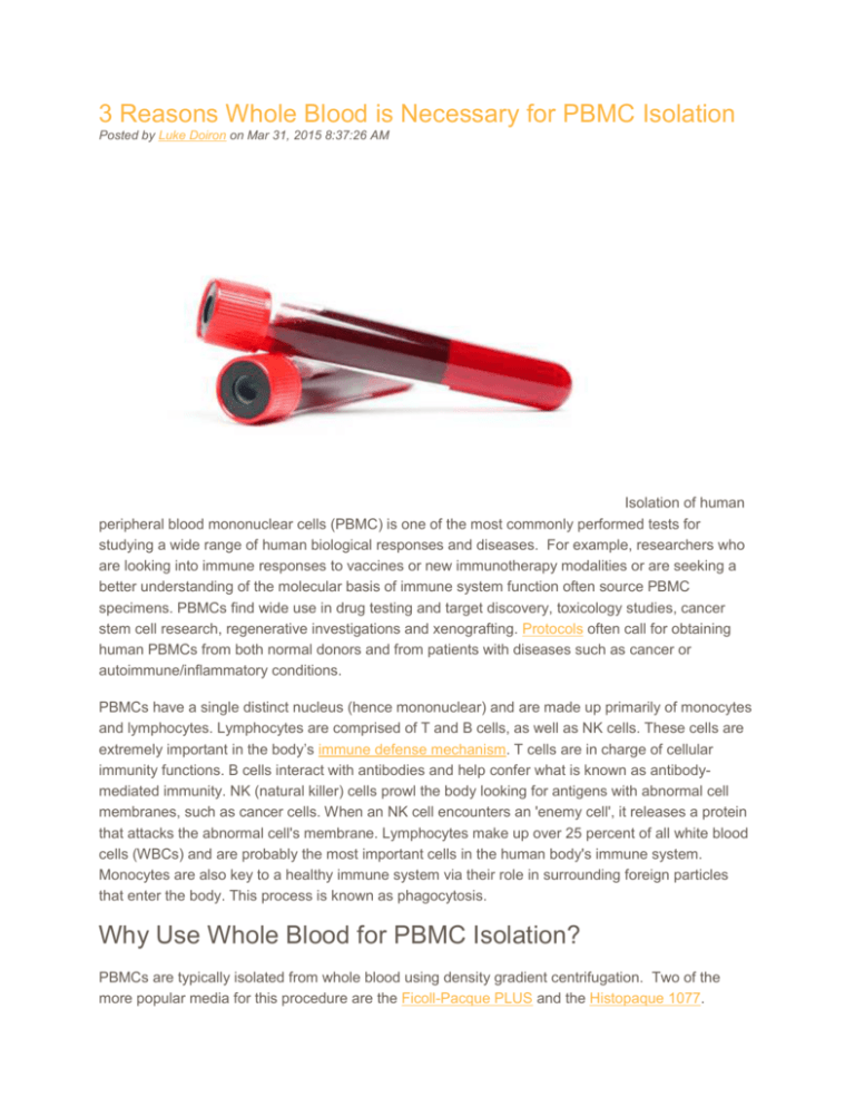
3 Reasons Whole Blood is Necessary for PBMC Isolation Posted by Luke Doiron on Mar 31, 2015 8:37:26 AM Isolation of human peripheral blood mononuclear cells (PBMC) is one of the most commonly performed tests for studying a wide range of human biological responses and diseases. For example, researchers who are looking into immune responses to vaccines or new immunotherapy modalities or are seeking a better understanding of the molecular basis of immune system function often source PBMC specimens. PBMCs find wide use in drug testing and target discovery, toxicology studies, cancer stem cell research, regenerative investigations and xenografting. Protocols often call for obtaining human PBMCs from both normal donors and from patients with diseases such as cancer or autoimmune/inflammatory conditions. PBMCs have a single distinct nucleus (hence mononuclear) and are made up primarily of monocytes and lymphocytes. Lymphocytes are comprised of T and B cells, as well as NK cells. These cells are extremely important in the body’s immune defense mechanism. T cells are in charge of cellular immunity functions. B cells interact with antibodies and help confer what is known as antibodymediated immunity. NK (natural killer) cells prowl the body looking for antigens with abnormal cell membranes, such as cancer cells. When an NK cell encounters an 'enemy cell', it releases a protein that attacks the abnormal cell's membrane. Lymphocytes make up over 25 percent of all white blood cells (WBCs) and are probably the most important cells in the human body's immune system. Monocytes are also key to a healthy immune system via their role in surrounding foreign particles that enter the body. This process is known as phagocytosis. Why Use Whole Blood for PBMC Isolation? PBMCs are typically isolated from whole blood using density gradient centrifugation. Two of the more popular media for this procedure are the Ficoll-Pacque PLUS and the Histopaque 1077. Density gradient centrifugation results in the lower density PBMCs migrating into a thin layer that is isolated from the higher density red blood cells and granulocytes. In some cases, the specimen provided for PBMC isolation is what is known as a buffy coat. Sometimes people use the terms buffy coat and PBMC interchangeably, but in fact there are differences which will impact the quality of the PBMC isolate. Consider these reasons why whole blood is the best type of specimen for obtaining high-quality PBMC isolate: 1. PBMCs are a subset of the buffy coat cells. The buffy coat will include granulocytes whereas PBMCs do not; granulocytes are not PBMCs and hence the purity of the buffy coat is lower than PBMC isolated from whole blood. 2. If not used in conjunction with PBMC density gradient preparations, the buffy coat really refers to the leukocyte layer which is obtained from a simple centrifugation of whole blood without using density gradient media. This whitishcolored (“buffy”) layer will separate directly above and will be in contact with the red blood cell (RBC)/granulocyte layer, which can make it difficult or even impossible to isolate pure PBMCs. 3. When hospitals or other specimen sources provide the buffy coat in bags, it typically contains a large amount of blood which has PBMCs but also RBCs. This type of sample requires additional steps to reduce the problem of aggregation, including additional dilution and separation of granulocytes. The end result can be much lower numbers of desired mononuclear cells. Additionally, buffy coats degrade rapidly, often within 48 hours, causing many dead cells in the sample.
