9_MHC
advertisement
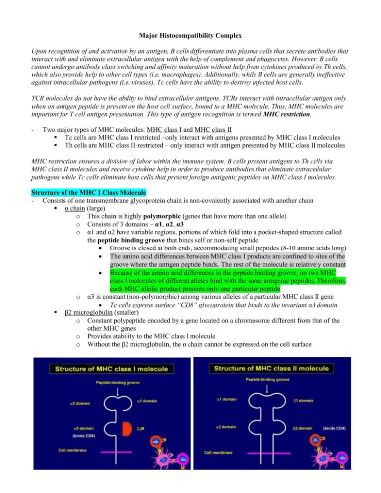
Major Histocompatibility Complex Upon recognition of and activation by an antigen, B cells differentiate into plasma cells that secrete antibodies that interact with and eliminate extracellular antigen with the help of complement and phagocytes. However, B cells cannot undergo antibody class switching and affinity maturation without help from cytokines produced by Th cells, which also provide help to other cell types (i.e. macrophages). Additionally, while B cells are generally ineffective against intracellular pathogens (i.e. viruses), Tc cells have the ability to destroy infected host cells. TCR molecules do not have the ability to bind extracellular antigens. TCRs interact with intracellular antigen only when an antigen peptide is present on the host cell surface, bound to a MHC molecule. Thus, MHC molecules are important for T cell antigen presentation. This type of antigen recognition is termed MHC restriction. - Two major types of MHC molecules: MHC class I and MHC class II Tc cells are MHC class I restricted –only interact with antigens presented by MHC class I molecules Th cells are MHC class II-restricted – only interact with antigen presented by MHC class II molecules MHC restriction ensures a division of labor within the immune system. B cells present antigens to Th cells via MHC class II molecules and receive cytokine help in order to produce antibodies that eliminate extracellular pathogens while Tc cells eliminate host cells that present foreign antigenic peptides on MHC class I molecules. Structure of the MHC I Class Molecule - Consists of one transmembrane glycoprotein chain is non-covalently associated with another chain α chain (large) o This chain is highly polymorphic (genes that have more than one allele) o Consists of 3 domains – α1, α2, α3 o α1 and α2 have variable regions, portions of which fold into a pocket-shaped structure called the peptide binding groove that binds self or non-self peptide Groove is closed at both ends, accommodating small peptides (8-10 amino acids long) The amino acid differences between MHC class I products are confined to sites of the groove where the antigen peptide binds. The rest of the molecule is relatively constant Because of the amino acid differences in the peptide binding groove, no two MHC class I molecules of different alleles bind with the same antigenic peptides. Therefore, each MHC allelic product presents only one particular peptide o α3 is constant (non-polymorphic) among various alleles of a particular MHC class II gene Tc cells express surface “CD8” glycoprotein that binds to the invariant α3 domain β2 microglobulin (smaller) o Constant polypeptide encoded by a gene located on a chromosome different from that of the other MHC genes o Provides stability to the MHC class I molecule o Without the β2 microglobulin, the α chain cannot be expressed on the cell surface Structure of the MHC II Class Molecule - Consists of two transmembrane glycoprotein chains that are non-covalently associated α chain o Has 2 domains – α1, α2 α1 has polymorphic amino acid residues α2 is constant β chain o Has 2 domains – β1, β2 β1 has polymorphic amino acid residues β2 is constant Peptide binding groove is formed by folding parts of the α1 and β1 domains o Polymorphic amino acid residues are located in and around the groove o Groove is open at both ends and can accommodate residues of 12-20 amino acids o Amino acid differences between MHC class II products are confined to the sites of the groove where the antigen peptide binds o No two MHC class II molecules of different alleles bind with the same antigenic peptides Th cells express “CD4” glycoprotein on their surface that binds to the invariant β2 domain of MHC II When there are no foreign antigens to present, peptide-binding grooves of MHC molecules are loaded with peptide fragments derived from the body’s own proteins, called self-antigens. The presentation of self-antigens is important during the development phase of T lymphocytes in the thymus. Developing T cells that react with self-antigens are eliminated in primary lymphoid organs. MHC Molecule Expression - MHC class I molecules are expressed on almost all nucleated cells Expressed most highly on lymphocytes and specialized APCs - MHC class II molecules are only expressed by specialized APCs Dendritic cells, B cells, macrophages, basophils, and thymic epithelial cells - MHC proteins play a role in T cell development in the thymus, T cell activation, and transplant rejection - Some MHC alleles are also associated with autoimmune or inflammatory diseases Organization of MHC Genes - MHC genes are organized into regions encoding three classes of molecules Genes that encode MHC class I Genes that encode MHC class II Genes that encode MHC class III o Are not membrane proteins o Code for complement components and other proteins o Functionally distinct from MHC I and II gene products and play no role in antigen presentation Genetic organization of equine MHC Genes The above figure depicts the genetic organization of equine MHC genes, which are present on chromosome 20. Equine ELA has two class I regions and two class II regions that have been characterized. MHC class I regions are designated as A and B, both with a single gene loci that encodes for the α-chain. The β2 microglobulin is encoded by a gene located on a different chromosome. The α-chains from each of the two loci (A and B) bind to the β2 microglobulin to form two MHC I molecules. MHC class II regions are designated as DQ and DR. The DQ region contains two loci, one that encodes for an αchain (DQA locus) and one that encodes for a β-chain (DQB locus). The DR region is organized similarly. The αchain from DQA locus pairs with the β-chain from the DQB locus to form one MHC class II molecule. Similarly, the α-chain from the DRA locus pairs with the β-chain from the DRB locus to form one MHC class II molecule. Locus Position of a gene on a chromosome (i.e. ELA-DRB, ELA-DRA) Allele One of two or more alternative forms of a gene occupying the same locus in a species. Alleles are generally differentiated numerically (i.e. ELA-A1, ELA-B15). The presence of multiple MHC alleles in a population at a give locus makes the MHC molecules highly polymorphic, a characteristic that appears to protect a species from a wide range of infectious diseases. Several species have very limited MHC gene polymorphism (i.e. cheetah), making it likely for a lethal disease to wipe out the entire species. Haplotype A complete set of MHC class I and MHC class II alleles on a single chromosome (either maternal or paternal). The term is generally used to classify inbred mice strains with respect to their MHC genes. Co-dominant expression of MHC molecules A diploid cell has two copies of each chromosome, one maternal and one paternal. All cells of vertebrates are diploid, with exception of reproductive cells (gametes). MHC genes are co-dominantly expressed, meaning both alleles (one on maternal chromosome and one on paternal chromosome) for each MHC gene are expressed on the cell surface. This cell is an APC. How many different MHC II molecules will be expressed? Co-dominant expression of MHC molecules For MHC I molecules, the α-chain gene on the ELA-A locus of a maternal chromosome and the α-chain gene on the ELA-A locus of a paternal chromosome are both expressed on the cell surface. Therefore, the surface of a horse cell can express four different MHC class I molecules – 2 allelic forms of an α-chain gene from the ELA-A locus and 2 allelic forms of α-chain gene from the ELA-B locus Cells expressing different alleles on all loci can express 8 different types of MHC class II molecules. This is because α-chain and β-chain alleles of both DQ and DR regions can combine either with alleles of the same parental derivation (maternal α-chain allele + maternal β-chain allele) or different parental derivation (maternal α-chain allele + paternal β-chain allele).
