Emily_Ryon_MD_Abstract_Final
advertisement
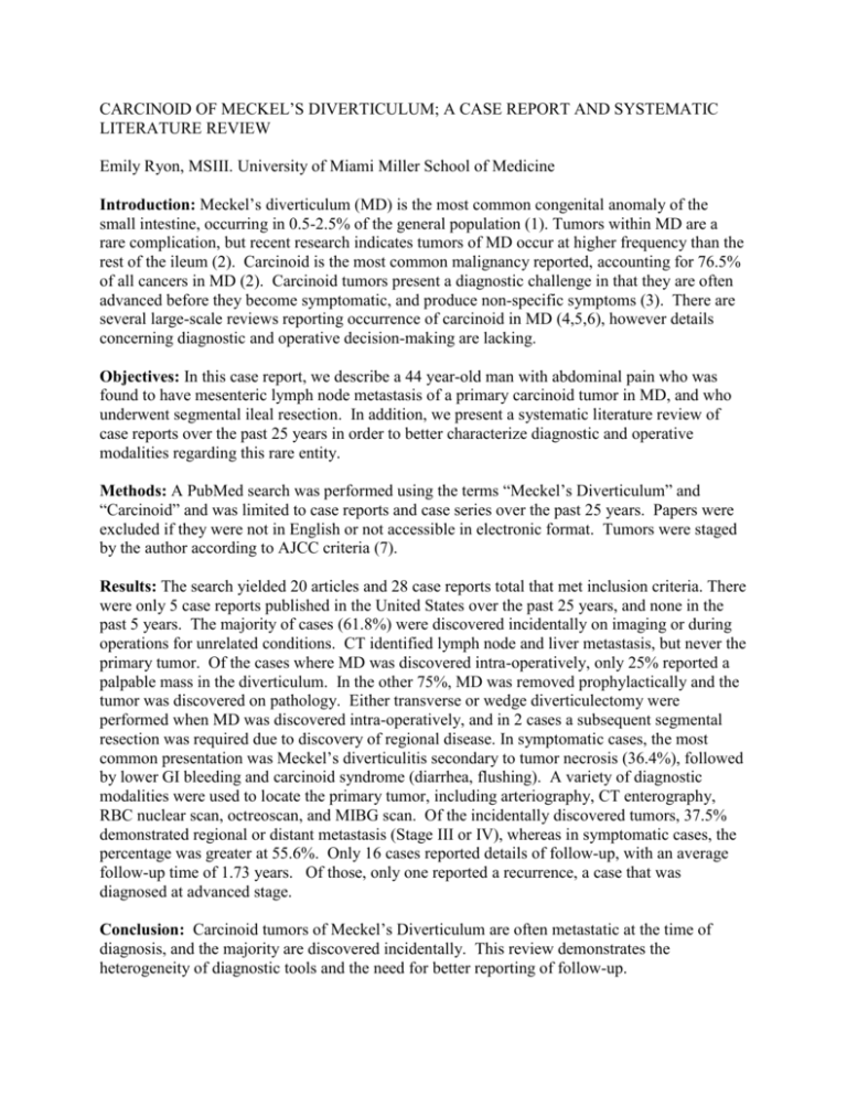
CARCINOID OF MECKEL’S DIVERTICULUM; A CASE REPORT AND SYSTEMATIC LITERATURE REVIEW Emily Ryon, MSIII. University of Miami Miller School of Medicine Introduction: Meckel’s diverticulum (MD) is the most common congenital anomaly of the small intestine, occurring in 0.5-2.5% of the general population (1). Tumors within MD are a rare complication, but recent research indicates tumors of MD occur at higher frequency than the rest of the ileum (2). Carcinoid is the most common malignancy reported, accounting for 76.5% of all cancers in MD (2). Carcinoid tumors present a diagnostic challenge in that they are often advanced before they become symptomatic, and produce non-specific symptoms (3). There are several large-scale reviews reporting occurrence of carcinoid in MD (4,5,6), however details concerning diagnostic and operative decision-making are lacking. Objectives: In this case report, we describe a 44 year-old man with abdominal pain who was found to have mesenteric lymph node metastasis of a primary carcinoid tumor in MD, and who underwent segmental ileal resection. In addition, we present a systematic literature review of case reports over the past 25 years in order to better characterize diagnostic and operative modalities regarding this rare entity. Methods: A PubMed search was performed using the terms “Meckel’s Diverticulum” and “Carcinoid” and was limited to case reports and case series over the past 25 years. Papers were excluded if they were not in English or not accessible in electronic format. Tumors were staged by the author according to AJCC criteria (7). Results: The search yielded 20 articles and 28 case reports total that met inclusion criteria. There were only 5 case reports published in the United States over the past 25 years, and none in the past 5 years. The majority of cases (61.8%) were discovered incidentally on imaging or during operations for unrelated conditions. CT identified lymph node and liver metastasis, but never the primary tumor. Of the cases where MD was discovered intra-operatively, only 25% reported a palpable mass in the diverticulum. In the other 75%, MD was removed prophylactically and the tumor was discovered on pathology. Either transverse or wedge diverticulectomy were performed when MD was discovered intra-operatively, and in 2 cases a subsequent segmental resection was required due to discovery of regional disease. In symptomatic cases, the most common presentation was Meckel’s diverticulitis secondary to tumor necrosis (36.4%), followed by lower GI bleeding and carcinoid syndrome (diarrhea, flushing). A variety of diagnostic modalities were used to locate the primary tumor, including arteriography, CT enterography, RBC nuclear scan, octreoscan, and MIBG scan. Of the incidentally discovered tumors, 37.5% demonstrated regional or distant metastasis (Stage III or IV), whereas in symptomatic cases, the percentage was greater at 55.6%. Only 16 cases reported details of follow-up, with an average follow-up time of 1.73 years. Of those, only one reported a recurrence, a case that was diagnosed at advanced stage. Conclusion: Carcinoid tumors of Meckel’s Diverticulum are often metastatic at the time of diagnosis, and the majority are discovered incidentally. This review demonstrates the heterogeneity of diagnostic tools and the need for better reporting of follow-up. 1. Mudatsakis N, Paraskakis S, Lasithiotakis K, Andreadakis E, Karatsis P. Acute appendicitis and carcinoid tumor in Meckel’s diverticulum. Three pathologies in one: a case report. Tech Coloproctol (2011) 15 (Suppl 1):S83–S85. 2. Thirunavukarasu P, Sathaiah M, Sukumar S, et al. Meckel's Diverticulum—A High-Risk Region for Malignancy in the Ileum: Insights From a Population-Based Epidemiological Study and Implications in Surgical Management. Ann Surg. 2011 February ; 253(2): 223–230. 3. Shebani KO, Souba WW, Finkelstein DM, et al. Prognosis and survival in patients with gastrointestinal tract carcinoid tumors. Ann Surg. 1999; 229:815–821. 4. Stone PA, Hofeldt MJ, Campbell JE, et al. Meckel Diverticulum: Ten-year Experience in Adults Southern Medical Journal 2004; 97: 1038-1041 5. Modlin IM, Shapiro MD, Kidd M. An Analysis of Rare Carcinoid Tumors: Clarifying These Clinical Conundrums. World J. Surg. 2005; 29: 92–101. 6. Park JJ, Wolff BG, Tollefson MK, et al. Meckel diverticulum: the Mayo Clinic experience with 1476 patients (1950–2002). Ann Surg. 2005; 241:529–533. 7. Edge SB, Byrd DR, Compton CC, et al., eds.: AJCC Cancer Staging Manual. 7th ed. New York, NY: Springer, 2010, pp 181-9. Table 1 Sex Male Female Table 2 Mean Age 51.4 58.3 Presentation Incidental Finding Appendectomy Colectomy Aortoiliac disease Cholecystectomy Sigmoid diverticulitis Hematuria Hernia repair Hysterectomy Intussusception LoA n (%) 21 (75) 7 (25) 17 (61.8) 3 3 2 2 2 1 1 1 1 1 Symptomatic 11 (39.2) Diverticulitis1 4 Bleeding2 3 Carcinoid Syndrome 3 Obstruction 1 _________________________________________________________ 1 On pathology, the tumor was identified as the source of necrosis and micro-perforation in 3 of 4 cases. 2 On pathology, the tumor was identified as the source of ulceration, not ectopic glandular tissue Table 4. Tumor size, procedure and followup by stage I IIA IIB IIIA IIIB IV NS Size 0.3 0.9 1.5 Divert. 3 2 4 SR 1 4 0 Ave. f/u (y) 3.5 3 3 1.6 2.2 1.0 12 0 13 5 5 1 2.7 2.8 Divert. – transverse or wedge diverticulectomy, SR – Segmental Ileal Resection, NS – not staged 1 In one case, subsequent segmental resection was recommended, but refused by patient 2 Wedge diverticulectomy with removal of solitary mesenteric lymph node 3 One report did not specify a method of resection. None of the un-staged cases reported follow-up time Symptomatic Cases Total 11 (100) Pre-op diagnosis of carcinoid 4 (36.4) Tumor palpable during operation 2 (18.2) Stage at diagnosis1 I IIA IIB IIIA IIIB IV Not staged Average tumor size (cm) _______ 1 (9.1) 3 (27.3) 2 (18.2) 3 (27.3) 2 (18.2) 1.22 1 Staged by author using pathology report and American Joint Committee on Cancer (AJCC) Criteria Table 3. Incidental Cases _______________ Total 17 (100) Diagnosis MD discovered at laparotomy LN discovered at laparotomy Liver met found on imaging MD causing intussusception 12 (70.6) 2 (11.8) 2 (11.8) 1 (5.8) Tumor palpable during operation 4 (23.5) Stage at diagnosis I IIA IIB IIIA IIIB IV Not staged Average tumor size (cm) 1 3 (17.6) 3 (17.6) 4 (23.5) 4 (23.5) 2 (11.8) 1 (5.9) 1.25 Staged by author using pathology report and American Joint Committee on Cancer (AJCC) Criteria

