Supplemental Data
advertisement
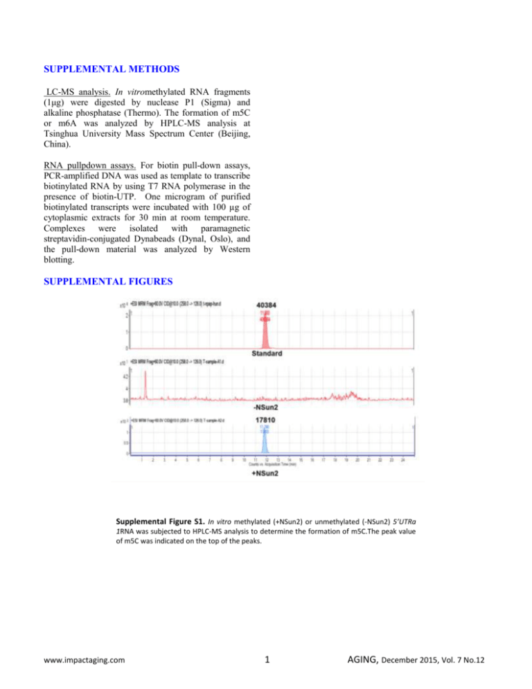
SUPPLEMENTAL METHODS LC-MS analysis. In vitromethylated RNA fragments (1μg) were digested by nuclease P1 (Sigma) and alkaline phosphatase (Thermo). The formation of m5C or m6A was analyzed by HPLC-MS analysis at Tsinghua University Mass Spectrum Center (Beijing, China). RNA pullpdown assays. For biotin pull-down assays, PCR-amplified DNA was used as template to transcribe biotinylated RNA by using T7 RNA polymerase in the presence of biotin-UTP. One microgram of purified biotinylated transcripts were incubated with 100 µg of cytoplasmic extracts for 30 min at room temperature. Complexes were isolated with paramagnetic streptavidin-conjugated Dynabeads (Dynal, Oslo), and the pull-down material was analyzed by Western blotting. SUPPLEMENTAL FIGURES Supplemental Figure S1. In vitro methylated (+NSun2) or unmethylated (-NSun2) 5’UTRa 1RNA was subjected to HPLC-MS analysis to determine the formation of m5C.The peak value of m5C was indicated on the top of the peaks. www.impactaging.com 1 AGING, December 2015, Vol. 7 No.12 Supplemental Figure S2. Translational repression of p27 by NSun2 is independent of HuR or CUBP1. (A) RNA pull-down assays using biotinylated p275’UTR, CR, 3’UTR, 5’UTRa, and 5’UTRb fragments and HeLa cell lysates. The presence of NSun2, HuR, and CUGBP1 in the pulldown materials was tested by Western blot analysis. A 10-µg aliquot of whole-cell lysates (Input) and bound GAPDH were included. (B) Biotinylated p275’UTR fragment was methylated in vitro (Met) by NSun2 or left unmethylated (Unmet). The methylated and unmethylated 5’UTR fragments then were subjected to RNA pulldown assays to assess the association of p27 5’UTR with NSun2, HuR, and CUGBP1, as described in Figure S2A.(C)HeLa cells were transfected with a siRNA targeting NSun2; 48 h later, cell lysates were prepared and subjected to RNA pulldown analysis as described in Figure S2A to assess the presence of HuR in the pulldown materials of p27 5’UTR and 3’UTR fragments (left) as well as the presence of CUGBP1 in the p27 5’UTRfragment (right). (D, E) HeLa cells were transfected with a siRNA targeting HuR (D) orCUGBP1 (E); 48 h later, cell lysates were prepared and subjected to RNA pulldown assays as described in Figure S2A to assess the association of NSun2 with the p27 5’UTR fragment.(F)HeLa cells were transfected with each of the pGL3-derived reporter vectors (left, Schematic)together with a pRL-CMV control reporter. Twenty-four h later, cells were further transfected with siRNA stargeting NSun2, HuR, or CUGBP1 and cultured for additional 48 h. Firefly luciferase activity was assayed relative to Renilla luciferase activity. Data represent the means ± SEM from 3 independent experiments. www.impactaging.com 2 AGING, December 2015, Vol. 7 No.12 Supplemental Figure S3. Constitutive expression of p53 and p16 in proliferating and senescent IDH4 cells. The levels of p53, p16, and GAPDH in proliferating (Y) and senescent (S) IDH4 cells were analyzed by Western blot analysis. The levels of proteins p53, p16, and GAPDH in mid-passage human diploid fibroblast (2BS, ~PDL 35) served as controls. Supplemental Figure S4. Modulating NSun2 levels in 2BS cells alters the cellular methylation levels of p27mRNA. (A, B) RNA isolated from cells described in Fig. 5A were subjected to methylation-specific PCR analysis to assess the levels of unmethylated and methylated p27mRNA. Data represent the means ± SEM from 3 independent experiments. www.impactaging.com 3 AGING, December 2015, Vol. 7 No.12
