Supplemental Materials
advertisement
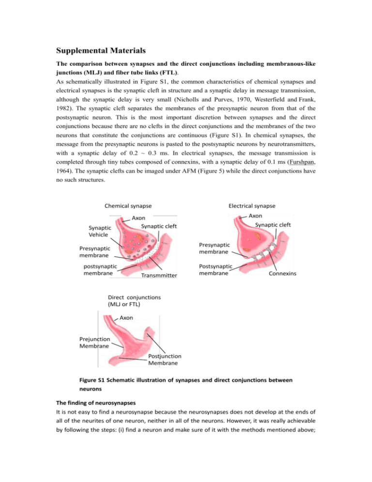
Supplemental Materials The comparison between synapses and the direct conjunctions including membranous-like junctions (MLJ) and fiber tube links (FTL). As schematically illustrated in Figure S1, the common characteristics of chemical synapses and electrical synapses is the synaptic cleft in structure and a synaptic delay in message transmission, although the synaptic delay is very small (Nicholls and Purves, 1970, Westerfield and Frank, 1982). The synaptic cleft separates the membranes of the presynaptic neuron from that of the postsynaptic neuron. This is the most important discretion between synapses and the direct conjunctions because there are no clefts in the direct conjunctions and the membranes of the two neurons that constitute the conjunctions are continuous (Figure S1). In chemical synapses, the message from the presynaptic neurons is pasted to the postsynaptic neurons by neurotransmitters, with a synaptic delay of 0.2 ~ 0.3 ms. In electrical synapses, the message transmission is completed through tiny tubes composed of connexins, with a synaptic delay of 0.1 ms (Furshpan, 1964). The synaptic clefts can be imaged under AFM (Figure 5) while the direct conjunctions have no such structures. Chemical synapse Synaptic Vehicle Electrical synapse Axon Synaptic cleft Axon Synaptic cleft Presynaptic membrane Presynaptic membrane postsynaptic membrane Transmmitter Postsynaptic membrane Connexins Direct conjunctions (MLJ or FTL) Axon Prejunction Membrane Postjunction Membrane Figure S1 Schematic illustration of synapses and direct conjunctions between neurons The finding of neurosynapses It is not easy to find a neurosynapse because the neurosynapses does not develop at the ends of all of the neurites of one neuron, neither in all of the neurons. However, it was really achievable by following the steps: (i) find a neuron and make sure of it with the methods mentioned above; (ii) find the axon of the neuron, which was the very neurite, at the end of which, neurosynapse was most possibly found; (iii) scanning the AFM tip from the beginning of the axon at the neuron soma and going along the axon to its end; (iv) if there were branches at the end of the axon, continue the scanning along the branches one after another and the neurosynapses was often found at the ends of these branches; (v) judging whether a joint structure was a neurosynapses by its morphology and structures and finally conformed by punctate staining of synapsin I. A neurosynapse should meet four standards: (1) Typical composition including pre- and postsynaptic neurites; (2) the end of the presynaptic neurite enlarged typically; (3) there was obvious synaptic cleft between the pre- and postsynaptic membranes; (4) the thickening of the pre- and postsynaptic membranes or neurites; (5) positive stained by synapsin I. During the scanning, the movement of the scanned areas should be gradually and the z-range should be kept at the optimum value, in avoid of the missing of the interesting neurite. References 1. Nicholls By J.G.and Purves D(1970). Monosynaptic chemical and electrical connexions between sensory and motor cells in tie central nervous system of the leech. J. Phyeiol., 209:647-667 2. Furshpan E.J (1964). Electrical transmission" at an excitatory synapse in a vertebrate brain. Science. 144(3620):878-80. 3. Westerfield M, Frank E (1982). Specificity of electrical coupling among neurons innervating forelimb muscles of the adult bullfrog. J Neurophysiol. 48(4):904-13
