BMED 2801 - Lecture 8: Study of one tissue in some detail
advertisement
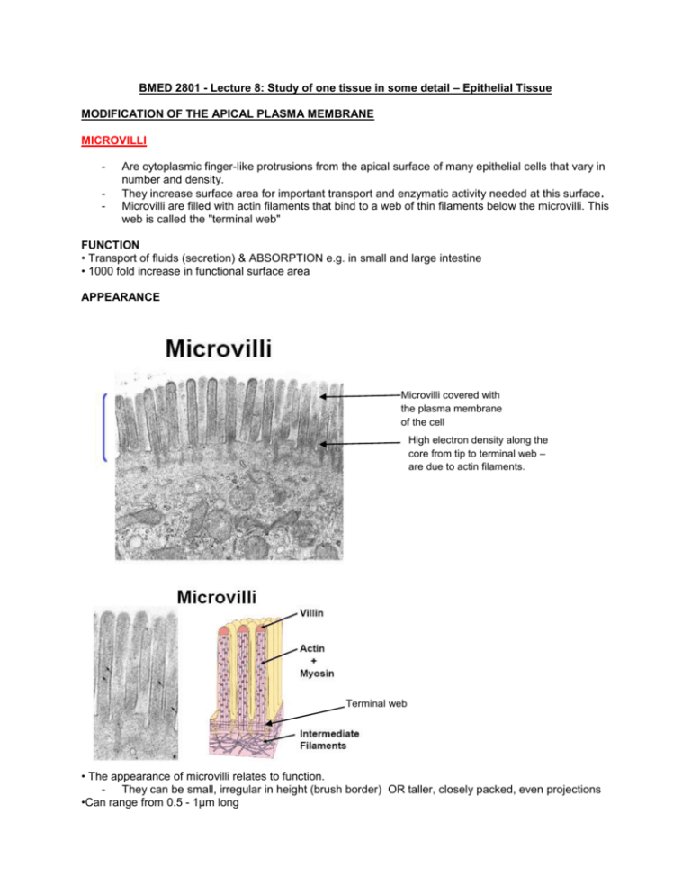
BMED 2801 - Lecture 8: Study of one tissue in some detail – Epithelial Tissue MODIFICATION OF THE APICAL PLASMA MEMBRANE MICROVILLI - Are cytoplasmic finger-like protrusions from the apical surface of many epithelial cells that vary in number and density. They increase surface area for important transport and enzymatic activity needed at this surface. Microvilli are filled with actin filaments that bind to a web of thin filaments below the microvilli. This web is called the "terminal web" FUNCTION • Transport of fluids (secretion) & ABSORPTION e.g. in small and large intestine • 1000 fold increase in functional surface area APPEARANCE Microvilli covered with the plasma membrane of the cell High electron density along the core from tip to terminal web – are due to actin filaments. Terminal web • The appearance of microvilli relates to function. - They can be small, irregular in height (brush border) OR taller, closely packed, even projections •Can range from 0.5 - 1μm long • The number & shape of microvilli correlates with their absorptive capacity. MICROVILLI ARE MOTILE • Sway slowly to increase absorptive power – by moving fluid along surface, allow for mixing/stirring allows for more absorption – increase functional ability of plasma membrane. STRUCTURE - Microvilli are covered with the plasma membrane – dark line along the edge - The core of actin filaments of cytoskeleton goes straight up through the microvilli - from tip to Terminal Web - Actin filaments – create the microvilli structure by pushing the plasma membrane forward. These embed into more actin filaments around the top of the cell – called the terminal web (web of actin filaments). - Amorphous capping proteins (villin) are at the tip of the microvillus to prevent depolymerisation. gives - rigidity + motility STEREOCILIA Very few, long, immotile (due to length- high energy demand), same structure as microvilli. 10-25μm long Location Epididymis + vas deferens absorption of sperm Sensory cells of ear Function Facilitate absorption Sensory receptors CILIA - MOTILE (sweeping action) cytoplasmic structures - Invagination of plasma membrane Image below: Respiratory tract - Pseudostratified columnar ciliated epithelium (all cells rest on basement membrane but not all reach the surface) Basement membrane LIGHT MICROSCOPE APPEARANCE • Short, fine, hair like • ½ length of cell • All the same length/cell • thin, dark staining band at base = Basal Bodies • each Cilia associated with a Basal Body FUNCTION Respiratory tract organised movement sweeping action towards mouth- sweep mucus + particulate matter from lungs Oviducts transport Ova towards uterus for implantation (ectopic pregnancy – cilia not functioning properly = implantation of Ova in the wall of the fallopian tube), move Ova from fimbri towards sperm. CROSS-SECTION OF CILIA ARRANGEMENT OF MICROTUBULES IN CILIA • Central core (Axoneme) consisting of 9 doublets of microtubules + 2 central microtubules • axoneme inserts into Basal Body Microtubule doublets = tubulin is attached to a motor protein - dynein arms (ATPase enzyme activity) Basal Body – slight change in microtubule structure = modified centriole • 9 microtubule triplets • form a short cylinder MOVEMENT OF CILIA Regular, synchronous & undulating Creates a WAVE motion SWEEPS across the apical epithelial surface MODIFICATION OF THE LATERAL PLASMA MEBRANE: Terminal Bar (Junctional Complex) IMPORTANT: Don’t confuse terminal bar (junctional complex) with terminal web (network of actin and intermediate filaments that microvilli insert into) Zonula Occludens (apical end) Terminal bar = Junctional Complex = Zonula Adherens Macula Adherens There are 3 different types of junctions: (1) zonnula occludens or tight junctions acts as a diffusion barrier - A region of occlusion (blocking) There are tight bonds holding two cells together (2) zonnula adherens (zones of adherence) & macula adherens (spots of adherence - also known as desmisomes) acts as an anchor via the cytoskeleton (3) Gap junctions or Nexus direct communication between adjacent cells (e.g. ionic communication) Other Structures: Plicae - Down the length of the plasma membrane Are morphological changes to lateral cell surface - Folds & processes create interdigitating cytoplasmic processes – help to adhere cells together. - Function: increase cell surface area Apical surface Epithelial cell 1 Epithelial cell 2 Lateral plasma membrane - (1) Junctional complexes are seen as regions of increased density around the plasma membrane of the two cells. ZONULA OCCLUDENS (TIGHT JUNCTIONS) Plasma membrane – appear as railway tracks Occluding proteins embedded on the plasma membrane = ring of plasma membrane union between neighboring cells - Multiple sites of membrane fusion involves a fusion of the two adjacent membranes with fibrous connections - The intercellular space between the two plasma membranes are not seen because they are so close together – but these are not stuck together. Location Lateral border, close to apical membrane Description • Multiple sites of membrane fusion • Intercellular space obliterated • Occluden (trans membrane protein) – integral part of the plasma membranes of the two cells– intertwine and connect to each other – pulling the two cells together associated within the cell with the actin filaments of the cytoskeleton – which help with drawing the two cells together. Function • Barrier junction • Completely seal the intercellular space off from lumen E.g. epithelium in bladder – very acidic urine sitting in the bladder – if these seep into cells that line epithelium = cause toxic damage to tissue and integrity of the organ itself -- >need tight junctions to hold them together. (2) ZONULA ADHERENS High electron density (fuzzy plaque) – due to presence of proteins + actin microfilaments of the cytoskeleton = ring around the cell Location It is located below the tight junctions/occludens Description • The structure reveals a 20-30nm intercellular space clearly visible extracellular space between the two cells. • The space contains E-cadherin (membrane Protein) + Ca2+ (form a bridge the two cells) • The cytoplasm contains 6nm actin microfilaments (Fuzzy Plaque) • It is continuous with Terminal Web Function - associate with the actin cytoskeleton - Involved in adhesion (sticking cells together or sticking cells to surfaces). - Transduce signals into and out of the cell, influencing a variety of cellular behaviours including proliferation, migration, and differentiation. (3) MACULA ADHERENS (DESMOSOME) – spot junctions Desmisomes – two cells attached together with the same structure that bring them together V. electron dense attachment plaque (protein structure on cytoplasmic side of plasma membrane) Intermediate Filaments of cytoskeleton V. electron denserepresents interlocking of cadherin proteins. - Paler region – cadherin just in intercellular space. = localized spot attachments around cell periphery VERY STRONG attachment Cadherin protein- located within the plasma membrane of the cell - Cadherin projects through the gap. Location Lateral plasma membrane Description Cytoplasmic side has an ATTACHMENT PLAQUE plaques on plasma membrane contain desmoplakins which are intermediate filament binding proteins. Intermediate filaments loop into the plaques connecting the desmosome with the cytoskeletal system of both plaques. • 10nm intermediate filaments (Tonofilaments) • anchor proteins • 400nm x 250nm x 10nm disc 30nm wide intercellular space • contains Cadherin protein molecules that connect one another via homophilic binding + Ca2+ (Binding requires calcium) arranged like a ZIPPER (4) HEMIDESMOSOMES = ½ desmosome Location • Basal plasma membrane Hemidesmisomes– a connection between one cell and connective tissue function: Link cell to basement membrane at basal surface • stratified squamous epithelium Function Connect epithelium TO basement membrane (5) FASCIA ADHERENS • located in Cardiac Muscle • cells attach at a BROAD FACE & not in a ring • Otherwise same as Z. Occludens + M. Adherens (6) GAP JUNCTIONS Plasma membrane Gap junction – between the two plasma membranes = broad areas of closely opposed plasma membranes (but do not fuse) Connexins - integral protein embedded on the plasma membrane embed through the plasma membrane of the opposing cell creating a hollow channel. Function •Some degree of intercellular adhesion • Intercellular exchange & communication • The physical travelling of ions, metabolites & regulatory molecules (epithelium) between cells. • The spread of excitation (cardiac muscle) Allow for the coordinated rhythmic contractions of ventricles so it acts as a singular pump irregular contraction – occurs when SA and AV nodes and action potentials are not created properly due to dysfunction of gap junctions e.g. toxins blocking the gap junction = this causes the heart to fibrillate – uncoordinated contractions as cells can not communicate with each other. • Gap junctions are dynamic structures – they change Visualized with FREEZE-FRACTURE and TEM Description 2 parallel plasma membranes separated by 2nm GAP 6 Connexin proteins arranged in circle form a hollow channel going form one cell to another 1 Channel per membrane Channels are aligned Action Changes in Connexin confirmation due to humeral or neural response received by the cell results in a Rapid change of arrangement either increasing/decreasing the lumen of the channel (open/closing controlled barrier) Channels can OPEN 2nm wide & CLOSE Lateral interdigitations: - all junctional complexes occur Basal-Lateral Interdigitations. - - Epithelia that are important in regulated transport also may have specializations along their basal surface. These look as if the cell has multiple "legs" like an octopus. The "legs" of different cells interdigitate with one another and form a space into which ions and water are transported. Even though these are at the basal surface, they are called "lateral interdigitations". Also, any other substances that are moved across the cell to the blood supply are secreted into this region. The interdigitations of the membranes increase surface area available for the ion and water transport. The membranes have important ion pumps, like sodium/potassium ATPases.
