Word docx
advertisement
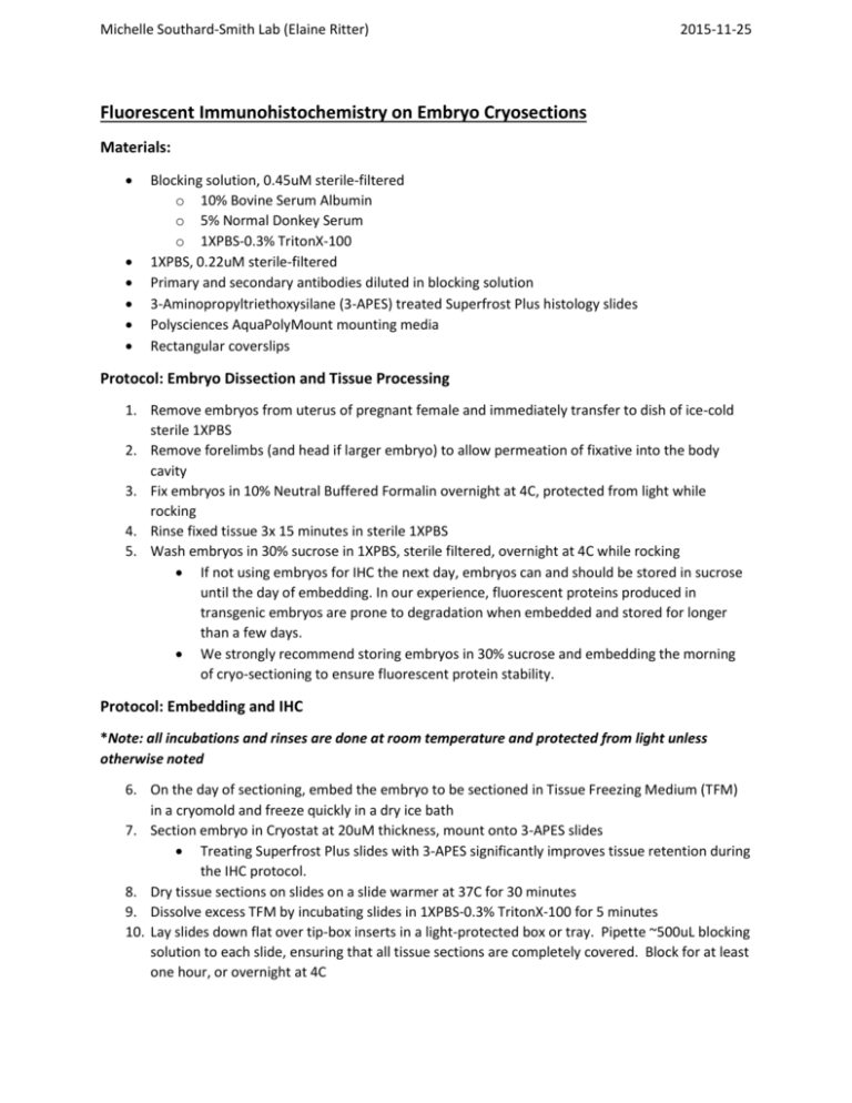
Michelle Southard-Smith Lab (Elaine Ritter) 2015-11-25 Fluorescent Immunohistochemistry on Embryo Cryosections Materials: Blocking solution, 0.45uM sterile-filtered o 10% Bovine Serum Albumin o 5% Normal Donkey Serum o 1XPBS-0.3% TritonX-100 1XPBS, 0.22uM sterile-filtered Primary and secondary antibodies diluted in blocking solution 3-Aminopropyltriethoxysilane (3-APES) treated Superfrost Plus histology slides Polysciences AquaPolyMount mounting media Rectangular coverslips Protocol: Embryo Dissection and Tissue Processing 1. Remove embryos from uterus of pregnant female and immediately transfer to dish of ice-cold sterile 1XPBS 2. Remove forelimbs (and head if larger embryo) to allow permeation of fixative into the body cavity 3. Fix embryos in 10% Neutral Buffered Formalin overnight at 4C, protected from light while rocking 4. Rinse fixed tissue 3x 15 minutes in sterile 1XPBS 5. Wash embryos in 30% sucrose in 1XPBS, sterile filtered, overnight at 4C while rocking If not using embryos for IHC the next day, embryos can and should be stored in sucrose until the day of embedding. In our experience, fluorescent proteins produced in transgenic embryos are prone to degradation when embedded and stored for longer than a few days. We strongly recommend storing embryos in 30% sucrose and embedding the morning of cryo-sectioning to ensure fluorescent protein stability. Protocol: Embedding and IHC *Note: all incubations and rinses are done at room temperature and protected from light unless otherwise noted 6. On the day of sectioning, embed the embryo to be sectioned in Tissue Freezing Medium (TFM) in a cryomold and freeze quickly in a dry ice bath 7. Section embryo in Cryostat at 20uM thickness, mount onto 3-APES slides Treating Superfrost Plus slides with 3-APES significantly improves tissue retention during the IHC protocol. 8. Dry tissue sections on slides on a slide warmer at 37C for 30 minutes 9. Dissolve excess TFM by incubating slides in 1XPBS-0.3% TritonX-100 for 5 minutes 10. Lay slides down flat over tip-box inserts in a light-protected box or tray. Pipette ~500uL blocking solution to each slide, ensuring that all tissue sections are completely covered. Block for at least one hour, or overnight at 4C Michelle Southard-Smith Lab (Elaine Ritter) 2015-11-25 11. Gently tap blocking solution off the slides but do not rinse. Incubate tissue in primary antibody, diluted in sterile blocking solution, for either: 4-6 hours at room temperature, or Overnight at 4C Note: optimal incubation times vary depending on antibody 12. To preserve diluted antibody for future use, pour antibody off the slide back into original storage tube Note: number of reuses varies depending on the antibody. When staining begins to weaken or change in any way, discard the dilution. If sterile technique is used and dilutions are stored properly, they can be reused many times for months to years. 13. Gently pour sterile 1XPBS over slides to rinse away excess antibody. To avoid damaging tissue sections, start pouring over the painted portion so it trickles down the length of the slide. Approximately 5 seconds of streaming PBS is sufficient for rinsing at this step. 14. Incubate tissue in secondary antibody, diluted in sterile blocking solution, for 1 hour 15. Gently pour sterile 1XPBS over slides to rinse away excess antibody. To avoid damaging tissue sections, start pouring over the painted portion so it trickles down the length of the slide. Approximately 20 seconds of streaming PBS is sufficient for rinsing at this step. Thorough but gentle rinsing is necessary to avoid fluorescent “speckling” background on tissue sections. 16. Incubate slides in 0.5mM CuSO4 in 50mM Sodium Acetate buffer, pH5.0 for 10 minutes to quench autofluorescence 17. Transfer slides to sterile water to cease CuSO4 quenching reaction 18. Mount slides with AquaPolyMount, coverslip and let dry before imaging Michelle Southard-Smith Lab (Elaine Ritter) 2015-11-25 Quenching Autofluorescence with Cupric Sulfate Buffer Materials: Cupric Sulfate, certified A.C.S. grade (Fisher Scientific, C493-500) Ammonium Acetate, crystalline HPLC grade (Fisher Scientific, A639-500) Sterile MilliQ water pH Meter 1N HCl for adjusting pH Sterile 1L storage bottle (such as Corning 430518, sterile polystyrene with plug seal cap) Procedure: In a sterilized 1L storage bottle, add the following: o 3.85g Ammonium Acetate o 0.07g Cupric Sulfate o 900mL Sterile MilliQ water Adjust pH to 5.0 with 1N HCl Add sterile MilliQ water to make 1L total volume Store at room temperature To use in immunohistochemistry to quench autofluorescence: o Following ALL staining procedures (unless mounting media contains DAPI), apply cupric sulfate buffer to tissue (either in wholemount or on slides) for 10 minutes at room temperature o Stop quenching reaction by rinsing with sterile water o Discard Cupric Sulfate after use – not suitable for reuse once it’s been applied to tissue.
