Testing for effects of resource base on DNA methylation levels
advertisement
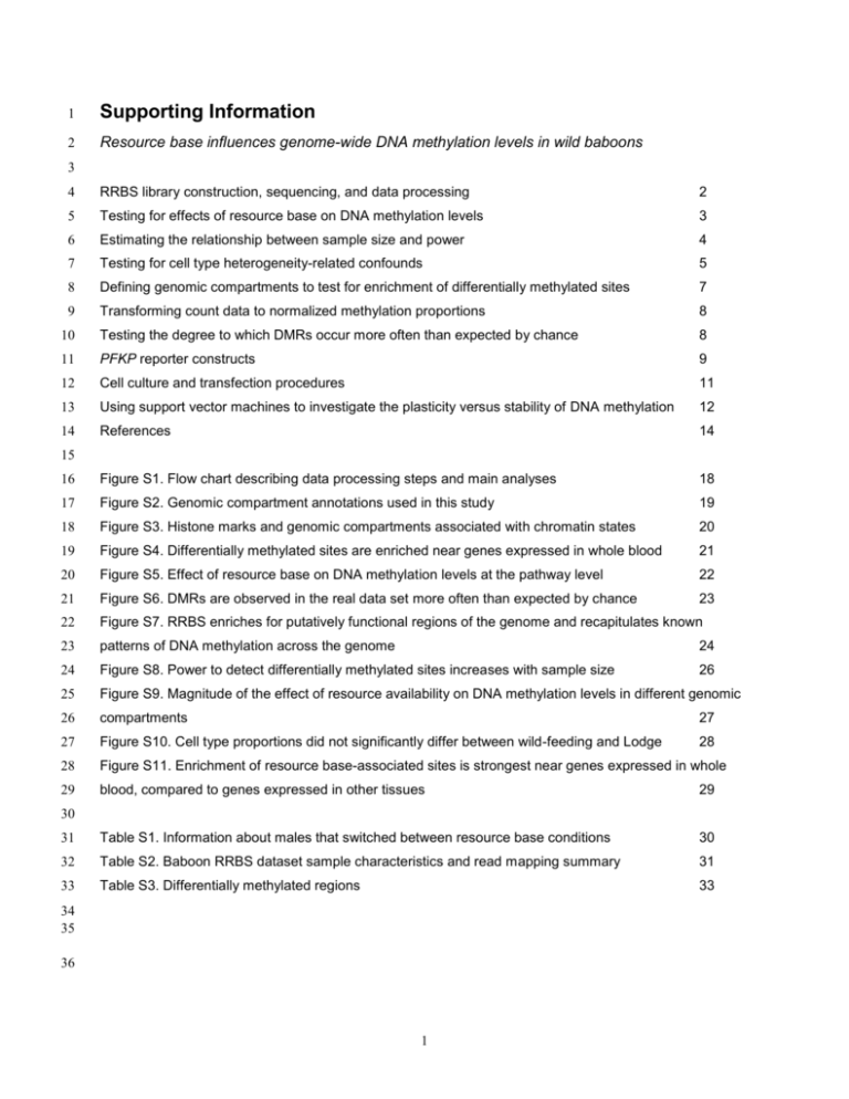
1 Supporting Information 2 Resource base influences genome-wide DNA methylation levels in wild baboons 3 4 RRBS library construction, sequencing, and data processing 2 5 Testing for effects of resource base on DNA methylation levels 3 6 Estimating the relationship between sample size and power 4 7 Testing for cell type heterogeneity-related confounds 5 8 Defining genomic compartments to test for enrichment of differentially methylated sites 7 9 Transforming count data to normalized methylation proportions 8 10 Testing the degree to which DMRs occur more often than expected by chance 8 11 PFKP reporter constructs 9 12 Cell culture and transfection procedures 11 13 Using support vector machines to investigate the plasticity versus stability of DNA methylation 12 14 References 14 16 Figure S1. Flow chart describing data processing steps and main analyses 18 17 Figure S2. Genomic compartment annotations used in this study 19 18 Figure S3. Histone marks and genomic compartments associated with chromatin states 20 19 Figure S4. Differentially methylated sites are enriched near genes expressed in whole blood 21 20 Figure S5. Effect of resource base on DNA methylation levels at the pathway level 22 21 Figure S6. DMRs are observed in the real data set more often than expected by chance 23 22 Figure S7. RRBS enriches for putatively functional regions of the genome and recapitulates known 23 patterns of DNA methylation across the genome 24 24 Figure S8. Power to detect differentially methylated sites increases with sample size 26 25 Figure S9. Magnitude of the effect of resource availability on DNA methylation levels in different genomic 26 compartments 27 27 Figure S10. Cell type proportions did not significantly differ between wild-feeding and Lodge 28 28 Figure S11. Enrichment of resource base-associated sites is strongest near genes expressed in whole 29 blood, compared to genes expressed in other tissues 29 31 Table S1. Information about males that switched between resource base conditions 30 32 Table S2. Baboon RRBS dataset sample characteristics and read mapping summary 31 33 Table S3. Differentially methylated regions 33 15 30 34 35 36 1 37 38 RRBS library construction, sequencing, and data processing To construct RRBS libraries, we extracted genomic DNA from whole blood samples 39 using the DNeasy Blood and Tissue kit (QIAGEN), according to the manufacturer’s instructions. 40 We then used 180 ng of genomic DNA per individual to generate adapter barcoded libraries for 41 each sample. Libraries were pooled together in sets of 9-12 samples, subjected to sodium 42 bisulfite conversion using the EpiTect Bisulfite Conversion kit (QIAGEN), and then PCR 43 amplified prior to sequencing on the Illumina HiSeq 2000 platform. Each pooled set of libraries 44 was sequenced in a single lane to a mean depth (± SD) of 27.25 ± 13.62 million reads per 45 sample (173.85 ± 12.87 million reads per lane; Table S2). To assess the efficiency of the 46 bisulfite conversion, 1 ng of unmethylated lambda phage DNA (Sigma Aldrich) was added to 47 each 180 ng sample prior to library construction. 48 Following read trimming for adaptor contamination and base quality, we mapped all 49 reads to a combined reference genome that included the olive baboon genome (Panu 2.0) and 50 the lambda phage genome. We used sequences that mapped to the lambda phage genome to 51 estimate the bisulfite conversion efficiency for each DNA sample. Specifically, for each sample, 52 we summed (i) the number of reads that mapped to lambda phage CpG sites and were read as 53 thymine (reflecting an unmethylated cytosine that was converted to thymine by sodium bisulfite); 54 and (ii) the total number of reads that mapped to any lambda phage CpG site. Because all CpG 55 sites in the lambda phage genome were originally unmethylated (and thus should have been 56 converted to thymine), the ratio of these two sums provides an estimate of bisulfite conversion 57 efficiency. Here, a ratio of 1 would represent perfect conversion of every unmethylated cytosine 58 to thymine. 59 For data generated from baboon DNA, we performed several additional filtering steps 60 before performing differential methylation analyses. First, following previous studies [1,2], we 61 excluded sites that were constitutively hypermethylated (average DNA methylation level > 0.90) 62 or hypomethylated (average DNA methylation level < 0.10). We also excluded sites in which the 2 63 standard deviation of DNA methylation levels fell in the lowest 5% of the overall data set, and 64 sites that fell in the lowest quartile of mean coverage depth (4.74 reads). The final filtered data 65 set contained 535,996 CpG sites. 66 Importantly, although we mapped reads generated from yellow baboons to the olive 67 baboon genome, biases associated with mapping to a heterospecific genome should be minimal 68 in the filtered dataset. The primary source of potential bias comes from C/T SNPs, and 69 specifically cases in which the olive baboon reference genome contains a CG site, but yellow 70 baboons carry TG at the same location in the genome. In these cases, BSMAP would 71 erroneously estimate the CpG site in our yellow baboon samples as completely unmethylated. 72 However, because we filtered out all sites with average methylation levels below 10%, sites 73 affected by this type of error should not be included in our final filtered dataset. 74 Testing for effects of resource base on DNA methylation levels 75 We used the software MACAU [3] to test for differences in DNA methylation levels 76 between wild-feeding and Lodge individuals at each CpG site in our filtered dataset. Specifically, 77 we fit a binomial mixed effects model for each CpG site across all individuals (i): 𝑦𝑖𝑗 = 𝐵𝑖𝑛(𝑟𝑖 , 𝜋𝑖 ) 78 79 where 𝑟𝑖 is the total read count for the ith individual; 𝑦𝑖 is the methylated read count for that 80 individual; and 𝜋𝑖 is an unknown parameter that represents the true proportion of methylated 81 reads for individual i. MACAU then uses a logit link to model 𝜋𝑖 as a linear function of several 82 parameters: 83 𝜋𝑖 𝑙𝑜𝑔 ( ) = 𝒘𝑇𝑖 𝜶 + 𝑥𝑖 𝛽 + 𝑔𝑖 + 𝑒𝑖 , 1 − 𝜋𝑖 84 𝒈 = (𝑔1 , ⋯ , 𝑔𝑛 )𝑇 ∼ 𝑀𝑉𝑁(0, 𝜎𝑔2 𝑲), 85 𝒆 = (𝑒1 , ⋯ , 𝑒𝑛 )𝑇 ∼ 𝑀𝑉𝑁(0, 𝜎𝑒2 𝑰), 3 86 where 𝒘𝑖 is a vector of covariates we wish to control for, as well as an intercept, and 𝜶 is an 87 identically sized vector of corresponding coefficients. In our case, 𝒘𝑖 contains the following 88 covariates: sex of the sampled individual, age of the sampled individual, age of the blood 89 sample (measured in years since date of sampling), and bisulfite conversion rate (estimated 90 from the lambda phage spike in). The ages of all individuals born in the Amboseli study 91 population (n = 51 in our data set) are known to within a few days’ error. For animals that 92 immigrate into the study population, ages are estimated from morphological features by trained 93 observers (n = 18 animals in our data set). 𝑥𝑖 denotes the foraging group of the ith individual 94 (Lodge or wild-feeding) and 𝛽 is the corresponding estimate of this effect. In addition, we 95 included a random genetic effect (𝑔𝑖 ). The variance of 𝑔𝑖 is influenced by pairwise relatedness 96 between individuals in the sample, given by the matrix 𝑲. Pairwise relatedness values were 97 calculated from previously collected microsatellite data (14 highly polymorphic loci) using the 98 program COANCESTRY [5–7]. In this model, 𝜎𝑔2 and 𝜎𝑒2 represent genetic and environmental 99 variance components for the DNA methylation trait, and 𝑒𝑖 denotes model error. For each model 100 (i.e., CpG site), we extracted the p-value associated with the effect of foraging group on DNA 101 methylation levels (𝛽). 102 Estimating the relationship between sample size and power to detect resource base- 103 associated sites 104 We used a subset of our data to estimate the relationship between sample size and the 105 number of true positive, resource-base associated sites detected in our analyses. Specifically, 106 for each CpG site on chromosome 1 (in our filtered dataset, n = 49,576 CpG sites), we 107 estimated the p-value for the resource base effect on DNA methylation levels in our full data set 108 (n = 61 lifelong wild-feeding or Lodge individuals, excluding switching individuals) and in 16 109 randomly subsampled data sets of n = 20, 30, 40, or 50 (four replicates for each sample size, 4 110 balancing the number of Lodge versus wild-feeding individuals to reflect the composition of the 111 original data set). We modeled resource base effects on DNA methylation levels in the 112 subsampled data sets using the same approach described in the main text Methods and in the 113 SI section on Testing for effects of resource base on DNA methylation levels. 114 Next, we modeled the relationship between sample size and p-value at each site using a 115 power law distribution (as described in Mukherjee et al. 2003). For sites truly associated with 116 resource base, we expect an inverse relationship between sample size and significance (the 117 larger the sample, the lower the p-value). We considered sites that met this criterion as putative 118 true positives, and estimated the p-values we would observe at these sites given data sets of 119 increasing sample size. In contrast, we considered sites that showed no relationship between 120 sample size and significance to be putative true negatives. Finally, we predicted our power to 121 detect the total set of putative true positive sites at a 10% FDR in data sets of increasing sample 122 size. The results of this analysis (Figure S8) show that we are underpowered to detect all true 123 resource base-associated sites in our current data set at a 10% FDR. Specifically, they suggest 124 that we detected ~9% of putative true positive sites with relatively large effects (sites that fall in 125 the upper 25% of the effect size distribution) and probably none of the true positive sites with 126 small effect sizes (bottom 25% of the effect size distribution). 127 Testing for cell type heterogeneity-related confounds 128 Our analyses focused on DNA methylation levels in whole blood, which is composed of 129 several cell types with distinct DNA methylation profiles [9]. Thus, if cell type composition 130 consistently differed between blood samples collected from Lodge versus wild-feeding animals, 131 differentially methylated sites could reflect these differences rather than changes in DNA 132 methylation levels within cells. To investigate this possibility, we used two complementary 133 approaches. First, we measured cell type proportions from wild-feeding animals (n = 25, 134 including 6 animals for whom we also collected DNA methylation data) and Lodge group 5 135 animals (n = 15, including 5 animals for whom we also collected DNA methylation data) based 136 on manual counts of Giemsa-stained blood smears collected at the time of blood sample 137 collection. These counts resulted in estimates of the proportional representation of eosinophils, 138 basophils, neutrophils, lymphocytes, and monocytes for baboons in both resource conditions. 139 We then used generalized linear models with a binomial link function (implemented in R via 140 ‘glm’) to test the hypothesis that foraging group influenced these proportions, controlling for the 141 effects of age, sex, and the identity of the person who counted the blood smear. We found that, 142 for all cell types tested, there was no significant differences in abundance between wild-feeding 143 and Lodge individuals (all p > 0.05, Figure S10). 144 Second, we drew on publicly available DNA methylation data generated from pure 145 populations of CD4+ (helper) T cells, CD8+ (cytotoxic) T cells, natural killer cells, B cells, 146 monocytes, and granulocytes from human whole blood [10]. Because these data were 147 generated on Illumina 450K Infinium arrays, relatively few CpG sites overlapped with our data 148 set. However, for the 7,425 CpG sites that were profiled in both the Jaffe data set and in our 149 samples, we were able to ask whether sites associated with resource base in our analysis (at a 150 10% FDR) were more likely to be differentially methylated between cell types than expected by 151 chance, using a Fisher’s Exact Test. We considered a CpG site to be differentially methylated 152 by cell type if (i) a significant (10% FDR) difference among cell types was reported by Jaffe 153 2015 (using an ANOVA), and (ii) average DNA methylation levels differed by at least 10% 154 between the two most prevalent cell types (granulocytes and CD4+ T cells, which together 155 account for greater than two-thirds of nucleated human blood cells on average [11]. To explore 156 the sensitivity of our findings to these cutoffs, we also repeated our analysis while varying both 157 the FDR threshold used to identify CpG sites associated with resource base (from 0 to 20%) 158 and the required methylation difference between the two most prevalent cell types (from 0 to 159 50%). The results of these analyses revealed that CpG sites that exhibit cell-type specific DNA 160 methylation patterns in whole blood were not more likely to be associated with resource base in 6 161 our analyses. Further, this result was robust across a range of cutoffs (p > 0.05 for 99.7% of 162 tests). 163 Defining genomic compartments to test for enrichment of differentially methylated sites 164 In the main text, we report analyses that investigated the enrichment of resource base- 165 associated sites in functionally coherent genomic compartments (gene bodies, promoters, CpG 166 islands and shores, enhancers, and regions with no known regulatory function, i.e., unannotated 167 regions). Here, we provide information about how we defined those compartments. 168 We defined gene bodies as the regions between the 5’-most transcription start site 169 (TSS) and 3’-most transcription end site (TES) of each gene using Panu 2.0 annotations from 170 Ensembl [12] and gene promoters as the 2 kb region upstream of the TSS (following Deng et al. 171 2009; Shulha et al. 2013). CpG islands were annotated based on the UCSC Genome Browser 172 track for baboon [15], and CpG island shores were defined as the 2 kb regions flanking either 173 side of the CpG island boundary (following Gu et al. 2011; Rönn et al. 2013; Hernando-Herraez 174 et al. 2013). Because no baboon-specific enhancer annotations are available, we defined 175 putative enhancer regions based on H3K4me1 ChIP-seq data generated by ENCODE from 176 human peripheral blood mononuclear cells [19]. Specifically, we took the list of all H3K4me1 177 ChIP-seq peak locations in the human genome, and projected these coordinates onto the olive 178 baboon genome using the UCSC Genome Browser liftover tool [20]. Finally, we defined CpG 179 sites that were not included in any of the above annotation categories as ‘unannotated’. Further 180 information about the location and functional role of these genomic compartments is provided in 181 Figure S2. 182 Transforming count data to normalized methylation proportions 183 184 For our main association analyses, we directly modeled counts of methylated reads and unmethylated reads for each CpG site and individual. However, two of the analyses we 7 185 performed (on DNA methylation levels near metabolism-related genes using the R package 186 GlobalTest: [21]; and on methylation patterns in individuals that switched resource base using 187 the R package kernlab: [22]) required continuous, normally distributed input data. For these 188 analyses, we transformed our count data into normalized proportions as follows. First, we 189 obtained the proportion of methylated reads for each individual and CpG site by dividing the 190 number of methylated reads by the total read count for each individual-site combination. We 191 then quantile normalized the resulting proportions for each CpG site across individuals to a 192 standard normal distribution, and imputed any missing data using the K-nearest neighbors 193 algorithm in the R package impute [23]. 194 Testing the degree to which DMRs occur more often than expected by chance 195 To identify resource base-associated DMRs in our data set, we focused on resource 196 base-associated sites that had at least one other measured CpG site within either 1 kb 197 upstream or downstream of the focal site. This criterion was met for 847 of the 1,014 resource 198 base-associated sites detected at a 10% FDR. For these 847 sites, we counted (i) the absolute 199 number of nearby (i.e., within 1 kb) sites that also exhibited evidence for differential methylation, 200 and (ii) the proportion of nearby sites that also exhibited evidence for differential methylation. 201 We considered a nearby site to show evidence for differential methylation if it was associated 202 with resource base at a relaxed 20% FDR threshold. 203 To understand whether DMRs occurred more often than expected by chance in our real 204 data, and to identify the cutoffs we ultimately used to define DMRs, we compared the 205 distributions of the number and proportion of differentially methylated nearby sites to 206 distributions created in an identical manner from permuted data. Specifically, we permuted 207 resource base label (wild-feeding or Lodge) across individuals in our data set and re-ran our 208 association analyses in MACAU. These permutations produced uniform p-value distributions, as 209 expected: see Figure 2 in the main text. We then replicated the procedure for identifying DMRs 8 210 using results obtained from the permuted data. Specifically, we (i) chose the 847 sites with the 211 smallest p-values (again focusing only on CpG sites with at least 1 other CpG site within a 1 kb 212 distance); and (ii) counted both the absolute number and proportion of nearby sites with 213 evidence for differential methylation. We performed this procedure using p-values obtained from 214 four independent permutations, and averaged the resulting distributions. To test whether DMRs 215 were more likely to occur in the real versus permuted data sets, we then compared the 216 distributions generated from the real versus permuted data sets using Kolmogorov-Smirnov 217 tests. 218 We found that differentially methylated sites occurred in spatial clusters more often than 219 expected by chance in the real data. CpG sites associated with resource base were surrounded 220 by a higher number and higher proportion of nearby sites that also were associated with 221 resource base, compared to chance expectations (Kolmogorov-Smirnov test for proportion of 222 surrounding sites: D = 0.113, p = 3.76 x 10⁻⁵; for number of nearby sites, D = 0.123, p = 5.69 x 223 10-6 ; Figure S6). Together, these results suggest that changes in DNA methylation associated 224 with resource base are consistently targeted to specific regions of the genome, and are thus 225 more likely to have functional effects on gene expression [24–26] 226 PFKP reporter constructs 227 We amplified 817 bp of the PFKP promoter region using the following primer pair: 5’- 228 TCCAGATCTTAAGCTCTTTGGATGCGCGTATTTC-3’ (forward) and 5’- 229 TCCATGGACTAAGCTGTGGGTTGGAGGACTCTGGT-3’ (reverse). These primers each 230 contain a 15 base-pair tail that is complementary to the pCpGL vector, which is necessary for 231 directional cloning via Gibson assembly. We amplified the PFKP promoter region by combining 232 1 U of AmpliTaq Gold, 1.5 mM MgSO4, 0.2 mM dNTPs, and 1 uM of the forward and reverse 233 primers in a 1X buffer solution. Water was added to bring the total reaction volume to 25 uL. 234 PCR cycling conditions were as follows: 94 °C for 3 minutes; followed by 35 cycles of 94 °C for 9 235 30 seconds, 53 °C for 30 seconds, and 72 °C for 1 minute; and a final 5 minutes at 72 °C. 236 Following PCR amplification, we confirmed correct amplification of the target region by gel 237 electrophoresis and Sanger sequencing. 238 We linearized the pCpGL vector with the restriction enzyme HindIII (New England 239 BioLabs), following the manufacturer’s instructions. We then purified both the PFKP amplicon 240 and the linearized plasmid via column cleanup (MinElute Reaction Cleanup Kit, QIAGEN), and 241 ligated the two fragments together (Gibson Assembly Master Mix, New England BioLabs). The 242 resulting plasmid DNA was chemically transformed into competent E. coli GT115 cells 243 (InvivoGen), selected and grown on LB agar plates in the presence of Zeocin, and purified using 244 the QIAprep Spin Miniprep Kit (QIAGEN). 245 We subjected the purified plasmid DNA (containing the pCpGL backbone and the PFKP 246 promoter) to three different treatments: (i) in vitro methylation by M.SssI methyltransferase in 247 the presence of the methyl donor S-adenosylmethionine (SAM), which results in methylation of 248 all CpGs; (ii) in vitro methylation by HhaI methyltransferase in the presence of SAM, which 249 results in methylation of the first cytosine (second base) in the sequence GCGC; and (iii) a 250 mock treatment using only SAM and no methyltransferase. Below, we report the sequence for 251 the PFKP promoter region we assayed, and show which CpGs were methylated in the 2 252 experimental treatment conditions. We confirmed that the PFKP promoter was methylated as 253 expected by digesting each treated plasmid with MspI, a methylation insensitive enzyme that 254 targets the sequence motif CCGG, and HpaII, an isoschizomer of MspI whose activity is blocked 255 by DNA methylation, followed by gel electrophoresis to visualize the resulting fragments. 256 All restriction enzymes and methyltransferases were obtained from New England 257 BioLabs. 258 259 260 261 PFKP promoter region sequence with color annotation for M.SssI condition: pink denotes CpGs methylated by M.SssI, yellow denotes primer sequence 10 262 263 264 265 266 267 268 269 270 271 272 273 274 275 276 277 278 279 280 281 282 283 284 285 286 287 288 289 290 291 292 293 294 295 296 297 298 CTTTGGATGCGCGTATTTCAGCACGTGGAGTTGGCCAGTGGAGCCGG CCGAGGTCGACACAGCGTACCGCAGCGCGCGTGTCCGTGGGGAGGAAGGG TCGTCCCGCCTCCCTCCCAGGCCAGTCCTGTTGCGCCTCCTCCTGCCCCA CTCTGACTTTCGCGCGCCCAGGTGTCTCTGAGGGTCGCGCCCCGATACCC TCCTCCACCCCAAGGCTTCTTCTCCCCAAGGGCCGGGCTGGGAGCCCAGG AGCGCCACAGCCCCCATGGGCCCAGGAAATTGCAGCTTCGTCATAGCTAA GCCCGCCGCGCTCAGCTTTTCCCACCACCTGAAGGATTCACTCTCCATCC GTCCTTCCCGACCTCAGGCTGCAGAGAAAACTTGGGGGCGGGGGTCGAGG AAGCCGCCCCCCGAGGCTCCCAGCGCAGCCCCAGGAGGAATCCTGCCGGC AACTAGAAGCTGTGCGCGTGGCCCGGCTCGGCGCCGACAACAGCCGGAGC GGCCGGGCCTGCAGCGAACCTCCGCATCCAGGTGGGGTCTCCGCGCCCCA ATTCCACCCCGCCCCGCCATCGGCACTCCCCGCAAGTGCGGGGTTTCCAC CCGCCCCGGCGTTGGCGCCCACTCACGGTCTCCCTCGCCCCCTCGGGAGA CGCTCCCCGCGTTCTATCCCGCCACGCCCTGGCGCGCCCCGCAAAAGATG TGACCCCGCCCCCCTGCGATCTCCGCTCCCCCGGACGCTGCCCCTGCCCC CACTTGGGGAGCTCCGGGCATCTCTAAAGCCCCCAACCAGAGTCCTCCAACCCAC 299 Cell culture and transfection procedures 300 PFKP promoter region sequence with color annotation for HhaI condition: pink denotes CpGs methylated by HhaI, yellow denotes primer sequence CTTTGGATGCGCGTATTTCAGCACGTGGAGTTGGCCAGTGGAGCCGG CCGAGGTCGACACAGCGTACCGCAGCGCGCGTGTCCGTGGGGAGGAAGGG TCGTCCCGCCTCCCTCCCAGGCCAGTCCTGTTGCGCCTCCTCCTGCCCCA CTCTGACTTTCGCGCGCCCAGGTGTCTCTGAGGGTCGCGCCCCGATACCC TCCTCCACCCCAAGGCTTCTTCTCCCCAAGGGCCGGGCTGGGAGCCCAGG AGCGCCACAGCCCCCATGGGCCCAGGAAATTGCAGCTTCGTCATAGCTAA GCCCGCCGCGCTCAGCTTTTCCCACCACCTGAAGGATTCACTCTCCATCC GTCCTTCCCGACCTCAGGCTGCAGAGAAAACTTGGGGGCGGGGGTCGAGG AAGCCGCCCCCCGAGGCTCCCAGCGCAGCCCCAGGAGGAATCCTGCCGGC AACTAGAAGCTGTGCGCGTGGCCCGGCTCGGCGCCGACAACAGCCGGAGC GGCCGGGCCTGCAGCGAACCTCCGCATCCAGGTGGGGTCTCCGCGCCCCA ATTCCACCCCGCCCCGCCATCGGCACTCCCCGCAAGTGCGGGGTTTCCAC CCGCCCCGGCGTTGGCGCCCACTCACGGTCTCCCTCGCCCCCTCGGGAGA CGCTCCCCGCGTTCTATCCCGCCACGCCCTGGCGCGCCCCGCAAAAGATG TGACCCCGCCCCCCTGCGATCTCCGCTCCCCCGGACGCTGCCCCTGCCCC CACTTGGGGAGCTCCGGGCATCTCTAAAGCCCCCAACCAGAGTCCTCCAACCCAC K562 cells were maintained in RPMI 1640 medium (Sigma Aldrich) with 1% antibiotic- 301 antimycotic and 10% fetal bovine serum. When cells were approximately 50% confluent, we 302 washed and seeded them in pools of 25,000 cells for the transfection assays. Transient 303 transfection was performed by adding 20 uL of OPTIMEM Reduced Serum Media (Gibco) 304 containing the following reagents: (i) 100 ng of methylated, partially methylated, or unmethylated 11 305 vector (4 replicates for each condition); (ii) 10 ng of Renilla control vector; (iii) 0.5 uL of 306 Lipofectamine; and (iv) 0.1 uL of the PLUS reagent (from the Lipofectamine 2000 system, Life 307 Technologies). 308 Cells were incubated for 24 hours following transfection, and subsequently assayed for 309 transgene luciferase expression with the Dual Luciferase Assay kit (Promega). Firefly luciferase 310 activity was normalized against Renilla activity to control for variation in the transfection 311 efficiency or total number of cells in each experimental replicate. 312 313 Using support vector machines to investigate the plasticity versus stability of DNA 314 methylation levels 315 To differentiate between the hypotheses presented in Figure 5, we used a machine 316 learning approach. First, we built an SVM classifier that could distinguish between individuals 317 that spent most or all of their lives in either a wild-feeding group or in Lodge group (n = 61 318 individuals) based on DNA methylation data alone. To do so, we used the ksvm function 319 implemented in R package kernlab, using a linear kernel and setting the penalty constant, C, to 320 100 (Karatzoglou et al. 2004; note that our results were robust across several orders of 321 magnitude of C). As predictive features for this model, we used the 334,840 CpG sites that were 322 not associated with age, sex, bisulfite conversion rate, or sample age at a nominal p-value of 323 0.05. Because SVMs cannot work on binomially distributed count data, we used data that had 324 been transformed to methylation proportions, transformed to a standard normal, and imputed to 325 remove missing values (see Supporting Information: Transforming count data to normalized 326 methylation proportions). 327 To assess the performance of the SVM model, we used leave-one-out cross-validation. 328 Specifically, we iteratively (i) removed one individual from the data set, (ii) trained the SVM on 329 DNA methylation data from equally sized samples of the remaining Lodge group individuals and 330 wild-feeding individuals (to avoid biased estimates as a result of differences in class size); and 12 331 (iii) used the resulting fitted model to predict the resource base of the originally removed test 332 case. We repeated this procedure 610 times, so that DNA methylation data from every 333 individual (n = 61) was used as the test case 10 times. We did not observe any consistent bias 334 in class assignments during this procedure (49.02% of all misclassification events involved data 335 from Lodge individuals, while 50.98% involved wild-feeding individuals). 336 Finally, to understand whether individuals that switched between resource bases more 337 closely resembled their pre-switch or post-switch conspecifics, we repeated the same 338 procedures described above, but used DNA methylation data from the 8 switching individuals as 339 the test set. In this case, we used a fitted model trained on data (i.e., the n = 334,840 CpG sites 340 not associated with any covariates) from the full set of Lodge individuals (minus switching 341 individuals sampled in Lodge group), as well as an equally sized random sample of wild-feeding 342 individuals (minus switching individuals sampled in a wild-feeding group). We used this fitted 343 model to predict the resource base of the 8 individuals that dispersed between groups, using 344 their DNA methylation data alone. We repeated this procedure 60 times to ensure that 345 subsampling from the wild-feeding individuals did not bias our model predictions. 13 346 References 347 348 349 1. Zou J, Lippert C, Heckerman D, Aryee M, Listgarten J. Epigenome-wide association studies without the need for cell-type composition. Nat Methods. 2014;11: 309–11. doi:10.1038/nmeth.2815 350 351 352 2. Lam LL, Emberly E, Fraser HB, Neumann SM, Chen E, Miller GE, et al. Factors underlying variable DNA methylation in a human community cohort. Proc Natl Acad Sci. 2012;109: 17253–60. doi:10.1073/pnas.1121249109 353 354 355 3. Lea A, Tung J, Zhou X. A flexible, efficient binomial mixed model for identifying differential DNA methylation in bisulfite sequencing data. bioRxiv. 2015; doi:http://dx.doi.org/10.1101/019562 356 357 358 4. Altmann J, Altmann S, Hausfater G. Physical maturation and age estimates of yellow baboons, Papio cynocephalus, in Amboseli National Park, Kenya. Am J Primatol. 1981;1: 389–399. doi:10.1002/ajp.1350010404 359 360 361 5. Alberts SC, Buchan JC, Altmann J. Sexual selection in wild baboons: from mating opportunities to paternity success. Anim Behav. 2006;72: 1177–1196. doi:10.1016/j.anbehav.2006.05.001 362 363 6. Buchan JC, Alberts SC, Silk JB, Altmann J. True paternal care in a multi-male primate society. Nature. 2003;425: 179–81. doi:10.1038/nature01866 364 365 366 7. Wang J. COANCESTRY: a program for simulating, estimating and analysing relatedness and inbreeding coefficients. Mol Ecol Resour. 2011;11: 141–5. doi:10.1111/j.17550998.2010.02885.x 367 368 369 8. Mukherjee S, Tamayo P, Rogers S, Rifkin R, Engle A, Campbell C, et al. Estimating dataset size requirements for classifying DNA microarray data. J Comput Biol. 2003;10: 119–142. doi:10.1089/106652703321825928 370 371 372 9. Reinius LE, Acevedo N, Joerink M, Pershagen G, Dahlén S-E, Greco D, et al. Differential DNA methylation in purified human blood cells: implications for cell lineage and studies on disease susceptibility. PLoS One. 2012;7: e41361. doi:10.1371/journal.pone.0041361 373 374 10. Jaffe AE. FlowSorted.Blood.450k: Illumina HumanMethylation data on sorted blood cell populations. R package version 1.5.1. 2015. 375 376 11. Jaffe AE, Irizarry R a. Accounting for cellular heterogeneity is critical in epigenome-wide association studies. Genome Biol. 2014;15: R31. doi:10.1186/gb-2014-15-2-r31 377 378 12. Cunningham F, Amode MR, Barrell D, Beal K, Billis K, Brent S, et al. Ensembl 2015. Nucleic Acids Res. 2014;43: D662–D669. doi:10.1093/nar/gku1010 379 380 13. Shulha HP, Cheung I, Guo Y, Akbarian S, Weng Z. Coordinated Cell Type–Specific Epigenetic Remodeling in Prefrontal Cortex Begins before Birth and Continues into Early 14 381 382 Adulthood. Ren B, editor. PLoS Genet. 2013;9: e1003433. doi:10.1371/journal.pgen.1003433 383 384 385 14. Deng J, Shoemaker R, Xie B. Targeted bisulfite sequencing reveals changes in DNA methylation associated with nuclear reprogramming. Nat Biotechnol. 2009;27: 353–360. doi:10.1038/nbt.1530.Targeted 386 387 388 15. Karolchik D, Barber GP, Casper J, Clawson H, Cline MS, Diekhans M, et al. The UCSC Genome Browser database: 2014 update. Nucleic Acids Res. 2014;42: 764–770. doi:10.1093/nar/gkt1168 389 390 391 16. Hernando-Herraez I, Prado-Martinez J, Garg P, Fernandez-Callejo M, Heyn H, Hvilsom C, et al. Dynamics of DNA methylation in recent human and great ape evolution. PLoS Genet. 2013;9: e1003763. doi:10.1371/journal.pgen.1003763 392 393 394 17. Gu H, Smith ZD, Bock C, Boyle P, Gnirke A, Meissner A. Preparation of reduced representation bisulfite sequencing libraries for genome-scale DNA methylation profiling. Nat Protoc. 2011;6: 468–81. doi:10.1038/nprot.2010.190 395 396 397 18. Rönn T, Volkov P, Davegårdh C, Dayeh T, Hall E, Olsson AH, et al. A six months exercise intervention influences the genome-wide DNA methylation pattern in human adipose tissue. PLoS Genet. 2013;9: e1003572. doi:10.1371/journal.pgen.1003572 398 399 400 19. Dunham I, Kundaje A, Aldred SF, Collins PJ, Davis C, Doyle F, et al. An integrated encyclopedia of DNA elements in the human genome. Nature. 2012;489: 57–74. doi:10.1038/nature11247 401 402 403 20. Hinrichs A, Karolchik D, Baertsch R, Barber G, Bejerano G, Clawson H. The UCSC Genome Browser Database: update 2006. Nucleic Acids Res. 2006;34: D590–D598. doi:10.1093/nar/gkj144 404 405 406 21. Goeman JJ, Van de Geer S, De Kort F, van Houwellingen HC. A global test for groups fo genes: Testing association with a clinical outcome. Bioinformatics. 2004;20: 93–99. doi:10.1093/bioinformatics/btg382 407 408 22. Karatzoglou A, Smola A, Hornik K, Zeileis A. kernlab -- An S4 Package for Kernel Methods in R. J Stat Softw. 2004;11: 1–20. 409 410 23. Hastie T, Tibshirani R, Narasimhan B, Chu G. Impute: imputation for microarray data. R package version 1.42.0. 2015. 411 412 413 24. Jaffe AE, Murakami P, Lee H, Leek JT, Fallin MD, Feinberg AP, et al. Bump hunting to identify differentially methylated regions in epigenetic epidemiology studies. Int J Epidemiol. 2012;41: 200–209. doi:10.1093/ije/dyr238 414 415 25. Lister R, Pelizzola M, Dowen R, Hawkins R. Human DNA methylomes at base resolution show widespread epigenomic differences. Nature. 2009; 15 416 417 418 26. Hansen KD, Timp W, Bravo HC, Sabunciyan S, Langmead B, McDonald OG, et al. Increased methylation variation in epigenetic domains across cancer types. Nat Genet. Nature Publishing Group; 2011;43: 768–75. doi:10.1038/ng.865 419 420 27. Tung J, Zhou X, Alberts SC, Stephens M, Gilad Y. The genetic architecture of gene expression levels in wild baboons. eLife. 2015;4: 1–22. doi:10.7554/eLife.04729 421 422 423 424 28. Peng X, Thierry-Mieg J, Thierry-Mieg D, Nishida a., Pipes L, Bozinoski M, et al. Tissuespecific transcriptome sequencing analysis expands the non-human primate reference transcriptome resource (NHPRTR). Nucleic Acids Res. 2014;43: D737–D742. doi:10.1093/nar/gku1110 425 426 427 29. Jostins L, Ripke S, Weersma RK, Duerr RH, McGovern DP, Hui KY, et al. Host-microbe interactions have shaped the genetic architecture of inflammatory bowel disease. Nature. 2012;491: 119–124. doi:10.1038/nature11582.Host-microbe 428 429 430 431 30. Franke A, McGovern DPB, Barrett JC, Wang K, Radford-Smith GL, Ahmad T, et al. Genome-wide meta-analysis increases to 71 the number of confirmed Crohn’s disease susceptibility loci. Nat Genet. Nature Publishing Group; 2010;42: 1118–1125. doi:10.1038/ng.717 432 433 434 435 31. Estrada K, Krawczak M, Schreiber S, van Duijn K, Stolk L, van Meurs JBJ, et al. A genome-wide association study of northwestern Europeans involves the C-type natriuretic peptide signaling pathway in the etiology of human height variation. Hum Mol Genet. 2009;18: 3516–3524. doi:10.1093/hmg/ddp296 436 437 438 32. Vithana EN, Khor C-C, Qiao C, Nongpiur ME, George R, Chen L-J, et al. Genome-wide association analyses identify three new susceptibility loci for primary angle closure glaucoma. Nat Genet. 2012;44: 1142–1146. doi:10.1038/ng.2390 439 440 441 33. Estrada K, Styrkarsdottir U, Evangelou E, Hsu YH, Duncan EL, Ntzani EE, et al. Genome-wide meta-analysis identifies 56 bone mineral density loci and reveals 14 loci associated with risk of fracture. Nat Genet. 2012;44: 491–502. doi:10.1038/ng.2249 442 443 444 445 34. Soranzo N, Spector TD, Mangino M, Kühnel B, Rendon A, Teumer A, et al. A genomewide meta-analysis identifies 22 loci associated with eight hematological parameters in the HaemGen consortium. Nat Genet. Nature Publishing Group; 2009;41: 1182–1190. doi:10.1038/ng.467 446 447 448 35. Gieger C, Kühnel B, Radhakrishnan a, Cvejic a, Serbanovic-Canic J, Meacham S, et al. New gene functions in megakaryopoiesis and platelet formation. Nature. Nature Publishing Group; 2011;480: 201–208. doi:10.1038/nature10659 449 450 451 36. Comuzzie AG, Cole S a., Laston SL, Voruganti VS, Haack K, Gibbs R a., et al. Novel Genetic Loci Identified for the Pathophysiology of Childhood Obesity in the Hispanic Population. PLoS One. 2012;7. doi:10.1371/journal.pone.0051954 452 453 454 37. Service SK, Verweij KJH, Lahti J, Congdon E, Ekelund J, Hintsanen M, et al. A genomewide meta-analysis of association studies of Cloninger’s Temperament Scales. Transl Psychiatry. 2012;2: e116. doi:10.1038/tp.2012.37 16 455 456 457 38. Divaris K, Monda KL, North KE, Olshan a. F, Lange EM, Moss K, et al. Genome-wide Association Study of Periodontal Pathogen Colonization. J Dent Res. 2012;91: S21–S28. doi:10.1177/0022034512447951 458 459 460 39. Scuteri A, Sanna S, Chen WM, Uda M, Albai G, Strait J, et al. Genome-wide association scan shows genetic variants in the FTO gene are associated with obesity-related traits. PLoS Genet. 2007;3: 1200–1210. doi:10.1371/journal.pgen.0030115 461 17 462 463 Figure S1. Flow chart describing data processing steps (light blue boxes) and main analyses (dark blue boxes). Raw reads from 69 Amboseli baboons (~27M per individual) Trim low quality bases, adaptor contamination, and artificially introduced cytosines (Trim Galore!) High quality reads without adaptor contamination or introduced cytosines Map to Panu 2.0 and the lambda phage genome; discard multiply mapping reads (BSMAP) Uniquely mapped reads (~19M per individual) Convert mapped reads to counts; combine counts from both strands (Python script packaged with BSMAP) Counts of C versus T reads for each individual and CpG site Pull out data for CpG sites mapping to baboon genome Pull out data for CpG sites mapping to lambda phage genome Files containing (i) cytosine counts and (ii) total reads counts for each individual and CpG site Bisulfite conversion rates for each individual sample Remove (i) low coverage sites, (ii) low variance sites; and (iii) sites where <50% of individuals were sequenced Matrices of (i) methylated read counts and (ii) total read counts for each individual (n=69) and filtered CpG site Convert counts to proportions; quantile-quantile normalize and impute missing values (R package: impute) Binomial mixed effects model (MACAU) to detect differential methylation (n=61 lifelong Lodge or wild-feeding animals) Matrix of normalized values for each individual (n=69) and filtered CpG site Associations between feeding regime and DNA methylation levels at individual CpG sites Test for differentially methylated regions Test for enrichment in regions with a given genomic annotation Focus on lifelong Lodge or wild-feeding animals (n=61) Test for global methylation differences at metabolism-related pathways Test for enrichment near genes involved in coherent biological pathways 464 18 Train SVM on lifelong animals (n=61); test SVM on switching animals (n=8) (R package: kernlab) Test the relative stability or plasticity of DNA methylation levels for individuals that switched between feeding regimes 465 466 467 468 469 470 471 472 473 474 475 476 477 478 479 480 Figure S2. Genomic compartment annotations used in this study. We tested for enrichment of resource-base associated sites in promoters, gene bodies, CpG islands, CpG island shores, enhancers, and unannotated regions of the genome. Below, we provide a cartoon depiction of these functional elements and their typical methylation status at an example gene. Methylated CpG sites are depicted as gray shaded lollipops, unmethylated CpG sites are depicted as white/unshaded lollipops, the gene body is depicted as a blue rectangle, and molecules that aid in transcriptional activation (e.g., transcription factors/activator proteins) are depicted as colored ovals. The promoter region is directly upstream of the gene body (defined as 2 kb upstream in our analyses), and is often associated with a CpG island (a dense cluster of CpG sites, usually unmethylated). CpG shores are defined as the 2 kb flanking CpG islands. Enhancer regions are short regions of DNA that often occur far from genes (although can also be found within or proximal to genes). Distal enhancers interact with promoter regions through DNA looping; they bind proteins (e.g., the green oval) that activate transcription. In our study, unannotated regions are defined as regions that do not fall into one of the five defined functional genomic compartments (promoters, gene bodies, CpG islands, CpG island shores, and enhancers). Such regions are generally hypermethylated. 19 Figure S3. Histone marks and genomic compartments associated with individual chromatin states. (A) Histone mark data generated by the NIH Roadmap Epigenomics Project were used to define the 15 chromatin states used in this study (also produced by the Roadmap Epigenomics project). Each chromatin state is defined by the presence (dark blue square = strongly enriched; light blue square = weakly enriched) or absence (white square) of individual histone modifications (x axis labels). (B) We overlaid the Roadmap Epigenomics chromatin state annotations for human peripheral blood mononuclear cells onto the CpG sites tested in our data set and the genomic compartment annotations described in Figure S2. Here, we show the degree to which different chromatin states are more likely to occur in specific genomic compartments (dark purple square = strongly enriched; light purple square = weakly enriched; white square = not enriched). 20 Enhancer Gene body Promoter Island shore H3K27me3 H3K9me3 Unannotated 494 495 496 497 498 H3K36me3 B H3K4me1 H3K4me3 A Chromatin state Active TSS Flanking active TSS Transcription at 5’ or 3’ end of gene Strong transcription Weak transcription Genic enhancers Enhancers ZNF genes + repeats Heterochromatin Bivalent/poised TSS Flanking bivalent/poised TSS Bivalent enhancer Repressed Polycomb Weak repressed Polycomb Quiescent/low CpG island 481 482 483 484 485 486 487 488 489 490 491 492 493 499 500 501 502 503 504 505 506 507 508 509 510 511 512 Figure S4. Differentially methylated sites are enriched near genes expressed in baboon whole blood. In the main text, we report that differentially methylated sites (10% FDR) are more likely to occur in or near genes that are expressed in baboon whole blood, compared to genes that are unexpressed in this tissue (where “in or near” is defined as within the gene body or within 10 kb of the transcription start site, TSS, or transcription end site, TES). Here we report parallel results using alternative definitions for assigning CpG sites to genes, which result in analyses of different subsets of the data. These alternative definitions correspond to sites that occur: (i) 2 kb upstream, defined as <2 kb upstream of the TSS only (i.e., the putative promoter region); (ii) 10 kb upstream, defined as <10 kb upstream of the TSS only; (iii) within gene bodies only; or (iv) in CpG islands near genes, defined as in CpG islands within the gene body or within 10 kb of the gene TES or TSS. Below, we show the odds ratio from a Fisher’s exact test, asking whether differentially methylated sites are enriched near blood-expressed genes. Significant tests (p<0.05) are marked with a red asterisk, and the number of sites tested in each case is shown in parentheses. 513 514 515 21 Figure S5. Effect of resource base on DNA methylation levels analyzed at the pathway level. We used the R package ‘GlobalTest’ [21] to test for a global effect of resource base on DNA methylation levels at CpG sites in or near genes in specific predefined pathways. We performed this test on 36 pathways related to the metabolism of food or to energy balance. Results of these analyses are shown here (blue = significant at a 10% FDR). For pathways that include CpG sites in or near PFKP, we also conducted a parallel analysis excluding all sites in or near this gene (denoted as red diamonds). When sites near PFKP are excluded from the analysis, the top three pathways do not show differential methylation at a 10% FDR threshold, and only the glycolysis and gluconeogenesis pathway remains significant at a nominal p-value of 0.05. Galactose met. Fructose and mannose met. Glycolysis and gluconeogenesis Insulin signaling pathway Glycine, serine, and threonine met. Propanoate met. Tryptophan met. Phenylalanine met. Arachidonic acid met. Selenoamino acid met. Retinol met. Pyrimidine met. Tyrosine met. Butanoate met. Alanine, aspartate, and glutamate met. Histidine met. Sphingolipid met. Glycerolipid met. Fatty acid met. Arginine and proline met. Alpha linolenic acid met. Purine met. Linoleic acid met. Glycerophospholipid met. Galactose met. Pyruvate met. met. Galactose Cysteine and methionine met. Fructose and mannose mannose met. Fructose and Amino sugar and nucleotide sugar met. met. Glycolysis and gluconeogenesis Starchgluconeogenesis and sucrose met. Glycolysis and Ascorbate and aldaratepathway met. Insulin signaling Insulin signaling Taurine and hypotaurinepathway met. Glycine,Glyoxylate serine,and and threonine met. dicarboxylate met. met. Glycine, serine, and threonine Ether lipid met. Propanoate met. Propanoate Riboflavin met. met. Tryptophan met. Beta alanine met. met. Tryptophan Glutathione met. Phenylalanine met. Phenylalanine Sulfur met. met. 2.5 2.0 1.5 1.0 0.5 0.0 Arachidonic acid acid met. met. Arachidonic Selenoamino acid acid met. met. Selenoamino Retinol met. met. Retinol Pyrimidine met. met. Pyrimidine Global test statistic statistic Tyrosine met. met. GlobalTest Tyrosine 526 Butanoate met. met. 527 Butanoate Alanine, aspartate, and glutamate met. Alanine, aspartate, and glutamate met. Histidine met. met. Histidine Sphingolipid met. met. Sphingolipid Glycerolipid met. met. Glycerolipid Fatty acid acid met. met. 22 Fatty Arginine and and proline proline met. met. Arginine Alpha linolenic linolenic acid acid met. met. Alpha 3.5 FDR < 0.10 N.S. 3.0 516 517 518 519 520 521 522 523 524 525 528 529 530 531 532 533 534 535 Figure S6. DMRs are observed in the real data set more often than expected by chance. We counted the number of sites that occur within 2 kb (i.e., ≤1 kb upstream or ≤1 kb downstream) of sites associated with resource base at a 10% FDR, considering only the 847 resource base-associated sites with at least 1 nearby site within the 2 kb window. The resulting distribution of sites is shown in blue; the distribution of values obtained from performing the same analysis on permuted data is shown in red. To obtain the permuted data distribution shown below, we analyzed four different permuted datasets and averaged the results. 536 537 23 538 539 540 541 542 543 544 545 546 547 548 549 550 551 552 553 554 555 556 557 Figure S7. RRBS enriches for putatively functional regions of the genome and recapitulates known patterns of DNA methylation across the genome. Here, we present quality control measures for RRBS data from the full sample set we analyzed (n=69 individuals; panels B, D, and F) and from the previously published data set (n=50 of the 69 total individuals; panels A, C, and E). (A-B) Proportion of total annotated features in the baboon genome for which a least one CpG site was analyzed. (C-D) Mean DNA methylation levels as a function of distance from the TSS, stratified by gene expression level quartiles obtained from whole blood RNA-seq for the same baboon population [27]. Only expressed genes (as identified by [27] were included in these analyses. As expected, more highly expressed genes exhibit lower levels of DNA methylation near the TSS. (E-F) Violin plots showing the distribution of average DNA methylation levels for CpG sites located in different genomic compartments. The white boxes indicate the interquartile range, and the black bars indicate the median DNA methylation level for each group of CpG sites. As expected, CpG islands, H3K3me1-marked enhancers and promoters tend to be lowly methylated, while gene bodies and the background set of all sites analyzed tend to be highly methylated. Note that the background set in RRBS data is highly biased towards functionally active regulatory elements, reducing mean/median methylation levels below true genome-wide values. 24 558 25 Figure S8. Power to detect differentially methylated sites (between lifelong wild-feeding and Lodge individuals) increases with sample size. Using data from individuals that spent all or the majority of their lives in one resource base condition (either wild-feeding or Lodge: n=61), we estimated the relationship between sample size and power to detect putative true positive sites (x-axis = sample size; y-axis = proportion of putative true positive sites detected at a 10% FDR). Results are stratified by quartiles of effect sizes (e.g., Q1 shows putative true positive sites with effect sizes in the the top 25% of our data set). There appear to be many true resource base-associated sites in our data set that do not pass genome-wide significance, especially for small effect sizes. Effect size 0.2 0.4 0.6 0.8 Q1 Q2 Q3 Q4 0.0 Proportion of true positives detected 1.0 559 560 561 562 563 564 565 566 567 60 80 100 120 140 Sample size 568 26 160 180 200 569 570 571 572 573 574 575 576 577 578 Figure S9. Magnitude of the effect of resource availability on DNA methylation levels in different genomic compartments. The cumulative distribution function is shown for betas (effect sizes) generated by MACAU. Each line represents the distribution of betas associated with the effect of resource base on DNA methylation levels in a given genomic compartment. Only CpG sites with a significant effect of resource base (10% FDR) are shown. If the direction of the effect of resource base was random (i.e., methylation levels increased in Lodge versus wild animals with equal probability), we would expect all lines to pass through the intersection of the black dotted lines (at x = 0 and y = 0.5). Instead, promoters and enhancers are somewhat more likely to show increased methylation in Lodge animals, while all other regions are more likely to show decreased methylation in Lodge animals. 579 27 580 581 582 583 584 585 586 587 588 Figure S10. Cell type proportions did not significantly differ between wild-feeding and Lodge individuals. The distribution of cell-type proportions, obtained from manual counts of Giemsa-stained blood smears, are shown below (n = 15 Lodge individuals and 25 wild-feeding individuals). For the five major cell types we measured, cell type proportions did not differ between the two resource bases (p-values are from a generalized linear model with a binomial link function, controlling for age, sex, and the identity of the individual who scored the blood slide). Note that lymphocytes and neutrophils make up by far the largest proportion of blood cell types in whole blood. p = 0.382 300 p = 0.116 60 200 40 100 20 0 0 0.000 0.005 0.010 0.015 0.00 0.02 Density (arbitrary units) Basophil proportion 30 p = 0.448 4 4 3 3 2 2 1 1 0 0 0.2 0.4 0.6 30 0.06 p = 0.237 0.2 0.4 Neutrophil proportion 20 0.04 Eosinophil proportion 0.6 p = 0.430 Lodge 20 NWild-feeding animals YLodge group animals 10 10 0 0.000 0.025 0.050 0.075 0.100 Monocyte proportion 589 590 0 0.000 0.025 0.050 0.075 0.8 Lymphocyte proportion 0.100 Monocyte proportion 28 591 592 593 594 595 596 597 598 599 600 601 602 Figure S11. Enrichment of resource base-associated sites is strongest near genes expressed in whole blood, compared to genes expressed in other tissues. We report in the main text that differentially methylated sites are enriched near genes expressed in whole blood. This result could be due to targeted regulation of blood-expressed genes, or arise simply because sites affected by diet fall near genes expressed across many tissues. Here, we used tissue specific RNA-seq data from olive baboons to identify genes expressed (FPKM > 1) or unexpressed (FPKM < 1) in a range of tissues [28]. We then asked, for each tissue, whether resource base-associated sites were enriched near expressed genes, using a Fisher’s Exact Test. We considered a CpG site to be near a gene if it fell in the gene body or within 10kb of the transcription start or end sites. The FET odds ratio is plotted for each tissue. Differentially methylated sites are significantly biased towards genes expressed in all tissues except skeletal muscle (at a nominal p-value of 0.05), but are most strongly biased for whole blood. 603 604 29 605 606 607 Table S1. Information about males that switched between resource base conditions (n=8 baboons). Individual 608 609 610 611 612 613 614 615 616 617 618 Resource base (natal/adult)1 Years in post-dispersal resource base condition2 Certainty level for years in post-dispersal condition3 AMB_01 AMB_05 AMB_17 AMB_30 AMB_38 AMB_42 AMB_62 L/W W/L L/W W/L W/L W/L W/L 3.07 4.90 2.22 4.92 5.03 4.94 4.94 Known Lower bound Known Lower bound Lower bound Lower bound Lower bound AMB_67 L/W 2.11 Known 1 L = Lodge group; W = wild-feeding group Number of years the male resided in the post-dispersal resource base group prior to blood sample collection 3 For individuals that switched from the Lodge group to a wild-feeding group, the timing of dispersal events and group residency are known. For individuals that switched from a wild-feeding group to the Lodge group, early histories were inferred (see main text) and the precise timing of their switch from wildfeeding to Lodge is unknown. For these individuals, we provide the number of years they were directly observed in the Lodge group, which serves as a lower bound for the total number of years they experienced the Lodge resource base prior to blood sampling. 2 30 619 620 Table S2. Baboon RRBS data set sample characteristics and read mapping summary. Individual Sex M Age of animal (years) 11.29 Bisulfite conversion rate1 0.9850 Sample age (years)2 8.39 Total reads generated (in millions) 37.023 Uniquely mapped reads (in millions) 25.100 Resource base (natal/adult)3 L/W AMB_01 AMB_02 F 10.06 0.9994 24.30 33.072 22.820 L/L AMB_03 M 7.67 0.9842 6.37 24.088 16.943 W/W AMB_04 M 5.40 0.9988 20.20 14.729 10.458 L/L AMB_05 M 18.01 0.9849 25.22 51.052 35.687 W/L AMB_06 M 6.39 0.9847 25.16 21.887 14.800 W/W AMB_07 M 6.85 0.9840 4.13 14.934 10.013 W/W AMB_08 M 7.92 0.9988 25.21 32.612 22.532 W/W AMB_09 M 5.16 0.9994 25.13 14.677 10.610 W/W AMB_10 M 6.25 0.9837 25.21 35.170 23.064 W/W AMB_11 F 14.56 0.9995 25.16 18.719 13.103 L/L AMB_12 M 3.98 0.9837 25.14 26.056 17.660 L/L AMB_13 M 6.01 0.9840 25.13 24.440 16.309 W/W AMB_14 M 3.76 0.9989 25.16 20.660 14.073 L/L AMB_15 F 9.53 0.9989 25.19 9.586 7.285 L/L AMB_16 F 7.84 0.9994 22.64 18.432 12.718 L/L AMB_17 M 11.01 0.9990 25.15 18.549 12.903 L/W AMB_18 M 15.79 0.9990 6.29 36.645 25.193 W/W AMB_19 M 3.04 0.9990 25.07 31.059 21.321 W/W AMB_20 M 4.50 0.9990 25.13 29.389 20.758 W/W AMB_21 F 6.71 0.9995 6.30 28.666 19.779 W/W AMB_22 F 5.23 0.9994 25.17 16.784 12.084 W/W AMB_23 M 9.79 0.9963 7.42 11.771 8.084 W/W AMB_24 M 4.27 0.9987 20.20 24.483 16.747 L/L AMB_25 M 6.00 0.9986 21.16 71.814 42.517 W/W AMB_26 M 1.76 0.9987 25.09 15.461 10.784 L/L AMB_27 M 5.98 0.9987 25.20 31.122 21.156 W/W AMB_28 M 8.29 0.9980 25.20 35.575 24.680 W/W AMB_29 M 4.80 0.9981 25.16 35.878 25.526 L/L AMB_30 M 14.01 0.9980 25.21 15.382 10.708 W/L AMB_31 M 2.90 0.9980 25.13 34.860 24.045 L/L AMB_32 M 14.30 0.9980 8.34 21.900 16.169 W/W AMB_33 F 5.03 0.9988 20.88 20.593 14.621 L/L AMB_34 F 6.13 0.9963 25.16 39.121 27.385 W/W AMB_35 F 3.96 0.9994 25.09 19.536 13.870 W/W AMB_36 M 6.76 0.9978 25.18 39.791 27.011 W/W AMB_37 M 6.11 0.9978 25.20 41.871 29.169 W/W AMB_38 M 14.01 0.9978 25.13 23.945 18.063 W/L AMB_39 F 8.10 0.9994 24.25 19.089 13.553 L/L 31 AMB_40 4.97 0.9995 25.18 22.715 15.673 L/L AMB_41 F 3.49 0.9988 24.25 37.165 26.015 L/L AMB_42 M 18.01 0.9977 25.22 30.003 21.484 W/L AMB_43 F 4.69 0.9994 24.28 27.103 18.972 L/L AMB_44 M 5.80 0.9990 25.22 23.953 16.974 L/L AMB_45 F 16.44 0.9995 23.27 16.226 11.683 L/L AMB_46 F 4.01 0.9964 25.13 53.669 37.032 W/W AMB_47 M 3.64 0.9990 25.08 30.674 20.747 W/W AMB_48 M 10.62 0.9991 6.30 37.266 26.408 W/W AMB_49 M 11.86 0.9987 5.92 29.500 20.155 W/W AMB_50 M 6.72 0.9988 24.30 79.784 54.079 L/L AMB_51 F 4.92 0.9989 4.58 13.903 9.732 W/W AMB_52 F 7.54 0.9988 25.16 30.061 22.546 L/L AMB_53 M 2.15 0.9843 25.11 29.804 20.001 L/L AMB_54 F 4.14 0.9964 25.13 16.493 11.545 W/W AMB_55 M 7.43 0.9855 7.40 13.354 9.737 W/W AMB_56 M 9.19 0.9955 8.30 11.922 8.275 W/W AMB_57 F 5.95 0.9995 6.62 9.902 7.228 W/W AMB_58 F 7.73 0.9966 4.15 33.865 23.028 W/W AMB_59 M 5.44 0.9988 2.28 11.851 8.533 W/W AMB_60 M 6.26 0.9990 4.96 29.918 20.598 W/W AMB_61 M 2.59 0.9988 24.23 22.170 15.360 W/W AMB_62 M 18.01 0.9981 25.22 36.660 25.248 W/L AMB_63 M 4.50 0.9994 25.16 11.387 8.623 W/W AMB_64 M 6.49 0.9977 5.64 17.274 11.996 W/W AMB_65 F 9.24 0.9995 7.39 27.200 19.040 W/W AMB_66 M 4.72 0.9990 25.18 61.599 41.574 L/L AMB_67 M 7.89 0.9990 25.13 17.518 12.255 L/W AMB_68 F 2.92 0.9995 24.29 18.857 13.200 W/W AMB_69 M 7.58 0.9987 7.29 17.789 11.953 W/W 7.45 0.9965 19.53 27.2465 18.8260 Mean 621 622 623 624 625 626 627 628 629 630 631 F Standard 4.01 0.0051 8.38 13.6195 8.9361 deviation 1 To calculate bisulfite conversion rates, we mapped the sequencing data, for each individual separately, to the lambda phage genome. We then summed (i) the number of reads that mapped to lambda phage CpG sites and were read as thymine (reflecting an unmethylated cytosine converted to thymine); and (ii) the total number of reads that mapped to lambda phage CpG sites. Because all CpG sites in the lambda phage genome were completely unmethylated (and should thus have been converted to thymine), the ratio of these two sums gives us the efficiency of the bisulfite conversion. Here, a ratio of 1 would represent perfect conversion of every unmethylated cytosine to thymine. 2 Years from collection of blood sample to RRBS library construction 3 L = Lodge group; W = wild-feeding group 32 632 633 Table S3. Differentially methylated regions. Differentially methylated sites (10% FDR) Closest gene (within 100kb) Phenotypes associated with genetic variation at this gene (in humans) 3 KAZN None 3 KAZN None Chr1: 114729892 - 114731900 2 DCLRE1B Crohn’s disease [29] Chr11: 128022967 - 128024967 1 UBAP2L None Chr11: 129099353 - 129101353 1 SCAMP3 Crohn’s disease [30] Chr11: 131300175 - 131302175 1 GALNT9 None Chr11: 131420650 - 131422650 1 GALNT9 None Chr11: 131484840 - 131486840 1 GALNT9 None 3 ALPP/DIS3L2 Height [31] 3 GAPDH None 1 CLHC1 None 1 ANKRD11 None 3 RXRB/COL11A1/C OL5A1 Glaucoma [32]; Bone mineral density [33] 5 CRB2 None 2 PLK5 None 12 TPM1 Mean platelet volume [34]; Platelet count [35] 2 NKPD1 None 1 BCAT2 None 3 KCNIP4 Obesity-related traits [36] 1 CSMD3 Temperament [37]; Periodontal microbiota [38] 1 ZNF705D None 2 PFKP Obesity [39] 30 PFKP Obesity [39] 1 KIAA1644 None DMR coordinates Chr1: 17489362 - 17491377 Chr1: 17497885 - 17499900 Chr12: 94220922 - 94876852 Chr13: 40548003 - 40550240 Chr13: 54397960 - 54399960 Chr13: 79975255 - 79977255 Chr15: 3447168 - 4033270 Chr15: 11338900 - 11341350 Chr19: 1287252 - 1289259 Chr19: 14706240 - 14708325 Chr19: 38896370 - 38898401 Chr19: 42063798 - 42065798 Chr5: 15905159 - 15907165 Chr8: 2948895 - 2950895 Chr8: 6806879 - 6808879 Chr9: 3192817 - 3194825 Chr9: 3209124 - 3213314 Chr10: 84590364 - 84592364 33 Chr20: 67076699 - 67078699 1 None NA Chr7: 157297300 - 157299300 1 MEG9 None Chr6: 173378126 - 173380126 1 RASGEF1C None 634 635 636 34
