a sample task
advertisement
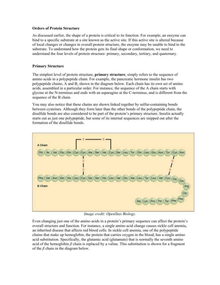
Orders of Protein Structure As discussed earlier, the shape of a protein is critical to its function. For example, an enzyme can bind to a specific substrate at a site known as the active site. If this active site is altered because of local changes or changes in overall protein structure, the enzyme may be unable to bind to the substrate. To understand how the protein gets its final shape or conformation, we need to understand the four levels of protein structure: primary, secondary, tertiary, and quaternary. Primary Structure The simplest level of protein structure, primary structure, simply refers to the sequence of amino acids in a polypeptide chain. For example, the pancreatic hormone insulin has two polypeptide chains, A and B, shown in the diagram below. Each chain has its own set of amino acids, assembled in a particular order. For instance, the sequence of the A chain starts with glycine at the N-terminus and ends with an asparagine at the C-terminus, and is different from the sequence of the B chain. You may also notice that these chains are shown linked together by sulfur-containing bonds between cysteines. Although they form later than the other bonds of the polypeptide chain, the disulfide bonds are also considered to be part of the protein’s primary structure. Insulin actually starts out as just one polypeptide, but some of its internal sequences are snipped out after the formation of the disulfide bonds. Image credit: OpenStax Biology. Even changing just one of the amino acids in a protein’s primary sequence can affect the protein’s overall structure and function. For instance, a single amino acid change causes sickle cell anemia, an inherited disease that affects red blood cells. In sickle cell anemia, one of the polypeptide chains that make up hemoglobin, the protein that carries oxygen in the blood, has a single amino acid substitution. Specifically, the glutamic acid (glutamate) that is normally the seventh amino acid of the hemoglobin β chain is replaced by a valine. This substitution is shown for a fragment of the β chain in the diagram below. Image credit: OpenStax Biology. What is most remarkable to consider is that a hemoglobin molecule is made up of two alpha chains and two beta chains, each consisting of about 150 amino acids, for a total of 600 amino acids in the whole protein. The difference between a normal hemoglobin molecule and a sickle cell molecule—which dramatically decreases life expectancy—is just one amino acid out of the 600. So, why should this small change have such a big effect? One reason is that valine and glutamic acid have side chains (R groups) with very different properties. Glutamic acid’s side chain is hydrophobic and bears a negative charge, while valine’s is hydrophilic and nonpolar. Thus, replacing a glutamic acid with a valine may be more likely to cause changes in function than, say, replacing glutamic acid with aspartic acid (which is negatively charged as well). In the case of sickle cell anemia, the amino acid change actually causes hemoglobin molecules to form long fibers that distort disc-shaped red blood cells into crescent shapes. These “sickled” cells, visible as small crescents in the blood sample below, get stuck as they attempt to pass through blood vessels. This impaired blood flow causes serious health problems, including breathlessness, dizziness, headaches, and abdominal pain, for sufferers of sickle cell disease. Credit: modification of work by Ed Uthman; scale-bar data from Matt Russell. Attribution: This article is a modified derivative of “Proteins,” by OpenStax Biology. Download the original article for free at http://cnx.org/contents/185cbf87-c72e-48f5-b51ef14f21b5eabd@9.85:13/Biology. Additional references: Raven, P. H., Johnson, G. B., Mason, K. A., Losos, J. B., and Singer, S. R. (2014). The chemical building blocks of life. In Biology (10 ed., AP ed., pp. 32-58). New York, NY: McGraw-Hill. th Reece, J. B., Urry, L. A., Cain, M. L., Wasserman, S. A., Minorsky, P. V., and Jackson, R. B. (2011). The structure and function of large biological molecules. In Campbell Biology (10 ed., pp. 66-91). San Francisco, CA: Pearson. th

