Supplementary Information
advertisement
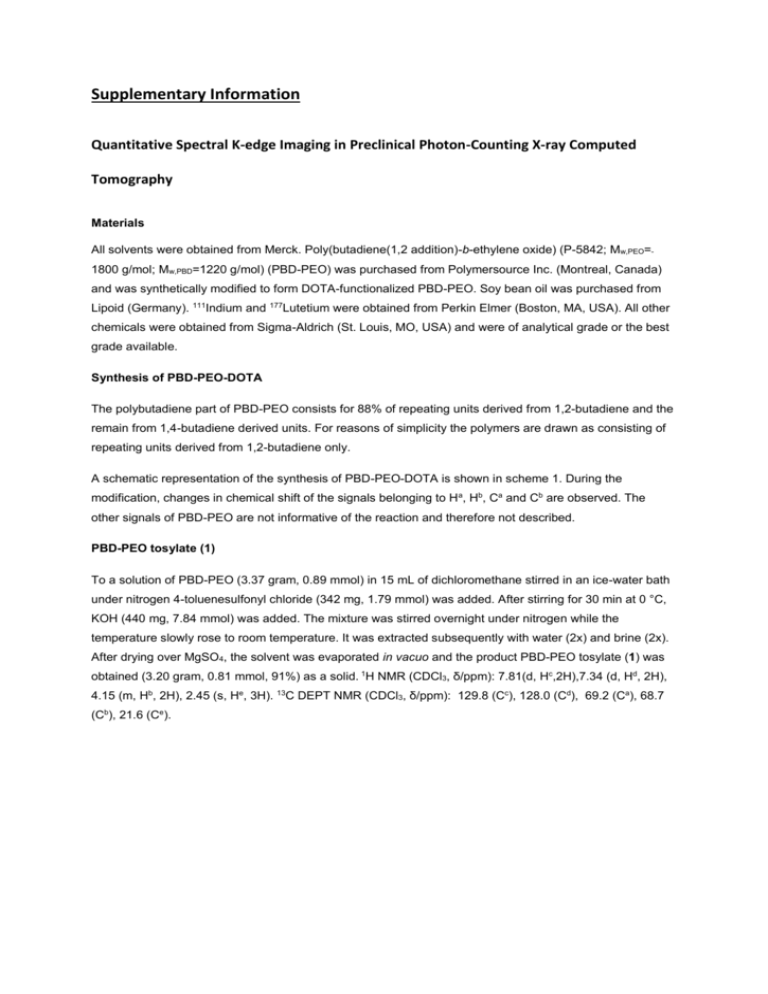
Supplementary Information Quantitative Spectral K-edge Imaging in Preclinical Photon-Counting X-ray Computed Tomography Materials All solvents were obtained from Merck. Poly(butadiene(1,2 addition)-b-ethylene oxide) (P-5842; Mw,PEO=1800 g/mol; Mw,PBD=1220 g/mol) (PBD-PEO) was purchased from Polymersource Inc. (Montreal, Canada) and was synthetically modified to form DOTA-functionalized PBD-PEO. Soy bean oil was purchased from Lipoid (Germany). 111Indium and 177Lutetium were obtained from Perkin Elmer (Boston, MA, USA). All other chemicals were obtained from Sigma-Aldrich (St. Louis, MO, USA) and were of analytical grade or the best grade available. Synthesis of PBD-PEO-DOTA The polybutadiene part of PBD-PEO consists for 88% of repeating units derived from 1,2-butadiene and the remain from 1,4-butadiene derived units. For reasons of simplicity the polymers are drawn as consisting of repeating units derived from 1,2-butadiene only. A schematic representation of the synthesis of PBD-PEO-DOTA is shown in scheme 1. During the modification, changes in chemical shift of the signals belonging to Ha, Hb, Ca and Cb are observed. The other signals of PBD-PEO are not informative of the reaction and therefore not described. PBD-PEO tosylate (1) To a solution of PBD-PEO (3.37 gram, 0.89 mmol) in 15 mL of dichloromethane stirred in an ice-water bath under nitrogen 4-toluenesulfonyl chloride (342 mg, 1.79 mmol) was added. After stirring for 30 min at 0 °C, KOH (440 mg, 7.84 mmol) was added. The mixture was stirred overnight under nitrogen while the temperature slowly rose to room temperature. It was extracted subsequently with water (2x) and brine (2x). After drying over MgSO4, the solvent was evaporated in vacuo and the product PBD-PEO tosylate (1) was obtained (3.20 gram, 0.81 mmol, 91%) as a solid. 1H NMR (CDCl3, δ/ppm): 7.81(d, Hc,2H),7.34 (d, Hd, 2H), 4.15 (m, Hb, 2H), 2.45 (s, He, 3H). 13C DEPT NMR (CDCl3, δ/ppm): 129.8 (Cc), 128.0 (Cd), 69.2 (Ca), 68.7 (Cb), 21.6 (Ce). Scheme 1. Synthesis of PBD-PEO-DOTA from PBD-PEO. PBD-PEO-NH2 (2) PBD-PEO tosylate (1) (3.20 gram, 0.81 mmol) dissolved in 12 mL toluene was divided into three ace pressure tubes (15 mL size) and ammonia in methanol (total 12 mL of 7N NH3 in MeOH, 84 mmol) was added. Closed tubes were heated at 50 °C for 63 hrs and after cooling down to room temperature the solvent was evaporated in vacuo. The residue was dissolved in DCM and was extracted subsequently with water (2x), brine(2x) and with a 10% NaHCO3 solution. After drying over MgSO4, the solvent was evaporated and the product pBd-PEO-NH2 (2) was obtained (1.19 gram, 0.31 mmol, 38%). 1H NMR (CDCl3, δ/ppm): 2.95-3.04 (Hb, 2H). 13C DEPT NMR (CDCl3, δ/ppm): , 41.3 (Cb) . PBD-PEO-DOTA (3) PBD-PEO-NH2 (2) (1.19 gram, 0.31 mmol) was dissolved in 12 mL dimethyl formamide, DOTA-mono-NHS ester (347 mg, 0.35 mmol) and triethylamine (0.9 mL, 6.47 mmol) were added. The mixture was stirred for 26 hrs at room temperature under nitrogen. The solvent was evaporated in vacuo and the product PBDPEO-DOTA (3) was obtained (1.36 gram, 0.30 mmol, 97%). A broad signal from 1.5-4.5 ppm was observed from the H-signals of the DOTA-moiety. 13C DEPT NMR (CDCl3, δ/ppm): The 13C signals of the DOTAmoiety could not be observed due to broadening of the peaks. Nanoparticle preparation and characterization Iodinated emulsions were prepared as previously described (23). The surfactant of the DOTAiodinated emulsions consisted of 5 mol% DOTA-PBD-PEO and 95 mol% of PBD-PEO. The hydrodynamic radius and polydispersity of the emulsions was determined using dynamic light scattering (DLS) (ALV/CGS-3 Compact Goniometer System, ALV-GmbH, Langen, Germany). Intensity correlation functions were measured at a scattering angle of θ=900 using a wavelength of 632.8 nm. Hydrodynamic radii and size distributions were derived from cumulant fits of the intensity autocorrelation function, applying the Stokes-Einstein relation. Cryogenic transmission electron microscopy (cryo-TEM) pictures were obtained with a FEI TECNAI F30ST electron microscope operated at an accelerating voltage of 300 kV in low dose mode. Samples for TEM were prepared by placing the emulsion (2 μL) on a 300-mesh carbon-coated copper grid and subsequently plungefreezing this grid into liquid ethane using a Vitrobot. The iodine concentration per sample was determined after Schöniger combustion using Inductively Coupled Plasma-Mass Spectrometry (ICPMS). Nanoparticles (32) were labelled with 111indium (111In) (Perkin Elmer, Boston, Ma, USA). In detail, 20 µL ammonium acetate buffer (2M NH4OAc, pH 4.5) was added to a 520 µL CT contrast agent solution together with 6 µL of 111In (60 MBq) and the pH was adjusted to 5.0. The sample was stirred at 60 °C for 60 min. Free DTPA (5 µL, 10 mM) was added to the reaction mixture for 15 min to scavenge free radionuclides. Labeling efficiency was checked on silica TLC using 200 mM EDTA as mobile phase and analyzed using a Phosphor Imager (FLA-7000, Fujifilm, Tokyo, Japan). Samples were purified using a PD-10 column (GE Healthcare). Pure fractions were collected and concentrated to its original concentration using a N2 flow. Radiochemical purities obtained were > 95% and the In content was measured using a -counting wizard (Wizard 1480 3" Wallac counter, Perkin Elmer, 111 Groningen, The Netherlands). After radioactive decay of 111In, the samples were analyzed using ICPMS to determine iodine concentrations.
