Cervical Evaluation Guide
advertisement
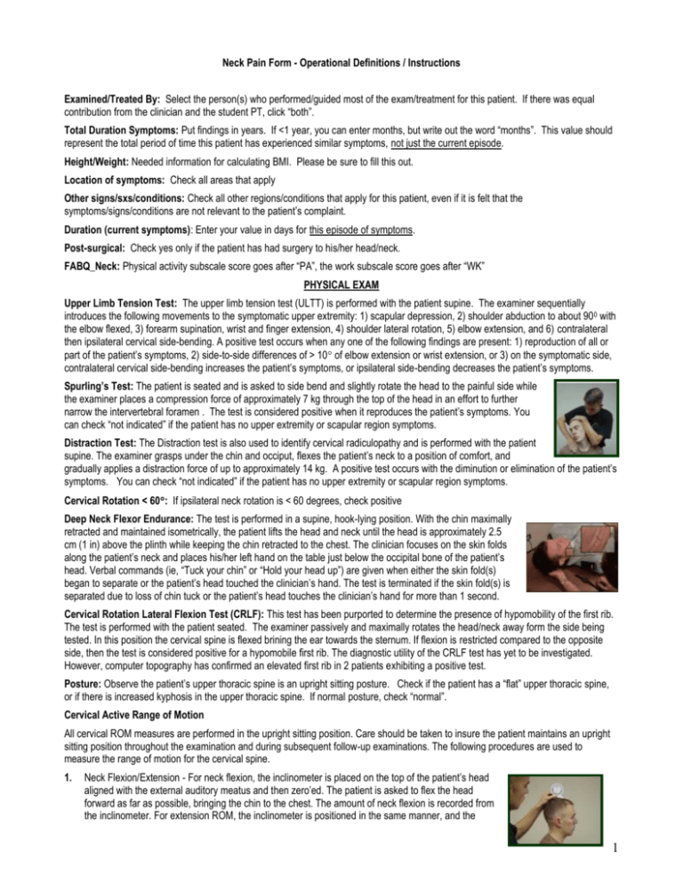
Neck Pain Form - Operational Definitions / Instructions Examined/Treated By: Select the person(s) who performed/guided most of the exam/treatment for this patient. If there was equal contribution from the clinician and the student PT, click “both”. Total Duration Symptoms: Put findings in years. If <1 year, you can enter months, but write out the word “months”. This value should represent the total period of time this patient has experienced similar symptoms, not just the current episode. Height/Weight: Needed information for calculating BMI. Please be sure to fill this out. Location of symptoms: Check all areas that apply Other signs/sxs/conditions: Check all other regions/conditions that apply for this patient, even if it is felt that the symptoms/signs/conditions are not relevant to the patient’s complaint. Duration (current symptoms): Enter your value in days for this episode of symptoms. Post-surgical: Check yes only if the patient has had surgery to his/her head/neck. FABQ_Neck: Physical activity subscale score goes after “PA”, the work subscale score goes after “WK” PHYSICAL EXAM Upper Limb Tension Test: The upper limb tension test (ULTT) is performed with the patient supine. The examiner sequentially introduces the following movements to the symptomatic upper extremity: 1) scapular depression, 2) shoulder abduction to about 90 0 with the elbow flexed, 3) forearm supination, wrist and finger extension, 4) shoulder lateral rotation, 5) elbow extension, and 6) contralateral then ipsilateral cervical side-bending. A positive test occurs when any one of the following findings are present: 1) reproduction of all or part of the patient’s symptoms, 2) side-to-side differences of > 10 of elbow extension or wrist extension, or 3) on the symptomatic side, contralateral cervical side-bending increases the patient’s symptoms, or ipsilateral side-bending decreases the patient’s symptoms. Spurling’s Test: The patient is seated and is asked to side bend and slightly rotate the head to the painful side while the examiner places a compression force of approximately 7 kg through the top of the head in an effort to further narrow the intervertebral foramen . The test is considered positive when it reproduces the patient’s symptoms. You can check “not indicated” if the patient has no upper extremity or scapular region symptoms. Distraction Test: The Distraction test is also used to identify cervical radiculopathy and is performed with the patient supine. The examiner grasps under the chin and occiput, flexes the patient’s neck to a position of comfort, and gradually applies a distraction force of up to approximately 14 kg. A positive test occurs with the diminution or elimination of the patient’s symptoms. You can check “not indicated” if the patient has no upper extremity or scapular region symptoms. Cervical Rotation < 60: If ipsilateral neck rotation is < 60 degrees, check positive Deep Neck Flexor Endurance: The test is performed in a supine, hook-lying position. With the chin maximally retracted and maintained isometrically, the patient lifts the head and neck until the head is approximately 2.5 cm (1 in) above the plinth while keeping the chin retracted to the chest. The clinician focuses on the skin folds along the patient’s neck and places his/her left hand on the table just below the occipital bone of the patient’s head. Verbal commands (ie, “Tuck your chin” or “Hold your head up”) are given when either the skin fold(s) began to separate or the patient’s head touched the clinician’s hand. The test is terminated if the skin fold(s) is separated due to loss of chin tuck or the patient’s head touches the clinician’s hand for more than 1 second. Cervical Rotation Lateral Flexion Test (CRLF): This test has been purported to determine the presence of hypomobility of the first rib. The test is performed with the patient seated. The examiner passively and maximally rotates the head/neck away form the side being tested. In this position the cervical spine is flexed brining the ear towards the sternum. If flexion is restricted compared to the opposite side, then the test is considered positive for a hypomobile first rib. The diagnostic utility of the CRLF test has yet to be investigated. However, computer topography has confirmed an elevated first rib in 2 patients exhibiting a positive test. Posture: Observe the patient’s upper thoracic spine is an upright sitting posture. Check if the patient has a “flat” upper thoracic spine, or if there is increased kyphosis in the upper thoracic spine. If normal posture, check “normal”. Cervical Active Range of Motion All cervical ROM measures are performed in the upright sitting position. Care should be taken to insure the patient maintains an upright sitting position throughout the examination and during subsequent follow-up examinations. The following procedures are used to measure the range of motion for the cervical spine. 1. Neck Flexion/Extension - For neck flexion, the inclinometer is placed on the top of the patient’s head aligned with the external auditory meatus and then zero’ed. The patient is asked to flex the head forward as far as possible, bringing the chin to the chest. The amount of neck flexion is recorded from the inclinometer. For extension ROM, the inclinometer is positioned in the same manner, and the 1 patient is asked to extend the neck backwards as far as possible. The amount of neck extension is recorded with the inclinometer. 2. Neck Side-Bending - The inclinometer is positioned in the frontal plane on the top of the patient’s head in alignment with the external auditory meatus. To measure right side-bending, the patient is asked to move the right ear to the right shoulder. The amount of side-bending is recorded with the inclinometer. The opposite is performed to measure left side-bending. Care should be taken to avoid concomitant rotation or flexion with the side-bending movement. 3. Neck Rotation –Rotation can be measured with a universal / standard goniometer. The patient is seated, looking directly forward with the neck a neutral position. The fulcrum of the goniometer is placed over the top of the head with the stationary arm aligned with the acromion process, and the moveable arm bisecting the patient’s nose. The patient is asked to rotate in each direction as far as possible. Similar to extension, cervical rotation may produce dizziness or nausea in patients with VBI. Centralizes: If you are able to centralize symptoms with either patient positioning or with repeated motions, check “centralizes”. If you are unable to centralize symptoms in the exam, check “cannot centralize”. If the patient has no upper extremity or scapular region symptoms, check “not applicable”. Note that centralization means that a movement, repeated movement, or position causes the pain and/or paresthesia to move from a distal to a more proximal area. NEUROLOGICAL EXAMINATION Note: You may check “not indicated” for dermatomes/myotomes/reflexes if the patient has no symptoms extending beyond the deltoid insertions bilaterally Dermatomes: Sensation is tested over key areas of dermatomes C5-T1 on each limb. After each limb is tested, the patient is asked; “Does that feel the same to you on each side?” If a difference is noted, the area should be explored further to map the extent of the sensory deficit. Results are recorded as normal or abnormal compared to the non-involved side. If a neuron exam is not indicated, check “not indicated”. Reflexes: The biceps brachii reflex tests the C5 nerve root. The reflex is tested by placing the patient’s arm in about 45 of flexion with the muscle relaxed. The examiner strikes the tendon in the cubital fossa, just proximal to its insertion. The thumb may be placed over the tendon to insure proper technique. The brachioradialis reflex primarily tests the C6 nerve root. The arm is positioned as for the biceps reflex. The examiner strikes the tendon at the distal aspect of the radius with the flat edge of the reflex hammer. The triceps reflex is used to test the C7 nerve root. The examiner supports the patient’s arm and strikes the triceps tendon just proximal to the olecranon. Each reflex is graded as Normal, Decreased, or Increased. Myotomes: Key muscles for each cervical nerve root (C5-T1) are tested. Each muscle test is graded as WNL or diminished. The examiner should also note if pain was produced during the muscle test. Muscle testing procedures are outlined in the table below. Key Muscles for MMT Dermatomal areas Key Muscle for MSRs C5 deltoid (shoulder in 90 abduction, resistance against lateral upper arm into adduction) Mid-deltoid biceps brachii (C5, C6) C6 biceps brachii (elbow at 90 flexion with forearm supinated, resistance against lower forearm into extension) radial aspect of 2nd metacarpal/ digit brachioradialis (C5, C6) extensor carpi radialis longus/brevis (wrist extended/ radially deviated with forearm pronated, resistance against dorsum of hand into flexion/ulnar deviation) C7 triceps (arm is placed overhead with elbow slightly flexed, resistance against forearm into flexion) dorsum of 3rd finger triceps (C7) flexor carpi radialis (wrist flexed/radially deviated with forearm supinated, resistance against thenar eminence into extension/ulnar deviation) C8 abductor pollicis brevis (thumb placed in abduction, resistance against proximal phalanx into adduction) medial aspect of 5th finger N/A T1 first dorsal interossei (index and middle finger are separated, resistance against the medial aspect of proximal phalanx of the index finger into adduction) medial forearm N/A *** The nerve root in bold is the primary nerve root assessed by the MSR. 2
