Methods of Polymer Molecular Weight Characterizations
advertisement

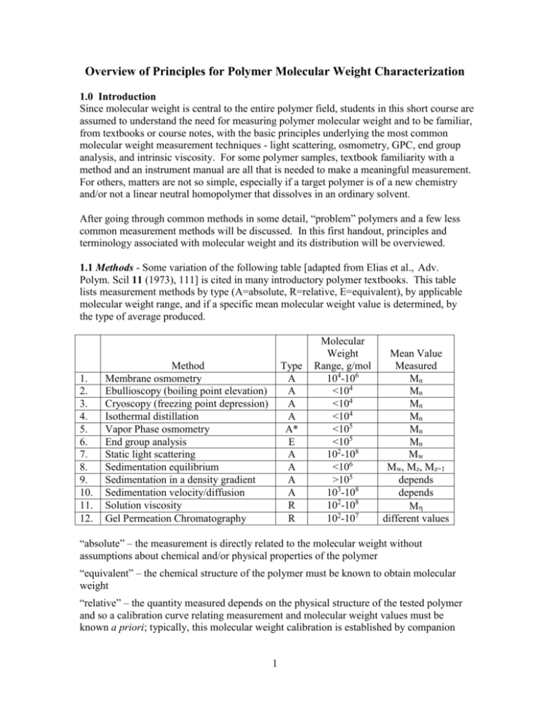
Overview of Principles for Polymer Molecular Weight Characterization 1.0 Introduction Since molecular weight is central to the entire polymer field, students in this short course are assumed to understand the need for measuring polymer molecular weight and to be familiar, from textbooks or course notes, with the basic principles underlying the most common molecular weight measurement techniques - light scattering, osmometry, GPC, end group analysis, and intrinsic viscosity. For some polymer samples, textbook familiarity with a method and an instrument manual are all that is needed to make a meaningful measurement. For others, matters are not so simple, especially if a target polymer is of a new chemistry and/or not a linear neutral homopolymer that dissolves in an ordinary solvent. After going through common methods in some detail, “problem” polymers and a few less common measurement methods will be discussed. In this first handout, principles and terminology associated with molecular weight and its distribution will be overviewed. 1.1 Methods - Some variation of the following table [adapted from Elias et al., Adv. Polym. Scil 11 (1973), 111] is cited in many introductory polymer textbooks. This table lists measurement methods by type (A=absolute, R=relative, E=equivalent), by applicable molecular weight range, and if a specific mean molecular weight value is determined, by the type of average produced. 1. 2. 3. 4. 5. 6. 7. 8. 9. 10. 11. 12. Method Membrane osmometry Ebullioscopy (boiling point elevation) Cryoscopy (freezing point depression) Isothermal distillation Vapor Phase osmometry End group analysis Static light scattering Sedimentation equilibrium Sedimentation in a density gradient Sedimentation velocity/diffusion Solution viscosity Gel Permeation Chromatography Type A A A A A* E A A A A R R Molecular Weight Range, g/mol 104-106 <104 <104 <104 <105 <105 102-108 <106 >105 103-108 102-108 102-107 Mean Value Measured Mn Mn Mn Mn Mn Mn Mw Mw, Mz, Mz=1 depends depends M different values “absolute” – the measurement is directly related to the molecular weight without assumptions about chemical and/or physical properties of the polymer “equivalent” – the chemical structure of the polymer must be known to obtain molecular weight “relative” – the quantity measured depends on the physical structure of the tested polymer and so a calibration curve relating measurement and molecular weight values must be known a priori; typically, this molecular weight calibration is established by companion 1 measurements on a series narrow molecular weight polydispersity standards of the same chemical and physical structure as the tested polymer I cannot vouch for the validity of this table, as I feel the authors specify molecular weight ranges that are overly broad; the table’s molecular weight limits seem to correspond to the most extreme values reported, not what is practical for the average polymer. Also, I deleted two rows from the original table, one for x-ray scattering and one for melt viscosity, neither of which would today be commonly considered a molecular weight measuring method. One might argue that any property sensitive to molecular weight could be used for molecular weight measurements assuming a theoretical or empirical expression for the molecular weight-property relationship is available. In practice, measurements must be robust (not unduly affected by polymer properties other than molecular weight), quick, cheap, and accurate. If only an average value of molecular weight is returned, the nature of the averaging process must be known. Methods based on melt viscosity and melt viscoelasticity are frequently used in industry to “index” molecular weight, even to discern the breadth of the molecular weight distribution, but such methods are not robust. They are applied empirically and can go much astray in the presence of unexpected impurities, chain branching, etc. “True” molecular weight measurements, excepting perhaps end group analysis, are practiced on dilute polymers, almost always dissolved in dilute solution, an environment in which deviations in properties from pure solvent reflect the behavior (or linearly added behaviors) of individual, isolated molecules and not the collective behavior of mutually interacting molecules. In addition to the methods listed, a huge number of techniques have been employed at one time or another to measure or infer molecular weight. Most of these methods don’t satisfy the criteria just listed, and at best, offer a molecular weight index, not a molecular weight value. A few such methods are: 1. membrane rejection [Higher molecular weight polymers don’t pass through welldefined pores of a single membrane or a stack of membranes while smaller molecular weight polymers do] 2. electron microscopy [(i) Individual polymers are deposited on a surface from a nonsolvent, or sprayed from such a solvent, and the size of the spherical single polymer “globules” then measured in the microscope. Knowing the polymer’s bulk density, a molecular weight can be calculated for each globule imaged. (ii) Individual polymers are deposited on a surface and imaged so that the chain contour can be measured], 3. AFM [(i) The two ends of a linear polymer are attached to the AFM tip and substrate, respectively, and the tip is then withdrawn until the polymer breaks. (ii). Individual polymers are deposited on a surface and imaged so that the chain contour is measurable.) 2 4. membrane translocation (The “blockage” time is measured as individual dissolved chains traverse the nanopores of a thin membrane.) 5. conformational relaxation time [Polymers are stretch/oriented in a field, and with the field subsequently switched off abruptly, recovery time is measured by a method such a flow or electric birefringence. The recovery time is coupled to molecular weight by a Rouse/Zimm chain description.) Although such esoteric methods are interesting from a theoretical perspective, few polymer scientists will ever practice them. There are, however, several practical methods not listed in the preceding table that I believe should be listed; some of these methods were developed since the table’s date, 1973, while others are older. I therefore offer a table of additions: 13. 14. 15. 16. 17. Method Electrophoresis (in gels, solutions) Mass spectrometry Diffusion coefficient Liquid chromatography Field flow fractionation Type E A R E A Molecular Weight Range, g/mol 102-109 <105 >103 <104 103-108 Mean Value Measured different values different values depends different values different values Indeed, I believe gel electrophoresis is the most commonly practiced method for measuring polymer molecular weight, albeit, not much in the context of many synthetic polymers. One can argue, for example, that DNA sequencing by electrophoresis is simply a polymer molecular weight separation with single repeat unit resolution. ”Counting up” from the peak for degree polymerization equal unity, the method become a molecular weight measurement that is “absolute” in terms of degree of polymerization Sequencing by gel and solution electrophoresis was the backbone of the Human Genome Project, a multibillion dollar activity. Many methods couple a separation by molecular weight or size (GPC, electrophoresis) with an absolute measurement of the molecular weight of the separated fractions (via light scattering, osmometry, intrinsic viscosity). The combined approach is termed a “hyphenated method”. In the early days of polymer science (<1965), the only way to measure the full molecular weight distribution of a synthetic polymer was to precipitate the material from solution with increasing concentrations of a nonsolvent, the precipitated fractions then examined in batch mode by an absolute method. This approach was exceedingly tedious. Today, the same basic concept is implemented as GPC-light scattering, the fractions created continuously by GPC monitored continuously for their molecular weight by light scattering. Field Flow Fractionation (FFF) is unique in that the FFF separation by molecular weight is so well defined that calibration is not needed; molecular weight can be theoretically calculated 3 directly from peak position. The difference of FFF from GPC or electrophoresis can be traced to the irregular, ill-characterized structure of the separation medium in the latter methods. Perhaps you can think of another method that should be added to the table. 1.2 Classification of Methods Calibration: The terms “absolute”, “relative”, and “equivalent” are not always distinct. GPC, for example, may be absolute or relative, depending on the method of calibration. Vapor phase osmometry, as presented in the method’s underlying theory, is an absolute method, but in actual practice, the instrument requires calibration with a standard compound of known molecular weight, making the method relative. Even an absolute method such as mass spectrometry is usually calibrated by molecular weight standards. Molecular Weight Range: Molecular weight ranges for each method are limited by constraints unique to that method. These limits will be discussed separately as methods are introduced. Molecular Weight Averages: The common molecular weight averages (Mn, Mw, Mz, Mz+1) are well understood by polymer students; they are associated with increasingly higher moments of the molecular weight distribution. However, students often do not grasp why different experimental methods are sensitive to different averages. 1.3 Why Different Methods Provide Different Molecular Weight Averages If the quantity measured by a given method directly manifests the number of polymer molecules - but not their molecular weights - this quantity can only be used to deduce Mn. As an example, consider vapor phase osmometry. In a thermodynamically ideal solution, vapor pressure is lowered by kT/V as each nonvolatile solute molecule of molecular mass M is added to volume V of volatile solvent. The lowered vapor pressure of N polymer solutes is thus NkT/V, a combination independent of M but dependent on N. Upon writing this product in terms of c, the mass concentration of the solutes, M merges as a measurable parameter, N c kT = kT V M so that c M = kT For the polydisperse molecular weight case, suppose that Ni polymers of molecular weight Mi are added to the solvent, each molecular weight fraction i in the mixture present at mass concentration ci. Only the total mass concentration c [=ci] is known and only 4 measured, the latter parameter summing contribution kT/V from each molecule irrespective of Mi, kT = Ni V Applying the formula previously analyzed for monodisperse M now yields ckT V ci Vci Ni Mi = = = = Mn Ni Ni Ni This is the “true” average molecular weight as the word “average” is used in nontechnical contexts. In essence, vapor phase osmometry allows a “count” of the number of molecules in a known mass of polymer. The same concept of molecular counting applies to all colligative property-based measurements (colligative properties are measured in osmometry, freezing point depression, boiling point elevation, etc.), which detect the solvent activity in the presence of solute. Contrarily, other methods manifest not just the number of dissolved molecules but also their molecular weight. Consider static light scattering. In the absence of optical interference, each polymer molecule dissolved in a fixed volume of solution contributes equally to the measured quantity, the reduced scattered intensity R (R=Rayleigh factor or ratio), R N c k = k V M where k is a molecular contrast factor (reflecting the optical contrast between polymer and solvent surroundings, the property ultimately responsible for all light scattering phenomena). Without inter- and intramolecular interference of scattered light, k is given by the Rayleigh scattering formula, 16 2 2no4 k 4 o where no is the solvent refractive index, o is the wavelength of incident light in vacuum, and is the molecular polarizability. If all subunits of a linear polymer (i.e., its repeat units) contribute equally to the polymer’s net polarizability, as expected for a homopolymer, is proportional to M: a longer polymer scatters more light than a shorter one. It then follows from the above formulas that the reduced scattered intensity R is proportional to the product of c and M. R=KcM 5 where the proportionality constant K is known as the optical constant; it has no dependence on M. By simple rearrangement, R M = Kc Contrasting vapor phase osmometry with light scattering, the contribution of individual molecules to the measured signals are quite distinct, N kT V vs. R N 2 M K V In light scattering, each molecule contributes to the overall measurement according to the square of its molecular weight. In osmometry, all molecules contribute equally, independently of molecular weight. Turning to the analogous polydisperse molecular weight case, and using the same notation as before, contributions to R by each molecular weight fraction simply add, R = KciMi The M formula for the monodisperse sample now yields R ci Mi Ni Mi2 = = = Mw Kc ci Ni Mi In essence, light scattering “sums” the product of the number of polymer molecules multiplied by the square of their molecular weight. One could naively imagine a measurement method exactly intermediate to the two just evaluated, i.e., a method based on a property sensitive to the product of N and M. This product, however, is simply the total mass of polymer; it could not be employed to calculate M. Because in vapor phase osmometry and similar techniques each molecule contributes to the overall measurement a constant, universal quantity independent of chemistry or structure, these techniques require, at least in theory, no calibration. As the previous discussion reveals, light scattering does require calibration, i.e., the value of K must be known to calculate M from R. K is typically obtained by measuring the refractive index increment dn/dc as polymer is added to solvent, and as a consequence, I would prefer the light scattering method instead be termed the light scattering-refractive index method. The standard formula for K is written, K = 4 2no2 o4 N A 6 2 dn dc This formula, offering K via measurement of dn/dc, is derived through an optical model that supposes a polymer consists of independent, identical, and isotropic scattering sites immersed an optically homogeneous medium of infinite extent. The scattering from such sites is then proportional to the square of the scattering site-solvent optical mismatch [~(dn/dc)2], while the contribution from such sites to solution refraction index is linearly proportional to the same quantity [~(dn/dc]. These assumptions, due to Debye, are far from obvious. The local symmetry of a polymer chain is cylindrical, not spherical (i.e., not isotropic); also, the interaction of light with a single scattering site could be influenced by neighboring scattering sites. If one represents the chain as an optically mismatched cylinder rather than a string of isotropic, optically independent scattering sites, a slightly different prefactor appears in the theoretical formula for K. The difference can be associated with depolarized light scattering, which fortunately, is usually small for high M polymers. Issues of the previous paragraph cast doubt on the categorization of light scattering as a absolute method of molecular weight determination. Calculated M depends on the adoption of a possibly imprecise optical model for the polymer chain. I would argue that the method is absolute, but subject to small correction. 1.4 Molecular Weight Distributions The number average is the arithmetic mean of the number fraction distribution, or stated more mathematically, it is this distribution’s first moment. The weight average is the mean of the weight fraction distribution, or more mathematically, the first moment of this distribution. The weight average is also equivalent to the second moment of the number fraction distribution. Surprisingly, despite the opposite belief of most polymer scientists, the number fraction distribution is not commonly reported. The normalized weight fraction wi of polymers of molecular weight Mi is given NiMi/NiMi; the function wi(Mi) [or the analogous continuous function w(M)] is the weight fraction distribution. When a technique such as GPC is used with a detector sensitive to the total mass concentration of repeat units (e.g., refractive index), the experimental result is wi(Mi). To get at the number fraction distribution xi(M) instead, one would have employ a GPC detector sensitive to the number concentration of molecules; on rare occasions, for example, osmometers have been used as GPC detectors (detector response by osmometers remains much too slow for routine use). Of course, each of these molecular weight distributions can be calculated from the other. Tablulated on the next page are real GPC data for a broad molecular weight distribution polystyrene sample. (Data taken from Yau, Kirkland, and Bly, Modern Size Exclusion Chromatography, 1979, Wiley & Sons, NY, sec. 10.3.) 7 Column variables are defined below: Columns 1-3 tabulate raw experimental quantities: Vi= elution volume of fraction i (preferred to elution time) hi = refractive index signal of fraction i (sensitive to the mass concentration of polystyrene) Mi = molecular weight derived from the Mi vs. Vi GPC calibration curve Columns 4-10 tabulate derived quantities: mi = normalized weight fraction eluting at Vi = hi/hi ni = normalized number fraction eluting at Vi = (hi/Mi)(hi/Mi) wi(Mi) = normalized weight fraction distribution wi = [(mi + mi+1)/2]/( Mi – Mi+1) Mi = [(Mi + Mi+1)/2] xi(Mi) = normalized number fraction distribution xi = [(ni + ni+1)/2]/( Mi – Mi+1) 1 2 Vi(ml) hi 20 21 22 23 24 25 26 27 28 29 30 31 32 33 34 35 36 37 38 39 40 0.0 0.0 0.8 3.5 16.8 42.4 67.9 81.5 81.4 71.0 57.0 43.0 30.0 19.0 12.2 9.0 4.0 2.6 2.0 1.0 0.0 3 Mi x10-6 (g/mol) 4.709 3.302 2.327 1.640 1.1555 0.8142 0.5738 0.4003 0.2821 0.1988 0.1410 0.09872 0.06887 0.04853 0.0342 0.0241 0.01698 0.01197 0.00843 0.00588 0.00414 4 5 mi ni 0.00 0.00 0.00147 0.00642 0.0308 0.0778 0.1244 0.1495 0.1494 0.1303 0.1046 0.0789 0.055 0.03486 0.02239 0.01651 0.00734 0.00477 0.00367 0.00183 0.00 0.00 0.00 0.0001 0.0005 0.0034 0.0121 0.0275 0.0474 0.672 0.0831 0.0947 0.1014 0.1014 0.0911 0.0830 0.0869 0.0548 0.0506 0.0552 0.0396 0.00 6 7 -6 miMix10 niMi x10-6 (g/mol) (g/mol) 0.0000 0.0000 0.0000 0.0000 0.0034 0.0002 0.0105 0.0008 0.0356 0.0039 0.0633 0.0099 0.0714 0.0158 0.0598 0.0190 0.0421 0.0189 0.0259 0.0165 0.0147 0.0133 0.0078 0.0100 0.0038 0.0070 0.0017 0.0044 0.0008 0.0028 0.0004 0.0021 0.0001 0.0009 0.0001 0.0006 0.000 0.0005 0.000 0.0002 0.000 0.0000 8 8 9 wi xi 0.000 0.0007 0.0057 0.0347 0.1591 0.4205 0.7893 1.2648 1.6783 2.000 2.2215 2.2429 2.2089 1.9979 1.9257 1.6756 1.2076 1.1955 1.0784 0.5260 0.0000 0.0001 0.0004 0.0040 0.0227 0.0824 0.2159 0.4848 0.9022 1.5145 2.3741 3.3970 4.7321 6.0747 8.4109 9.9508 10.5190 14.9858 18.5882 11.4451 10 Mi x10-6 (g/mol) 4.0055 2.8145 1.9835 1.3985 0.98485 0.694 0.48705 0.3412 0.24045 0.16945 0.11945 0.0838 0.0587 0.0414 0.02915 0.02054 0.0145 0.0102 0.0072 0.0050 Molecular weight averages are determined by summing columns 6 and 7: Mn = niMi = 126,900g/mol Mw = miMi = 341.400 g/mol PDI = Mw/Mn = 2.7 [Many GPC software packages calculate the z-average molecular weight Mz, defined and calculated here as Mz = (miMi2)/miMi = 595,300 g/mol This practice is fraught with error. For example, if the uncertainty in hi is 1% of the maximum peak height, a good signal-to-noise condition in GPC with refractive index detection, error in the calculated value of Mz is of the order 0.01(Mi2)/Mw. Here, this error estimate comes out to 1.1x10⁶ g/mol, twice the calculated value of Mz itself. The lesson is that the higher moments of any experimental distribution must be measured by a technique directly sensitive to the moments in question.] Consider ways that this molecular weight distribution might be graphically presented. Most commonly, the distribution is shown as the raw GPC trace, which is given next. A single, well-defined molecular weight peak is noted. [To define the molecular weight distribution better, there should be more data points spread over the peak; I didn’t want to do more hand calculations, so the table has only enough data to illustrate trends clearly.] 100 75 hi 50 25 0 0 10 20 30 40 50 Vi (ml) Despite this sample’s large PDI, the peak looks relatively narrow. This trace, termed the chromatogram, is by itself is pretty meaningless, since we don’t know how Mi and Vi are related: the poorer the molecular weight separation, for example, the narrower is the 9 chromatogram peak. Notwithstanding this trivial fact, raw GPC traces are frequently cited when arguing for low polydispersity. In this instance, the relationship between Mi and Vi is known by a calibration with polystyrene standards, so we can do much better than the chromatogram. Given below are the weight and number fraction distributions derived above. 20 15 x(M) w(M), x(M) 10 5 0 0.00 w(M) 0.25 0.50 0.75 1.00 1.25 M x10-6 (g/mol) These bear no resemblance to the raw trace, and the significant polydispersity of the sample is amply evident. Indeed, the x(M) curve shows that the most common polymer species has a molecular weight of 7,200 g/mol even though Mn and Mw both exceed 100,000 g/mol. Approximately 10% of the molecules by weight have molecular weights in excess of 800,000 g/mol, while 10% by weight have molecular weights less than 50,000 g/mol. The value of Mn falls slightly above the maximum of w(M), whereas the value of Mw lies well above this maximum, a fact commonly mentioned in introductory polymer texts. The difference between x(M) and w(M) simply manifests that it takes many small chains to balance the mass of a few larger chains. By number, the short chains dominate, while by weight, the converse is true. Surveying a whole year of Macromolecules, one is likely to never see a x(M) curve and just a handful of w(M) curves. In most polymer situations, greatest interest lies in the logarithmic spread of M. This is the case, for example, if we are interested in the power law exponents of polymer physics, where an increase from 10,000 g/mol to 100,000 g/mol leads to property changes comparable to those for an increase from 100, 000 g/mol to 1,000,000 g/mol. Unfortunately, the logarithmic spread of M is too often incorrectly displayed in a plot that simply switches the x-axis variable from M to log M, leaving the y-axis magnitude unchanged. [Unfortunately, my name is listed on a paper with such an error; I didn’t see the paper until it was too late to make a correction.] 10 To make the logarithmic plot properly, one must recognize that the plotted functions are distributions. Thus, the y-axis variable reflects the number of occurrences per unit change of x-axis variable. Here, for example, w(M)dM represents the number of chains with M in the range between M and M+dM. In logarithmic form, the y-axis variable must represent the number of chains with log M in the range between log M and log M+dlogM. The y-axis variable in a logarithmic plot of a distribution thus has a different functional form than the y-axis variable in a linear plot of this same distribution. To avoid the spontaneous creation of chains in converting plotting formats, the new y-axis variable y must be related to the old y-axis variable y by, y dx = y dlog(x) Solving, y = yx Thus, to present the weight fraction distribution in its logarithmic form, one must plot w(M)M vs. log M. Similarly, x(M), the number fraction distribution, must be plotted as x(M)M vs. log M. Don’t simply check the log(x) box in the plotting software! For the distribution of the example, these plots are given next. 0.5 w(M)M 0.4 0.3 0.2 x(M)M 0.1 0.0 0.0 0.0 0.1 1.0 M x10-6 (g/mol) The peak in the logarithmic weight fraction distribution is at 430,000 g/mol, well above Mw. This value nicely corresponds to the “peak molecular weight” Mp defined by the maximum of the GPC trace. When plotted in linear rather than logarithmic form, the peak is at 80,000 g/mol, which is quite different. Mp, although often used in GPC characterization, has no fundamental significance unless the distribution of molecular weights is very narrow. Loose definition of Mp creates confusion. This parameter usually represents the molecular weight position of the peak in the GPC trace. 11 Because the calibration curve in GPC offers an almost semi-logarithmic relationship between Vi and Mi, the function w(M)M, the weight fraction distribution in its logarithmic form, appears similar to a horizontal reflection of the original GPC trace. 1.5 Polydispersity Index and Narrow Molecular Weight Distributions In absence of the full molecular weight distribution, polydispersity is often assessed solely through the PDI, calculated from separate measurements of Mw and Mn (typically, osmometry and light scattering). As the following plot indicates, this practice has pitfalls. Three very different molecular weight distributions are displayed, corresponding to identical values of Mn, Mw, and Mz. Polymers made by living polymerizations often have relatively narrow molecular weight distributions; sometimes these polymers are even described as “monodisperse”. Most commercial polymer standards are said to have PDI values less than 1.10. How broad are these “model” distributions? The second figure of the next page illustrates three Poisson weight fraction distributions, the functional form predicted for an ideal living polymerization; actual living polymerization don’t quite meet the ideal and are broader. [from Yau et al., op. cit., p 12] 12 [from P. Munk and T. Aminabhavi, Introduction to Macromolecular Science, 2nd ed., p 89] For a Poisson distribution, the PDI is given Pn = 1 + Pw Pn Pn 12 where Pn and Pw are the number and weight average degrees of polymerization P. From this formula, the three PDI values are 1.08, 1.04, and 1.02. Such PDIs would be considered very low. Yet, the distributions are visually quite broad. The standard deviation of a Poisson distribution is equal to the square root of the mean. Therefore, with PDI=1.08 as in the first case, only ~65% of the polymers have P within 30% of the mean (1 std. dev.). Via an ideal polymerization, to get ~ 95% of the molecular weights within 5% of the mean ( 2 std. dev.) requires Pn in excess of 400. The corresponding PDI value would be 1.0025. For these reasons, even being generous, I would not consider a distribution “narrow” unless the PDI is less than 1.01. GPC can’t determine such low PDIs, so the method is not really an appropriate method to characterize the polydispersity of narrow distribution polymers. The synthetic polymer community often does not recognize this difficulty. 13 In distributions of everyday life (people’s weight, people’s height, test scores, etc.), distributions comparable to those of synthetic polymer molecular weight would be considered extremely broad. There is a clear lesson here: synthetic polymers never have true narrow molecular weight distributions, just distributions that are relatively narrow. On the other hand, biopolymers may possess PDI=1 if their sequence is genetically coded. 14


