Answer Key For Microscopy Quiz
advertisement
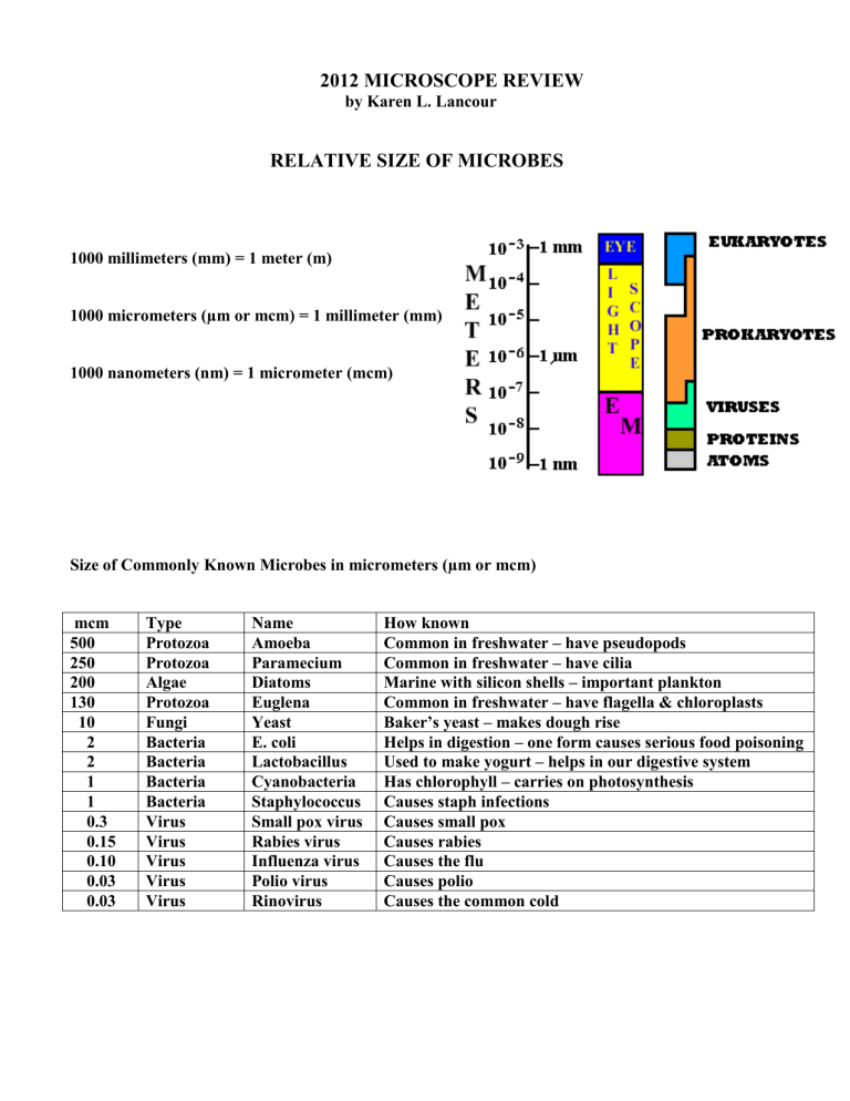
2012 MICROSCOPE REVIEW by Karen L. Lancour RELATIVE SIZE OF MICROBES 1000 millimeters (mm) = 1 meter (m) 1000 micrometers (µm or mcm) = 1 millimeter (mm) 1000 nanometers (nm) = 1 micrometer (mcm) Size of Commonly Known Microbes in micrometers (µm or mcm) mcm 500 250 200 130 10 2 2 1 1 0.3 0.15 0.10 0.03 0.03 Type Protozoa Protozoa Algae Protozoa Fungi Bacteria Bacteria Bacteria Bacteria Virus Virus Virus Virus Virus Name Amoeba Paramecium Diatoms Euglena Yeast E. coli Lactobacillus Cyanobacteria Staphylococcus Small pox virus Rabies virus Influenza virus Polio virus Rinovirus How known Common in freshwater – have pseudopods Common in freshwater – have cilia Marine with silicon shells – important plankton Common in freshwater – have flagella & chloroplasts Baker’s yeast – makes dough rise Helps in digestion – one form causes serious food poisoning Used to make yogurt – helps in our digestive system Has chlorophyll – carries on photosynthesis Causes staph infections Causes small pox Causes rabies Causes the flu Causes polio Causes the common cold TYPES OF MICROSCOPES Visible Light is the source of illumination: (up to 2000X) Bright Field Microscope (Lab – Light Compound Microscope) Most widely used Forms its image when light is transmitted through the specimen The specimen produces an image that is darker than the surrounding illuminated field Can be used with live, unstained and preserved, stain specimens Dark Field Microscope A bright-field microscope can be adapted to a dark-field microscope by adding a stop to the condenser The stop blocks all light from entering the objective lens except for peripheral light The specimen produces an image that is brightly illuminated against a dark field Effective for visualizing living cells that would be distorted by drying or heat or that can’t be stained with usual methods Does not allow for visualization of fine internal details of cells Phase Contrast Microscope Used for live specimens Specimen is contrasted against a gray background Excellent for internal cell details Differential - Interference Microscope Allows for detailed view of live, unstained specimens Includes two prisms that add contrasting colors to the image The image is colorful and three-dimensional Ultraviolet rays are the source of illumination: (up to 2000X) Fluorescence Microscope Includes a UV radiation source and a filter that protects the viewer’s eyes Used with dyes that show fluorescence under UV rays Forms a colored image against a black field Used in diagnosing infections caused by specific bacteria, protozoa, and viruses using fluorescent antibodies Confocal Microscope Allows for viewing cells at higher magnifications using a laser beam of light to scan various depths in the specimen Most often used on fluorescently stained specimens Electron beam forms image of specimen: Originally developed for studying nonbiological materials Biologists began using it in the early 1930s Forms an image with a beam of electrons o Electrons travel in wavelike patterns 1,000 times shorter than visible light waves o This increases the resolving power tremendously Magnification can be extremely high (between 5,000X and 1,000,000X for biological specimens) Allows scientists to view the finest structure of cells Transmission Electron Microscope - TEM (up to 100,000X) Often used to view structures of cells and viruses Electrons are transmitted through the specimen The specimen must be very thin (20-100 nm thick) and stained to increase image contrast dark areas of a TEM image represent thicker or denser parts TEM of HIV virus Scanning Electron Microscope - SEM (up to 650,000X) Creates an extremely detailed three-dimensional view of all kinds of objects Electrons bombard the surface of a whole metal-coated specimen Electrons deflected from the surface are picked up by a sophisticated detector The electron pattern is displayed as an image on a television screen Contours of specimens resolved with SEM are very revealing and surprising The images may be computer enhances to give them color SEM of HIV virus Note: Color can be added to the TEM & SEM images by computer MICROSCOPY REVIEW – Light Compound Microscope Parts of the microscope and their function: The number in front of each part of the microscope represents its number on the diagram. 1. ocular - magnifies the image formed by the objective. 2. nosepiece - holds the objectives. 3. objectives - lenses that receive the light from the field of view and form the first image. 4. stage - supports the slide and the specimen. 5. stage clips - hold the slide in place. 6. base - foundation which supports the scope & keeps it stable. 7. diaphragm - controls the amount of light reaching the specimen. 8. illuminator - source of light. 9. course adjustment - used for initial or low power adjustment. 10. fine adjustment - used for fine tuning & high power focusing. 11. arm - supports the ocular, objectives and body tube. 12. body tube - tube or barrel between the ocular and the objectives. PRINCIPLES OF MICROSCOPY A. Appearance of objects 1. Inverted and reversed (upside-down & backwards) 2. If an "e" is placed in the stage in its normal position, it will appear as an " ". 3. Only a thin layer of the specimen is in focus at any level (depth of focus). B. Movement of specimens 1. Actual movement is opposite to appeared direction of movement. 2. If an organism is actually moving ( ), it will appear to be moving ( ). C. Total magnification 1. Multiply ocular magnification times objective magnification. 2. Oculars are normally 10X or 12X. 3. Objectives are typically as follows: a. scanning power - 4X or 5X or 6X b. low power - 10X or 12X c. high power - 40X or 43X or 45X 4. Sample problem: If the ocular is 10X and the objective is 43X, the total magnification is 430. D. Changing objectives 1. When changing objectives from scanning power to lower power to high power, the following changes will occur: a. the size of the field of view decreases. b. the field of view is darker. c. the size of the image increases.. d. the resolution (ability to separate small details) increases. e. the working distance (distance between coverslip & objective) decreases. f. the depth of focus (thickness of the specimen which may be seen in focus) is reduced. 2. The relationships of magnification and the fields of view diameter and area ratios are approximately (10 X ocular and listed objective) objective scanning 5X low 10X high 40X total magnification 50 100 400 diameter 1/2 scanning 1/4 low area 1/4 scanning 1/16 low Principles of advanced microscopy A. Measuring the diameter of the field of view. 1. Place a transparent millimeter ruler on the stage, hold it down with the stage clips, and observe the ruler in the desired field of view - scanning or low. (See Fig. 1) 2. Focus on the metric edge of the ruler. Hint: applying gentle pressure to the free end of the ruler will help adjust for the thin ruler and allow for better focus. 3. Place the center of one millimeter marking at the left edge of the field of view (see Figure 2) and measure the diameter of the field in millimeters. For reference: Scanning power (50X) is about 3.0 to 3.2 mm and low power (100X) is about 1.5 to 1.6 mm. Sizes will vary with changes in magnification and manufacturer. 4. Convert the measurement in millimeters to micrometers by multiplying by 1000. Samples: low power field 1.6 mm X µm/mm = 1600 µm or mcm scanning power field 3.2 mm X 1000 µm/mm = 3200 µm or mcm 5. The diameter of the high power field is less than one millimeter. It can be calculated from the diameter of the low power field by using the following formula: high power field diameter = low power magnification lower power field diameter high power magnification Sample: h.p. field = 100 1600 µm 400 to h.p. field = 100 X 1600 = 400 µm or mcm 400 B. Estimating the size of an object. 1. Determine the number of cells or objects that would fit across the diameter of the field of view. 2. Estimate the size of the object by dividing the diameter of the field by the number of the objects that would fit across the field. Sample: a. diameter of the field is 2 mm x 1000 µm/mm = 2000 µm b. length of cell "x" is 2000 µm/ 3 cells = 660 µm/cell DEPTH OF FOCUS EXERCISE Below are four objects located between a slide and a coverslip. The actual objects would of course be 3-D. The objects are left to right; a cone, a cylinder, a sphere and a cube. As one focuses down through the various levels a two dimensional representation will be visible. At each level (represented by the dotted lines to the slide diagram) draw what two dimensional shapes would be present and give their proper location on the slide diagram. ANSWER KEY FOR DEPTH OF FOCUS EXERCISE Below are four objects located between a slide and a coverslip. The actual objects would of course be 3-D. The objects are left to right; a cone, a cylinder, a sphere and a cube. As one focuses down through the various levels a two dimensional representation will be visible. At each level (represented by the dotted lines to the slide diagram) draw what two dimensional shapes would be present and give their proper location on the slide diagram. MICROSCOPY QUIZ I. PARTS OF THE MICROSCOPE: For each of the following parts of the microscope, give the letter representing it's function and the number representing it's location. PART ocular coarse adjustment fine adjustment arm nosepiece objectives stage stage clips diaphragm illuminator base FUNCTION A. B. C. D. E. F. G. H. I. J. K. holds slide in place foundation to keep scope stable controls the amount of light to specimen supports slide and specimen lens that form initial image of specimen holds objectives - allows changing power used for initial & low power focusing supports ocular, objectives & body tube source of light magnifies image formed by objective used for fine tuning & high power focusing LOCATION II. MICROSCOPY SKILLS: 1. A student prepares a slide of the letter "d" and positions the slide on the stage of the microscope so the letter is in the normal reading position. When viewed through the microscope, the image of the letter will appear as A. d B. b C. q D. p 2. An organism viewed under the microscope appears to be moving " ". The organism is actually moving A. B. C. D. 3. A student observes a specimen under high (400X) power and then switches back to low (100X) power. How will the appearance of the image change when going from high power to low power? A. larger and darker B. smaller and darker C. smaller and brighter D. larger and brighter 4. A microscope is equipped with a 10X ocular and two objectives - one is 10X and the other is 43X. What is the highest total magnification possible with this microscope? Questions 5 - 9 are based upon the following diagram: 5. The diagram represents a field of view through a compound light microscope. What is the diameter of the field of view in millimeters (mm)? 6. What is the diameter of this field of view in micrometers (µm)? 7. What is the approximate length of the organism in micrometers ( µm)? 8. This diagram represents the field of view under low power with a total magnification of 100X. If the high power field is 400X, what would be the diameter of the high power field in micrometers? 9. When you switch from low power to high power, what happens to depth of focus? A. it will be greater B. it will be less C. it will remain the same D. it will be nonexistent ANSWER KEY FOR MICROSCOPY QUIZ I. PARTS OF THE MICROSCOPE: For each of the following parts of the microscope, give the letter representing it's function and the number representing it's location. PART 1. J ocular 9. G coarse adjustment 10.K fine adjustment 11.H arm 2. F nosepiece 3. E objective 4. D stage 5. A stage clips 7. C diaphragm 8. I illuminator 6. B base FUNCTION A. holds slide in place B. foundation to keep scope stable C. controls the amount of light to specimen D. supports slide and specimen E. lens that forms initial image of specimen F. holds objectives - allows changing power G. used for initial & low power focusing H. supports ocular, objectives & body tube I. source of light J. magnifies image formed by objective K. used for fine tuning & high power focusing LOCATION II. MICROSCOPY SKILLS: D 1. A student prepares a slide of the letter " d " and positions the slide on the stage of the microscope so the letter is in the normal reading position. When viewed through the microscope, the image of the letter will appear as A. d B. b C. q D. p B 2. An organism viewed under the microscope appears to be moving " ". The organism is actually moving A. B. C. D. C 3. A student observes a specimen under high (400X) power and then switches back to low (100X) power. How will the appearance of the image change when going from high power to low power? A. larger and darker B. smaller and darker C. smaller and brighter D. larger and brighter 430 4. A microscope is equipped with a 10X ocular and two objectives - one is 10X and the other is 43X. What is the highest total magnification possible with this microscope? Questions 5 - 9 are based upon the following diagram: 1.4 - 1.5 mm 5. The diagram represents a field of view through a compound light microscope. What is the diameter of the field of view in millimeters (mm)? 1400-1500 µm 6. What is the diameter of this field of view in micrometers ( µm)? 600 µm 7. What is the approximate length of the organism in micrometers? 350-375 µm 8. This diagram represents the field of view under low power with a total magnification of 100X. If the high power field is 400X, what would be the diameter of the high power field in micrometers? B 9. When you switch from low power to high power, what happens to depth of focus? A. it will be greater B. it will be less C. it will remain the same D. it will be nonexistent


