Pediatric Respiratory Distress Simulation Scenario
advertisement
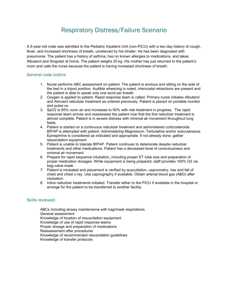
Respiratory Distress/Failure Scenario A 6-year-old male was admitted to the Pediatric Inpatient Unit (non-PICU) with a two day history of cough, fever, and increased shortness of breath, unrelieved by his inhaler. He has been diagnosed with pneumonia. The patient has a history of asthma, has no known allergies to medications, and takes Albuterol and Singulair at home. The patient weighs 20 kg. His mother has just returned to the patient’s room and calls the nurse because the patient is having increased shortness of breath. General code outline 1. Nurse performs ABC assessment on patient. The patient is anxious and sitting on the side of the bed in a tripod position. Audible wheezing is noted, intercostal retractions are present and the patient is able to speak only one word per breath. 2. Oxygen is applied to patient. Rapid response team is called. Primary nurse initiates Albuterol and Atrovent nebulizer treatment as ordered previously. Patient is placed on portable monitor and pulse-ox. 3. SpO2 is 85% room air and increases to 90% with neb treatment in progress. The rapid response team arrives and reassesses the patient now that the first nebulizer treatment is almost complete. Patient is in severe distress with minimal air movement throughout lung fields. 4. Patient is started on a continuous nebulizer treatment and administered corticosteroids. BIPAP is attempted with patient. Administering Magnesium, Terbutaline and/or subcutaneous Epinephrine is considered as indicated and appropriate. If not already done, gather resuscitation equipment. 5. Patient is unable to tolerate BIPAP. Patient continues to deteriorate despite nebulizer treatments and other medications. Patient has a decreased level of consciousness and minimal air movement. 6. Prepare for rapid sequence intubation, including proper ET tube size and preparation of proper medication dosages. While equipment is being prepared, staff provides 100% O2 via bag-valve-mask. 7. Patient is intubated and placement is verified by auscultation, capnometry, rise and fall of chest and chest x-ray. Use capnography if available. Obtain arterial blood gas (ABG) after intubation. 8. Inline nebulizer treatments initiated. Transfer either to the PICU if available in the hospital or arrange for the patient to be transferred to another facility. Skills reviewed: ABCs including airway maintenance with bag/mask respirations General assessment Knowledge of location of resuscitation equipment Knowledge of use of rapid response teams Proper dosage and preparation of medications Reassessment after procedures Knowledge of recommended resuscitation guidelines Knowledge of transfer protocols Event/Assessment Action Required Mother calls nurse into room because patient is having increased respiratory distress. Nurse performs assessment including ABCs. The patient is anxious and sitting on the side of the bed in a tripod position. Audible wheezing is noted, intercostal retractions are present and the patient is able to speak only one word per breath. Oxygen applied to patient. Patient placed on portable monitor and pulse-ox. Rapid response team is called. Primary nurse initiates Albuterol and Atrovent nebulizer treatment as ordered previously. Rapid response team arrives and reassesses the patient now that first neb treatment is almost completed. SpO2 is 85% room air and increases to 90% with neb treatment in progress. Patient with diminished breath sounds bilaterally, increased work of breathing, intercostal retractions. Patient is started on a continuous nebulizer treatment and administered corticosteroids. Attempt CPAP or BIPAP. Consider administering Magnesium, Terbutaline and/or subcutaneous Epinephrine as indicated and appropriate. If utilized, participants should calculate and prepare medications. Patient is unable to tolerate BIPAP. Patient continues to deteriorate despite nebulizer treatments and other medications. Patient has decreased level of consciousness, minimal air movement and tracheal tugging. Prepare for rapid sequence intubation. Participants should prepare all equipment needed for intubation and calculate and prepare medication dosages. While equipment is being prepared, staff provides 100% O2 via bag-valve-mask. Patient is intubated. Verify placement by visualizing the ETT passing through the cords, auscultation of lung sounds bilaterally, the absence of gastric sounds, capnometry, equal rise and fall of the chest and chest x-ray. Obtain ABG after intubation. Use capnography if available. Placement of the ETT is verified by equal but diminished breath sounds with expiratory wheezes throughout, colorimetric end tidal CO2 detector, and equal rise and fall of the chest. Chest x-ray shows placement of ETT is appropriate. Inline nebulizer treatments initiated. Transfer either to the PICU if available in the hospital or arrange for the patient to be transferred to another facility.
