comparison of surgical techniques for pterygium using
advertisement
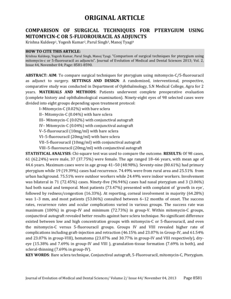
ORIGINAL ARTICLE COMPARISON OF SURGICAL TECHNIQUES FOR PTERYGIUM USING MITOMYCIN-C OR 5-FLUOROURACIL AS ADJUNCTS Krishna Kuldeep1, Yogesh Kumar2, Parul Singh3, Manoj Tyagi4 HOW TO CITE THIS ARTICLE: Krishna Kuldeep, Yogesh Kumar, Parul Singh, Manoj Tyagi. “Comparison of surgical techniques for pterygium using mitomycin-c or 5-fluorouracil as adjuncts”. Journal of Evolution of Medical and Dental Sciences 2013; Vol. 2, Issue 44, November 04; Page: 8581-8590. ABSTRACT: AIM: To compare surgical techniques for pterygium using mitomycin-C/5-fluorouracil as adjunct to surgery. SETTINGS AND DESIGN: A randomized, interventional, prospective, comparative study was conducted in Department of Ophthalmology, S.N Medical College, Agra for 2 years. MATERIALS AND METHODS: Patients underwent complete preoperative evaluation (complete history and ophthalmological examination). Ninety-eight eyes of 98 selected cases were divided into eight groups depending upon treatment protocol: I–Mitomycin-C (0.02%) with bare sclera II– Mitomycin-C (0.04%) with bare sclera III– Mitomycin-C (0.02%) with conjunctival autograft IV– Mitomycin-C (0.04%) with conjunctival autograft V–5-fluorouracil (10mg/ml) with bare sclera VI–5-fluorouracil (20mg/ml) with bare sclera VII–5-fluorouracil (10mg/ml) with conjunctival autograft VIII–5-fluorouracil (20mg/ml) with conjunctival autograft STATISTICAL ANALYSIS: Chi-square test was used to compare the outcome. RESULTS: Of 98 cases, 61 (62.24%) were male, 37 (37.75%) were female. The age ranged 18‒66 years, with mean age of 44.6 years. Maximum cases were in age group 41‒50 (48.98%). Seventy-nine (80.61%) had primary pterygium while 19 (19.39%) cases had recurrence. 74.49% were from rural area and 25.51% from urban background. 75.51% were outdoor workers while 24.49% were indoor workers. Involvement was bilateral in 71 (72.45%) cases. Ninety-five (96.94%) cases had nasal pterygium and 3 (3.06%) had both nasal and temporal. Most patients (73.47%) presented with complaint of ʿgrowth in eyeʾ, followed by redness/congestion (16.33%). At reporting, corneal involvement in majority (64.28%) was 1–3 mm, and most patients (53.06%) consulted between 6–12 months of onset. The success rates, recurrence rates and ocular complications varied in various groups. The success rate was maximum (100%) in group-IV and minimum (72.73%) in group-V. Within mitomycin-C groups, conjunctival autograft revealed better results against bare sclera technique. No significant difference existed between low and high concentration groups with mitomycin-C or 5-fluorouracil, and even the mitomycin-C versus 5-fluorouracil groups. Groups IV and VIII revealed higher rate of complications including graft-injection and retraction (46.15% and 23.07% in Group-IV, and 61.54% and 23.07% in group-VIII), hematoma (23.07% and 30.77% in group-IV and VIII respectively), dryeye (15.38% and 7.69% in group-IV and VIII ), granulation-tissue formation (7.69% in both), and scleral-thinning (7.69% in group-IV). KEY WORDS: Bare sclera technique, Conjunctival autograft, 5-Fluorouracil, mitomycin-C, Pterygium. Journal of Evolution of Medical and Dental Sciences/ Volume 2/ Issue 44/ November 04, 2013 Page 8581 ORIGINAL ARTICLE INTRODUCTION: The word pterygium is derived from a Latin word ʿPteronʾ meaning a wing. It is a horizontally-oriented, triangular, wing-shaped fibrovascular growth onto the cornea that is continuous with the conjunctiva. Pterygia are benign lesions that can be found on either side of the cornea but more often nasally.[1] A fully developed pterygium consists of three parts: head, neck and the body. The exact cause of pterygium is not well understood. However, long-term exposure to sunlight, especially ultraviolet rays and chronic eye irritation from dry, dusty conditions seem to play an important role.[2,3] Pterygium is an asymptomatic condition in the early stages except for cosmetic intolerance. However, these lesions may induce astigmatism, chronic irritation, and impaired vision. Large and thick pterygium may cause diplopia owing to limitation of abduction and adduction of eyeball.[4,5] A small pterygium with mild symptoms of photophobia and redness can often be managed with the use of topical preservative-free lubricants, vasoconstrictors and a mild steroid to prevent progression. Even the use of ultraviolet-blocking spectacles has been advocated.[4] The decision to remove a pterygium depends on the patient's willingness to tolerate symptoms and interest in cosmetic improvement. Although, the surgical treatment of pterygium is more than 3000 years old, number of techniques have been reported which include simple excision, simple excision with bare sclera, transposition of head of pterygium, mucous membrane grafting, lamellar keratoplasty, limbal autograft and use of adjunctive therapy which includes use of various drugs (e.g topical corticosteroids, thiotepa, mitomycin-C), beta-radiation, argon laser and excimer laser etc.[4,5] Despite several techniques for the treatment of pterygium, recurrence still remains an ophthalmic enigma. Recently, use of mitomycin-C (MMC)/5-fluorouracil (5-FU) after the excision of pterygium has been popularized to reduce the recurrence rate significantly for primary as well as recurrent pterygium. The present study was an attempt to find out easier, safe and effective approach to come out of the problem. Mitomycin-C/5-fluorouracil was applied at the stump of excised pterygium to prevent its recurrence. MATERIALS AND METHODS: The present study was conducted in the Department of Ophthalmology, S.N Medical College, Agra for 2 years (January 2004‒December 2005). Inclusion criteria: The cases diagnosed with pterygium reaching beyond the limbus and involving the cornea Progressive pterygia characterized by vascular engorgement and progressive growth, primary or recurrent, causing visual or cosmetic problems or chronic irritation Patients convinced for treatment and given written consent Exclusion criteria: Atrophic pterygium Cases of recurrent pterygium with previous use of the same drugs Patients having ocular infection, other ocular surface diseases, and dry eye were excluded from the study Patients who lost follow up Group distribution: Ninety-eight eyes of 98 selected cases were divided randomly into eight groups (Gps) depending upon treatment protocol.[Table 2] Pre-operative evaluation: Detailed history of each selected case was taken and they were subjected to local examination with special reference to vision, refraction, keratometry, and slit lamp Journal of Evolution of Medical and Dental Sciences/ Volume 2/ Issue 44/ November 04, 2013 Page 8582 ORIGINAL ARTICLE examination to know the size and grade of pterygium, horizontal extent, depth of involvement, vascularity of pterygium, Stocker’s line and pupillary encroachment. Patients with recurrent pterygium were looked for ocular motility, diplopia, symblepharon, condition of fornices and availability of conjunctival autograft. Tests for dry eye (Schirmer’s test and tear film break-up time) were done. Written informed consent was taken. Pre-operative medication: Topical antibiotic eye drops were instilled in the eye four times a day (q.i.d) for a minimum period of 24 hours. Surgical procedure: Cases selected for study were subjected to surgical excision followed by application of either MMC or 5FU with/without conjunctival autograft (CA) transplantation. After cleaning with povidone iodine solution and draping the eye, the palpebral fissure was widened using eye speculum. Topical anaesthesia was achieved by 4% xylocaine drops, and 2% xylocaine with adrenaline was injected in the body of pterygium. The head of pterygium was removed from the cornea by doing partial keratectomy with disposable blade. The subconjunctival pterygial tissue was separated from sclera, 5-6mm from the limbus and was excised along with 4-5mm of overlying conjunctiva. MMC (0.02 or 0.04%)/5FU (10mg/ml or 20mg/ml) soaked sponge was applied on the bare sclera for 2 minutes and washed vigorously with ringer lactate to remove excess amount. In groups I, II, V, VI bare sclera technique was used. In groups III, IV, VII, and VIII, desired size of conjunctival graft measured with caliper was taken from superotemporal bulbar conjunctiva and placed over the bare sclera and was sutured with adjoining conjunctiva by four to six interrupted sutures using 10-0 monofilament nylon suture. Eye was bandaged after application of antibiotic and steroid eye ointment. Post-operative management Oral antibiotics, anti-inflammatory and multivitamin-antioxidant preparations were given. Topical antibiotic with steroid eye drops q.i.d and lubricant eye drops q.i.d prescribed for 4– 6 weeks. Follow-up visits were scheduled for day 1,7,15 and 30 post-operatively, and then every month till minimum of ten months. In each post-operative visit, any complaint of pain, irritation, photophobia, redness, watering was asked and visual acuity, movement of eyeball, slit-lamp examination of cornea and conjunctiva to examine the graft were done. The complications such as graft injection, hematoma, graft necrosis, graft retraction, granulation tissue, symblepharon and recurrence were looked for. Recurrence was considered when fibrovascular tissue was found encroaching through the limbus onto the clear cornea in the area of previous pterygium excision. Statistical analysis: Chi-square test was used for comparison of outcome between various groups. Journal of Evolution of Medical and Dental Sciences/ Volume 2/ Issue 44/ November 04, 2013 Page 8583 ORIGINAL ARTICLE RESULTS: Out of all the outdoor patients who visited the ophthalmic department in the span of 2 years (January 2004- December 2005), 651 were diagnosed with pterygium, and 98 patients were selected and have given consent for the procedure. Complete clinico-demographic profile has been tabulated.[Table 1] The success rates, recurrence rates and ocular complications varied in various groups. The maximum success rate was observed in group IV (100%), while groups IV and VIII revealed higher rate of complications.[Table 2, 4] DISCUSSION: India is one such country where pterygium is of common occurrence, and despite several techniques for the treatment of pterygium; recurrence still remains the most enigmatic complication. Although the last word in the surgery for pterygium which can bring down the postoperative recurrence of pterygium to zero-percent is yet to come, an effort to find an effective surgical (adjuvant) method for the management of pterygium which may reduce its recurrence has been made. The efficacy and complications of MMC and 5-FU in cases of primary as well as recurrent pterygium were studied. Pterygia are reported to occur more frequently in males as compared to females [4], though the selected group in a study by Altay et al has more number of females probably seeking treatment for cosmetic reasons.[5] Although the prevalence of the lesion is known to increase with age, the highest incidence occurs between the ages of 20 and 49. Patients younger than the age of 15 rarely acquire a pterygium.[4] In the present study, males outnumbered females as latter are least involved in outdoor activities, and majority of cases belonged to middle age group ranging 41-50 years (48.98%) followed by 31-40 years (28.57%).[Table 1] In our study, only one case (1.02%), aged 18, was found below the age of 20 years. Maximum patients (75.51%) were farmers, laborers and rickshaw pullers etc. i.e., those who were having more outdoor activities, and hence were constantly exposed to atmospheric irritants.[Table 1] Almost 74.49% patients were from rural area and 25.51% were from urban background [Table 1], which is in concurrence with other studies. [4,6] Climatic conditions such as humid weather, dry, hot and dusty atmosphere, wind, sunlight especially ultraviolet light have been claimed to have a definite role in pterygium formation.[3,4,6,7,8] Pterygium may affect one or both the eyes in same individual. Simultaneous bilateral growth is reported to occur in 4%‒5%, although sequential bilateral ocular involvement may occur in up to one-third of patients with pterygia.[9] In this study, bilateral eye involvement (72.45%) was more common than unilateral (27.55%), probably because most patients seek treatment later. Moreover, it was observed that the patients felt problem only in one eye while in the other eye they were symptom-free, probably because the pterygium was in the initial stage of development. In 96.94% cases, there was only nasal involvement slightly below the horizontal meridian, and in 3.06% cases, it was nasal as well as temporal [Table 1], although the temporal lesion had developed later. A temporal pterygium without nasal is rarity, although occasional case has been reported. [10] No case was detected having only the temporal involvement. The higher nasal involvement in this study is in conformity with the findings of D’ Ombrains[7], who pointed out the anatomical configuration of medial side of palpebral aperture as cul-de-sac and physiological process of scavenging the dust on its lateral side by fresh tears as the possible explanation. A recent publication suggests a fiberoptic type of transmission of ultraviolet light from the temporal side of the cornea, through the stroma Journal of Evolution of Medical and Dental Sciences/ Volume 2/ Issue 44/ November 04, 2013 Page 8584 ORIGINAL ARTICLE onto the nasal aspect of the eye, perhaps partially explaining why these lesions are more commonly found nasally.[11] The majority of cases in the present study reported when pterygium has involved the cornea; 64.28% having corneal involvement of 1–3 mm. Reporting time of patients from onset varied from 3 months–2 year, with most of the cases presenting within 6 month to 1 year.[Table 1] Patients of pterygium may present with varying complaints; symptoms of burning, irritation, lacrimation, and foreign body sensation may accompany the growth of a pterygium onto the cornea. Significant astigmatism may be induced either with or against the rule as sectoral corneal steepening occurs. The astigmatism is often irregular and may lead to decreased vision.[4] In the present study, the commonest complaint was presence of growth (73.47%) followed by redness, watering and irritation of eye.[Table 1] Only four cases had diminution of vision due to error of astigmatism. When it encroaches the pupillary area, it may obstruct the peripheral and even central field of vision, but it was not found in the present study in the selected group. Approach to mild symptoms can be conservative, but the main treatment of pterygium is surgical excision. However, the recurrence of pterygium after its surgical excision is a challenge for years. The commonest cause of recurrence is vascularization, and fibroblastic and epithelial proliferation. The fleshiness of the pterygium is a significant risk factor for recurrence. 50% to 97% recurrences occur in 4 –12 months period respectively following surgery.[2] Simple excision of the pterygium alone has a very high rate of recurrence, about 30–70%. Even conjunctival autografting to cover the bare sclera is associated with recurrence rates of 2%–39%.[3] Recently MMC/5-FU after the excision of pterygium has been popularized to reduce the recurrence rate significantly for primary as well as recurrent pterygium. It also prevents the mechanical extraocular movement restrictive abnormalities caused by conjunctival and tenon’s tissue scarring after simple pterygial removal. Mitomycin C (MMC) is an antibiotic isolated from Streptomyces caespitosus. It selectively inhibits DNA, RNA and protein synthesis, and has antineoplastic action with radiomimetic properties. Adjunctive MMC in pterygium surgery was first described in Japan by Kunitomo and Mori in1963.[3] Its effects on local fibroblast appear to be very long lived and perhaps irreversible. Recurrence rates by the use of intraoperative and postoperative MMC were between 0 to 38%.[5] The concentration of intraoperative MMC application used in most of the studies range from 0.01 to 0.04%, with 0.02% applied for 3 min being the commonest dosage used.[12] Among our study groups (I-IV), in which MMC was used the recurrence rates were significantly lesser (χ2 =5.14, p<0.05) in the groups III and IV with conjunctival autograft (CA) against groups I and II with the bare sclera technique. The difference in the recurrence rates was not statistically significant when concentration of MMC varied from 0.02% to 0.04% [Gp I vs Gp II (χ2 =0.08, p>0.05), and Gp III vs Gp IV (χ2 =0.90, p>0.05)], although the time for appearance of recurrence increased slightly with the use of higher concentration.[Table 2] Moreover, complications were much higher with the higher concentration group (Gp IV against Gp III). The recurrence rates of 15%, 15.9%, 6.6%, and 5.76%, have been observed by Altay et al, Young et al, Frucht-Pery et al, and Akinci et al, respectively, when MMC (0.02%) was used with bare sclera[5,13,14,15], while recurrence rates of 0%, 4.6%, 9%, and 11.8% have been observed by Frucht-Pery et al, Narsani et al, Wong et al, and Bekibele et al, respectively, when MMC (0.01% or 0.02%) was used as adjunct to CA.[14,16,17,18] The results in present study groups I-II, and III-IV [Table 2] are comparable to the respective studies mentioned above. Journal of Evolution of Medical and Dental Sciences/ Volume 2/ Issue 44/ November 04, 2013 Page 8585 ORIGINAL ARTICLE 5-Fluorouracil (5FU) is a pyrimidine analogue which decreases fibroblast proliferation in vitro and in animal models. It blocks DNA synthesis and RNA processing, and is thus effective inhibitor of fibroblast proliferation and contraction. Rahman et al, Yesim et al, and Josephine et al revealed recurrence Srates of 33.33%, 20%, and 27.8%, respectively with 5-FU (25mg/ml or 50mg/ml) as adjunct to bare sclera[2,5,19], while recurrence of 8.7% has been observed by Bekibele with 5-FU (50mg/ml) as adjunct to CA.[18] The present study revealed the recurrence rates of 27.27% and 25% in 5-FU groups with bare sclera technique (Gp V and GpVI), and 8.33% and 7.69% with CA (Gp VII and GpVIII).The results of the various groups were comparable to respective studies mentioned. Moreover, success rates (χ2 =2.82, p<0.1) with CA (Gp VII and VIII) were higher than bare sclera technique (Gp V and VI), no significant difference have been observed between low or high dose groups [Gp V versus Gp VI (χ2 =0.01, p>0.05), and Gp VII versus Gp VIII (χ2 =0.01, p>0.05)], carrying lesser complications with low dose (VII) against high dose group (VIII) of 5-FU with CA. The time for appearance of recurrence was longer in 5-FU (4‒10 months) groups against MMC (3‒5 months) groups. As has also been suggested by Bekibele that 5-FU compares favorably with low dose MMC[18], this study did not reveal any statistically significant difference in terms of success/recurrence rates between the MMC groups and 5-FU groups (χ2 =0.43, p>0.05), though Altay et al concluded that application of 0.02% MMC for 5 min is more effective than 25 mg/ml 5-FU for same time.[5] The early postoperative period was uneventful in few patients, while others had minor complaints [Table 3], which were relieved by postoperative medications. Although various complications such as scleral ulceration, scleral calcification, corneoscleral and vitreo-retinal toxicity, uveitis, glaucoma, and endophthalmitis have been reported with topical use of MMC [5,12], no serious complication has been encountered by its intraoperative application.[5,17] Similarly, Rahman et al and Altay et al have not recorded any serious complication by the use of 5-FU[2,5], but yet few surgeons have encountered corneal opacity, conjunctivitis, scleral granuloma, and conjunctival necrosis.[20] In the present study, no serious sight-threatening complication was observed in MMC or 5-FU groups in the follow ups, except one case with mild scleral thinning (Gp IV), which was handled successfully by conjunctival flap, similar complication has also been encountered by Ahmad et al [21]. A few graft related complications were encountered in Gp III, IV, VII, and VIII, which were handled with topical steroids and antibiotics. Self-resolved hematoma, little reddish soft granulation tissue (proven histologically) which was excised completely, and dry eye treated with lubricants, were found in Gps IV, VII and VIII. Two cases had symblepharon, one in Gp II and other in Gp V, one case was treated surgically (symblepharon release and grafting) while other one lost the follow up later.[Table 4] The variability in the success rates/recurrence rates and complications faced in various comparable clinical trials (studies) might be due to different inclusion/exclusion criteria of primary/recurrent pterygium cases, mode, dosage and time of application of drugs, surgeons’ variability in skills, experience, and expertise in technique, and variable follow-up periods. CONCLUSION: Single intra-operative use of MMC/5-FU can be used as adjunct to conjunctival autograft for pterygium surgery to prevent its recurrence, with low dose MMC (0.02%) being efficacious and safer. Journal of Evolution of Medical and Dental Sciences/ Volume 2/ Issue 44/ November 04, 2013 Page 8586 ORIGINAL ARTICLE LIMITATIONS: Smaller groups could be studied; a still larger study population is required to support the results. Longer follow up in all the cases is desired to look for late complications. REFERENCES: 1. Farjo QA, Sugar A. Conjunctival and corneal degenerations. In. Ophthalmology, 2nd edition, Editors: Yanoff M, Duker JS. Mosby 2004. p.446-7. 2. Rahman LU, Baig MA, Islam QU. Prevention of pterygium recurrence by using intra-operative 5-fluorouracil. Pakistan Armed Force Medical Journal. 2008;1 (March). 3. Hussain Z, Rehman HU, Bilal M. Comparison of Preoperative Injection vs. Intra-operative application of Mitomycin C in Recurrent Pterygium Ophthalmology Update 2013;11(1): 21-4. 4. Waller SG, Adamis AP. Pterygium. In Duane’s Ophthalmology CD ROM edition, chapter 35, Editors: Tasman W, Jaeger EA. Lippincott Williams and Wilkins, Philadelphia 2006. 5. AltayY, Petricli S, Ugurlu N. Intra-operative application of 5-Fluorouracil and Mitomycin C in primary pterygium surgery and its effect on the fibroblast counts of conjunctival biopsies. Journal of Clinical and Analytical Medicine Published online DOI: 10.4328/JCAM.1184. 6. Marmamula S, Khanna RC, Gullapalli RN. Population based assessment of prevalence and risk factors for pterygium in south Indian state of Andhra Pradesh: the Andhra Pradesh Eye Disease Study (APEDS). Invest. Ophthalmol. Vis. Sci. July 16, 2013 IOVS-13-12529. 7. Arthur D’Ombrains. The surgical treatment of pterygium. Br J Ophthalmol 1948;32(2):65–71. 8. Cameron ME. Pterygium throughout the world. Springfield,IL, Thomas CC, 1965, p.141. 9. Greiner RH, Pajic B, Kutzner J. Radiotherapy for non-malignant disorders. Medical radiology 2008; 501-12. 10. Awan KJ. The clinical significance of a single unilateral temporal pterygium. Canadian J of Oph 1975; 10(02):222-6. 11. Rocha G. Surgical management of pterygium. Techniques in Ophthalmology 2003;1(1):22-8. 12. Ang LPK, Chua JLL, Tan DTH. Current concepts and techniques in pterygium treatment. Current Opinion in Ophthalmology 2007;18:308-13. 13. Young AL, Leung GYS, Wong AKK, Cheng LL, Lam DSC. A randomised trial comparing 0.02% mitomycin C and limbal conjunctival autograft after excision of primary pterygium. Br J Ophthalmol 2004; 88: 995-7. 14. Frucht-Pery J, Raiskup F, Ilsar M, Landau D, Orucov F, Solomon A. Conjunctival autografting combined with low-dose Mitomycin C for prevention of primary pterygium recurrence. Am J Ophthalmol 2006;141:1044-50. 15. Akinci A. Zilelioglu O. Comparison of limbal-conjunctival autograft and intraoperative 0.02% mitomycin-C for treatment of primary pterygium. Int Ophthalmol 2007;27(5):281-5. 16. Narsani AK, Nagdev PR, Memon MN. Outcome of recurrent pterygium with intraoperative 0.02% mitomycin C and free flap limbal conjunctival autograft. J Coll Physicians Surg Pak. 2013;23(3):199-202. 17. Wong VA, Law FC. Use of mitomycin C with conjunctival autograft in pterygium surgery in Asian-Canadians. Ophthalmology. 1999;106(8):1512-5. 18. Bekibele CO, Ashaye A, Olusanya B, Baiyeroju A, Fasina O, Ibrahim AO et al. 5-Fluorouracil versus mitomycin C as adjuncts to conjunctival autograft in preventing pterygium recurrence. International Ophthalmology 2012;32(1):3. Journal of Evolution of Medical and Dental Sciences/ Volume 2/ Issue 44/ November 04, 2013 Page 8587 ORIGINAL ARTICLE 19. Josephine Ubah N, Oluwatosin Gbadegesin G. A comparison of recurrence rates of pterygium after excision with conjunctival autograft and 5-Fluorouracil. International Journal of Biology and Biological Sciences 2013;2(5):88-90. 20. Bekibele CO, Baiyeroju AM, Ajayi BG. 5-fluorouracil vs. beta-irradiation in the prevention of pterygium recurrence. Int J Clin Pract 2004;58(10): 920-3. 21. Ahmad I, Untoo RA, Ahmad SS. Complications following Use of Intra-operative Mitomycin-C in Pterygium Surgery. JK science 2004;6(1):34-6. Clinico-demographic Variate Age groups 10-20 21-30 31-40 41-50 51-60 >60 Type of lesion Primary Recurrent Sex Male Female Laterality Bilateral Unilateral Location Nasal Nasal and temporal Work place Outdoor Indoor Residence Rural Urban Duration of occurrence 0-6 month 6-12month 12-18 months 18-24 months Chief complaint Growth No. of cases/eyes(%) 1 (1.02) 5 (5.10) 28 (28.57) 48 (48.98) 12 (12.24) 4 (4.08) 79(80.61) 19(19.39) 61(62.24) 37(37.75) 71(72.45) 27(27.55) 95(96.94) 3(3.06) 74(75.51) 24(24.49) 73(74.49) 25(25.51) 5(5.10) 52(53.06) 24(24.49) 17(17.35) 72(73.47) P=59(60.20) R=12(12.24) Journal of Evolution of Medical and Dental Sciences/ Volume 2/ Issue 44/ November 04, 2013 Page 8588 ORIGINAL ARTICLE Redness/congestion 16(16.33) P=13(13.26) R=3(3.06) Irritation/watering 7(7.14) P=5(5.10) R=2(2.04) Diminution of vision 3(3.06) P=2(2.04) R=2(2.04) Corneal involvement from limbus < 1mm 21(21.43) P=15(15.30) R=6(6.12) 1-3mm 63(64.28) P=51(52.04) R=12(12.24) >3mm 14(14.28) P=13(13.26) R=1(1.02) Table 1: Clinico-demographic profile of cases P: primary pterygium R: recurrent pterygium Groups I II III IV V VI VII VIII Successful (%) Duration of Surgical technique recurrence (months) 10 MMC (0.02%) with bare sclera 8(80) 2(20) 3-4 12 MMC (0.04%) with bare sclera 9 (75) 3 (25) 3-4 15 MMC (0.02%) with CA 14(93.33) 1(6.67) 3-4 13 MMC (0.04%) with CA 13(100) 0(0) 4-5 11 5FU (10mg/ml) with bare sclera 8(72.73) 3(27.27) 4-5 12 5FU (20mg/ml) with bare sclera 9(75) 3 (25) 5-6 12 5FU (10mg/ml) with CA 11(91.67) 1(8.33) 6-7 13 5FU (10mg/ml) with CA 12(92.31) 1(7.69) 9-10 Table 2: Outcome of surgical techniques in various treatment protocol groups Total Cases Recurrence (%) MMC: Mitomycin-C CA: conjunctival autograft 5-FU: Fluorouracil Treatment Group I II III IV V VI VII VIII Congestion Watering Irritation Pain Photophobia No. (%) No. (%) No. (%) No. (%) No. (%) 8 (80) 7 (70) 5 (50) 7 (70) 2 (20) 8 (66.67) 7 (58.33) 7 (58.33) 5 (41.67) 3 (25) 9 (60) 9 (60) 8 (53.33) 7 (46.67) 3 (20) 11 (84.61) 10 (76.92) 11 (84.61) 10 (76.92) 5 (38.46) 8 (72.72) 8 (72.72) 5 (45.45) 7 (63.63) 5 (45.45) 8 (66.67) 7 (58.33) 6 (50) 5 (41.67) 2 (16.67) 9 (75) 8 (66.67) 8 (66.67) 5 (41.67) 2 (16.67) 10 (76.92) 8 (61.53) 9 (69.23) 5 (38.46) 2 (15.38) Table 3: Complaints in immediate postoperative period Journal of Evolution of Medical and Dental Sciences/ Volume 2/ Issue 44/ November 04, 2013 Page 8589 ORIGINAL ARTICLE Complications Groups (%) Symblepharon I - II 1(8.33) III IV V 1(9.09) VI - - - Graft injection - - 6(40) 6(46.15) Graft retraction - - 4(26.67) 3(23.07) Hematoma - - - Granulation - - Dry eye - Scleral thinning Graft necrosis VII VIII - - - 6(50) 8(61.54) - - 3(25) 3(23.07) 3(23.07) - - 2(16.67) 4(30.77) - 1(7.69) - - - 1(7.69) - - 2(15.38) - - - 1(7.69) - - - 1(7.69) - - - - - - - - - - - - Table 4: Postoperative complications in various groups AUTHORS: 1. Krishna Kuldeep 2. Yogesh Kumar 3. Parul Singh 4. Manoj Tyagi PARTICULARS OF CONTRIBUTORS: 1. Associate Professor, Department of Ophthalmology, S.N. Medical College, Agra, Uttarpradesh, India. 2. Assistant Professor, Department of Ophthalmology, Shri Guru Ram Rai Institute of Health & Medical Sciences, Dehradun, Uttarakhand, India. 3. Assistant Professor, Department of Ophthalmology, VCSG Government Medical Sciences Research Institute, Srinagar, Uttarakhand, India. 4. Assistant Professor, Department of Ophthalmology, VCSG Government Medical Sciences Research Institute, Srinagar, Uttarakhand, India. NAME ADDRESS EMAIL ID OF THE CORRESPONDING AUTHOR: Dr. Yogesh Kumar, Assistant Professor, Department of Ophthalmology, Shri Guru Ram Rai Institute of Health & Medical Sciences, Dehradun, Uttarakhand, India. Email – eyeologist@gmail.com Date of Submission: 13/10/2013. Date of Peer Review: 15/10/2013. Date of Acceptance: 26/10/2013. Date of Publishing: 30/10/2013 Journal of Evolution of Medical and Dental Sciences/ Volume 2/ Issue 44/ November 04, 2013 Page 8590
