a comparative study of recurrence rate of pterygium with bare sclera
advertisement
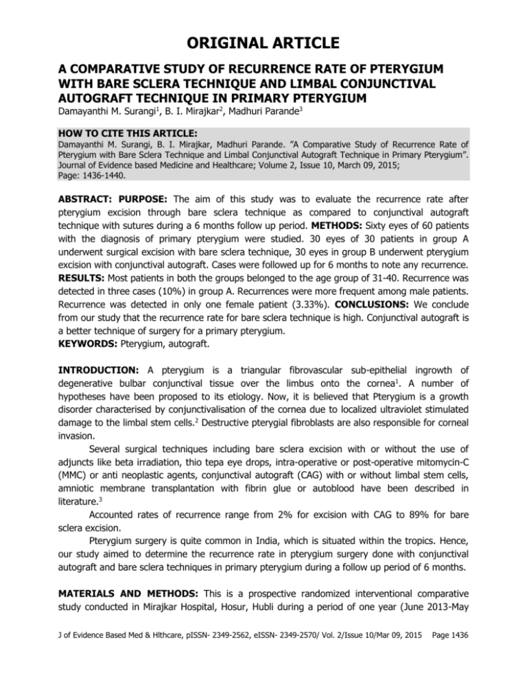
ORIGINAL ARTICLE A COMPARATIVE STUDY OF RECURRENCE RATE OF PTERYGIUM WITH BARE SCLERA TECHNIQUE AND LIMBAL CONJUNCTIVAL AUTOGRAFT TECHNIQUE IN PRIMARY PTERYGIUM Damayanthi M. Surangi1, B. I. Mirajkar2, Madhuri Parande3 HOW TO CITE THIS ARTICLE: Damayanthi M. Surangi, B. I. Mirajkar, Madhuri Parande. ”A Comparative Study of Recurrence Rate of Pterygium with Bare Sclera Technique and Limbal Conjunctival Autograft Technique in Primary Pterygium”. Journal of Evidence based Medicine and Healthcare; Volume 2, Issue 10, March 09, 2015; Page: 1436-1440. ABSTRACT: PURPOSE: The aim of this study was to evaluate the recurrence rate after pterygium excision through bare sclera technique as compared to conjunctival autograft technique with sutures during a 6 months follow up period. METHODS: Sixty eyes of 60 patients with the diagnosis of primary pterygium were studied. 30 eyes of 30 patients in group A underwent surgical excision with bare sclera technique, 30 eyes in group B underwent pterygium excision with conjunctival autograft. Cases were followed up for 6 months to note any recurrence. RESULTS: Most patients in both the groups belonged to the age group of 31-40. Recurrence was detected in three cases (10%) in group A. Recurrences were more frequent among male patients. Recurrence was detected in only one female patient (3.33%). CONCLUSIONS: We conclude from our study that the recurrence rate for bare sclera technique is high. Conjunctival autograft is a better technique of surgery for a primary pterygium. KEYWORDS: Pterygium, autograft. INTRODUCTION: A pterygium is a triangular fibrovascular sub-epithelial ingrowth of degenerative bulbar conjunctival tissue over the limbus onto the cornea1. A number of hypotheses have been proposed to its etiology. Now, it is believed that Pterygium is a growth disorder characterised by conjunctivalisation of the cornea due to localized ultraviolet stimulated damage to the limbal stem cells.2 Destructive pterygial fibroblasts are also responsible for corneal invasion. Several surgical techniques including bare sclera excision with or without the use of adjuncts like beta irradiation, thio tepa eye drops, intra-operative or post-operative mitomycin-C (MMC) or anti neoplastic agents, conjunctival autograft (CAG) with or without limbal stem cells, amniotic membrane transplantation with fibrin glue or autoblood have been described in literature.3 Accounted rates of recurrence range from 2% for excision with CAG to 89% for bare sclera excision. Pterygium surgery is quite common in India, which is situated within the tropics. Hence, our study aimed to determine the recurrence rate in pterygium surgery done with conjunctival autograft and bare sclera techniques in primary pterygium during a follow up period of 6 months. MATERIALS AND METHODS: This is a prospective randomized interventional comparative study conducted in Mirajkar Hospital, Hosur, Hubli during a period of one year (June 2013-May J of Evidence Based Med & Hlthcare, pISSN- 2349-2562, eISSN- 2349-2570/ Vol. 2/Issue 10/Mar 09, 2015 Page 1436 ORIGINAL ARTICLE 2014). Inclusion criteria included patients within age group of 20-65 years coming with primary pterygium willing to come for follow up for a period of 6 months. Exclusion criteria included recurrent pterygium, age less than 20 yrs and poor patient compliance, unwillingness to follow up. Informed Consent was obtained from all patients. All the patients were examined under slit lamp and the type, grade of Pterygium was noted. Visual acuity, refraction, intraocular pressure, ocular motility potency of lacrimal passages and fundus examination findings were noted. Routine investigations like random blood sugar, blood pressure, syringing was done preoperatively. SURGICAL TECHNIQUE: All patients were operated under local anesthesia, instillation of 4% xylocaine and subjconjunctival injection of 2% xylocaine into the body of pterygium. GROUP A: The complete excision of the head of the Pterysium from the Cornea and the body of the Pterygium dissected & excised. The abnormal tissues cleared from the Sclera. The remaining area including the healthy Conjunctiva was left as it is. GROUP B: Excision of Pterygium was done followed by limbal Conjunctival autograft taken from upper temporal quadrant, sutured on bare Sclera using 8/0 Vicryl suture. Post-operative follow up: Post-operatively topical steroid antibiotic eye drops were used every 2 hours for the first operative week and then tapered over the subsequent 5-6 weeks. Systemic antibiotics and analgesics were prescribed for 5 days. Follow up visits were scheduled for post-operative days 1st, 7th, 1stmonth, 3rd month & 6th month of operation. Additional visits were made as and when required. The surgical site was examined under slit lamp on every visit to note any recurrence or any complications. Presence of fibrovascular tissue past the limbus onto the clear cornea in the area of previous pterygium was noted as recurrence. RESULTS: A total of 60 eyes of 60 patients were randomly grouped into groups A & B of 30 eyes each. Group A underwent pterygium surgery with bare sclera technique. Group B underwent pterygium excision with conjunctival autograft. In both the groups, majority of patients belonged to the age group of 31-40 (13/30, 43. 33% in group A, 15/30, 50% in group B). There were 66. 66% males in patients undergoing bare sclera technique and 60% males in patients undergoing conjunctival autograft technique. In group A, i. e. in bare sclera group, among the 30 eyes operated, right eye was operated in 18 cases and left eye in 12 cases. 3 patients (10%) had recurrence at the end of 6 monthly follow up. In group B, i. e. in the conjunctival autograft group, right eye was operated in 14 cases and left eye in 16 cases. One patient (3. 33%) had recurrence during the follow up period. J of Evidence Based Med & Hlthcare, pISSN- 2349-2562, eISSN- 2349-2570/ Vol. 2/Issue 10/Mar 09, 2015 Page 1437 ORIGINAL ARTICLE DISCUSSION: Pterygium is a worldwide condition, typically develops in patients who have been living in hot climates-may represent a response to ultraviolet exposure and possibly to other factors such as chronic surface dryness1. India, being a tropical country, provides suitable conditions for pterygium formation. Pterygium surgery is quite a simple procedure but ironically, simple excision is associated with a high recurrence rate (around 80%). The recurrent lesion may be more aggressive than the initial one1. Recurrent pterygium is a challenging ocular surface disorder that is often resistant to conventional surgeries. There have been various efforts to optimize pterygium surgery with wide range of techniques being in use. The aim of this study was to evaluate the recurrence rate after pterygium excision through bare sclera technique as compared to conjunctival autograft technique with sutures during a 6 months follow up period. The recurrence rate in bare sclera technique was 10% compared to 3. 33% in conjunctival autograft technique. The recurrence rate observed in our study is lower compared to other studies – in both techniques. A recurrence rate of 6. 6% was observed in a study conducted in Vellore, where BST was done on 135 patients4. In a study conducted by Kocamis et al. recurrence was detected in 3 cases (8%) in the study group of 36 who underwent CAT5. Prabhasawat et al. study found a recurrence rate of 10. 9% in primary pterygium patients who underwent CAT.6 Recurrence occurred in 6 (6. 38%) eyes out of 100 CAT cases in a study by Salagar et al.7 In the present study, of the total 4 recurrences in both techniques, 3 were less than 45 yrs of age (2 in BST and 1 in CAT). One 53 yr old patient who underwent BST had recurrence. Also, males showed more propensity towards recurrence (total 3 males; all 3 underwent BST). CONCLUSION: Conjunctival autograft technique of pterygium surgery had a definite upper hand in reducing the recurrence, when compared to the bare sclera technique. Conjunctival autograft is a better technique of surgery for a primary pterygium. BIBLIOGRAPHY: 1. Kanski JJ, Bowling B. “Clinical Ophthalmology”. 7th ed. Elsevier Limited: 2011. p. 163. 2. Dushku N, Reid TW. Immunohistochemical evidence that human pterygia originate from an invasion of Vimentin expressing altered limbal epithelial basal cells. Curr. Eye Res. 1994; 13: 473–81. 3. Hirst LW. The treatment of Pterygium. Surv. Ophthalmology. 2003; 45: 145–80. 4. Quraishy M S, Ebenezer R. Recurrence of pterygium. Review of 135 cases-excision technique. Indian J Ophthalmol 1972; 20: 171-2. 5. Kocamis O, Bilgec M. Evaluation of the recurrence rate for pterygium treated with conjunctival autograft. Graefes Arch Clin Exp Ophthalmol. 2014 May; 252(5): 817-20. 6. Prabhasawat P, Barton K, Burkett G, Tseng SC. Comparison of conjunctival autografts, amniotic membrane grafts, and primary closure for pterygium excision. Ophthalmology. 1997 Jun; 104(6): 974-85. 7. Salagar KM, Biradar KG. Conjunctival Autograft in Primary and Recurrent Pterygium: A Study J of Evidence Based Med & Hlthcare, pISSN- 2349-2562, eISSN- 2349-2570/ Vol. 2/Issue 10/Mar 09, 2015 Page 1438 ORIGINAL ARTICLE Fig. 1 Fig. 2 Fig. 3 J of Evidence Based Med & Hlthcare, pISSN- 2349-2562, eISSN- 2349-2570/ Vol. 2/Issue 10/Mar 09, 2015 Page 1439 ORIGINAL ARTICLE AUTHORS: 1. Damayanthi M. Surangi 2. B. I. Mirajkar 3. Madhuri Parande PARTICULARS OF CONTRIBUTORS: 1. Assistant Professor, Department of Ophthalmology, KIMS, Hubli. 2. Consultant, Department of Ophthalmology, Mirajkar Hospital, Hosur, Hubli. 3. Post Graduate, Department of Ophthalmology, KIMS, Hubli. NAME ADDRESS EMAIL ID OF THE CORRESPONDING AUTHOR: Dr. Damayanthi M. Suranagi, Anugrah, 13th cross, Nirmal Nagar, Dharwad-3. E-mail: damayanthisurangi@gmail.com Date Date Date Date of of of of Submission: 24/02/2015. Peer Review: 25/02/2015. Acceptance: 27/02/2015. Publishing: 04/03/2015. J of Evidence Based Med & Hlthcare, pISSN- 2349-2562, eISSN- 2349-2570/ Vol. 2/Issue 10/Mar 09, 2015 Page 1440
