LAB EXERCISE #2
advertisement
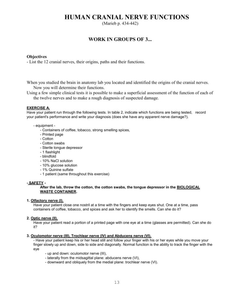
HUMAN CRANIAL NERVE FUNCTIONS (Marieb p. 434-442) WORK IN GROUPS OF 3... Objectives - List the 12 cranial nerves, their origins, paths and their functions. When you studied the brain in anatomy lab you located and identified the origins of the cranial nerves. Now you will determine their functions. Using a few simple clinical tests it is possible to make a superficial assessment of the function of each of the twelve nerves and to make a rough diagnosis of suspected damage. EXERCISE A. Have your patient run through the following tests. In table 2, indicate which functions are being tested, record your patient's performance and write your diagnosis (does she have any apparent nerve damage?). - equipment - Containers of coffee, tobacco, strong smelling spices, - Printed page - Cotton - Cotton swabs - Sterile tongue depressor - 1 flashlight - blindfold - 10% NaCl solution - 10% glucose solution - 1% Quinine sulfate - 1 patient (same throughout this exercise) - SAFETY After the lab, throw the cotton, the cotton swabs, the tongue depressor in the BIOLOGICAL WASTE CONTAINER. 1. Olfactory nerve (I). Have your patient close one nostril at a time with the fingers and keep eyes shut. One at a time, pass containers of coffee, tobacco, and spices and ask her to identify the smells. Can she do it? 2. Optic nerve (II). Have your patient read a portion of a printed page with one eye at a time (glasses are permitted). Can she do it? 3. Oculomotor nerve (III), Trochlear nerve (IV) and Abducens nerve (VI). - Have your patient keep his or her head still and follow your finger with his or her eyes while you move your finger slowly up and down, side to side and diagonally. Normal function is the ability to track the finger with the eye - up and down: oculomotor nerve (III), - laterally from the midsagittal plane: abducens nerve (VI), - downward and obliquely from the medial plane: trochlear nerve (VI). 13 - Pupillary reflex is indicative of the normal function of the oculomotor nerves (III). Have your patient sit with her eyes closed and covered with a blindfold for 2 minutes. Ask her to open her eyes and, as she opens them, flash a light into her eyes. The normal pupillary response is constriction of the pupils. Absence of reflex indicates disorder in the brain, in the afferent nerve (II, optic nerve) or in the efferent nerve (III, oculomotor). 4. Trigeminal nerve (V). - Test for motor function: Have your patient clench his or her teeth. Firmly press your hand under your patient's lower jaw and ask your patient to open her mouth. Normal action allows clenching the teeth and opening the jaw against resistance. - Test for sensory function: 1) Touching patient's cornea with wisp of cotton should elicit blinking (corneal reflex). 2) Have patient close her eyes and whisk a piece of cotton dipped into warm water over the mandibular, maxillary and ophthalmic areas of face. Then wet the cotton with cold water and repeat. Patient should be able to perceive temperature changes in these areas. 5. Facial nerve (VII). - Test for motor function: Have your patient wrinkle his or her forehead, raise one or both eyebrows, inflate cheeks and smile and show teeth. A lack of symmetry reveals abnormal function. - Test for sensory function: Touch the tip of your patient's tongue with a cotton swab dipped in 10% NaCl solution. Repeat using a 10% glucose solution. Your patient should be able to tell the difference. 6. Vestibulocochlear nerve (VIII). - Test for the cochlear portion of the VIII: Whisper a sentence to your patient and ask her to repeat it. - Test for the vestibular portion of the VIII: Ask your patient to look straight ahead and walk a straight line placing the heel of one foot directly in front of the toe of the other foot. 7. Glossopharyngeal nerve (IX) and Vagus nerve (X). - Have your patient say "aaah" while you observe the position of the uvula and the movement of the soft palate. Use a flashlight if necessary. The movement should be the same on both sides so that the uvula remains in the midline. Poor muscular action could indicate faulty innervation by the glossopharyngeal or vagus on the side showing little movement. - Test for motor function of the glossopharyngeal nerve: ask your patient to swallow (swallowing reflex). - Test for sensory function of the glossopharyngeal: test your patient's ability to taste on the posterior third of the tongue: apply water and a weak quinine solution to the back of your partner tongue. Can she feel the difference? 8. Spinal accessory nerve (XI). Have your patient sit and push gently but firmly on her shoulders. Ask her to raise them against this resistance (note that this does not mean to stand up). This will test the strength of the trapezius muscles. Now place your hands firmly on either side of your patient's head. Ask her to turn her head both way against this resistance. This tests the strength of the sternocleidomastoid muscles. Both the trapezius and sternocleidomastoid are innervated by the spinal accessory nerve. 9. Hypoglossal nerve (XII). Ask your patient to stick out her tongue. Normal action is the ability to keep the tongue straight. Deviation of the tongue toward one side indicates injury to the nerve on that side. 14 Table 2: Clinical tests for cranial nerve functions. PATIENT NAME: STAFF: NAME & SIGNATURE: TES T# CRANIAL NERVES & FUNCTIONS TESTED (write in this column which function is tested) 1 (I): Olfactory 2 (II): OptIc 3 (III): Oculomotor (IV): Trochlear (VI): Abducens 4 (V): Trigeminal 5 (VII): Facial 6 (VIII): Vestibulo-cochlear 7 (IX): Glossopharyngeal (X): Vagus 8 (XI): Spinal accessory 9 (XII): Hypoglossal 15 OBSERVATIONS DIAGNOSIS (nerve damage?)


